

Reproductive System, Male
Testis - Mineralization
Narrative
Comment:
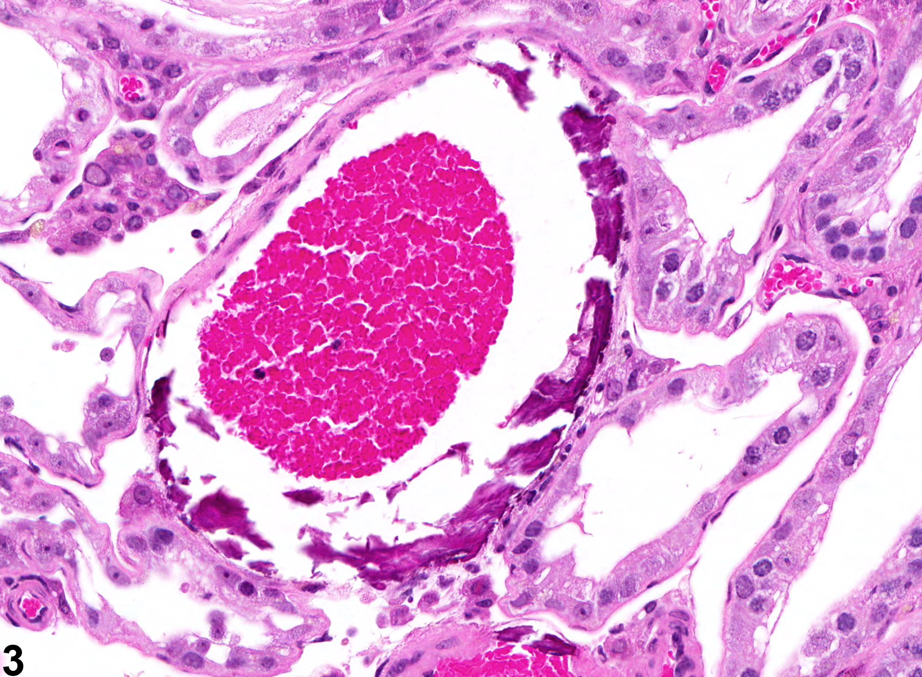
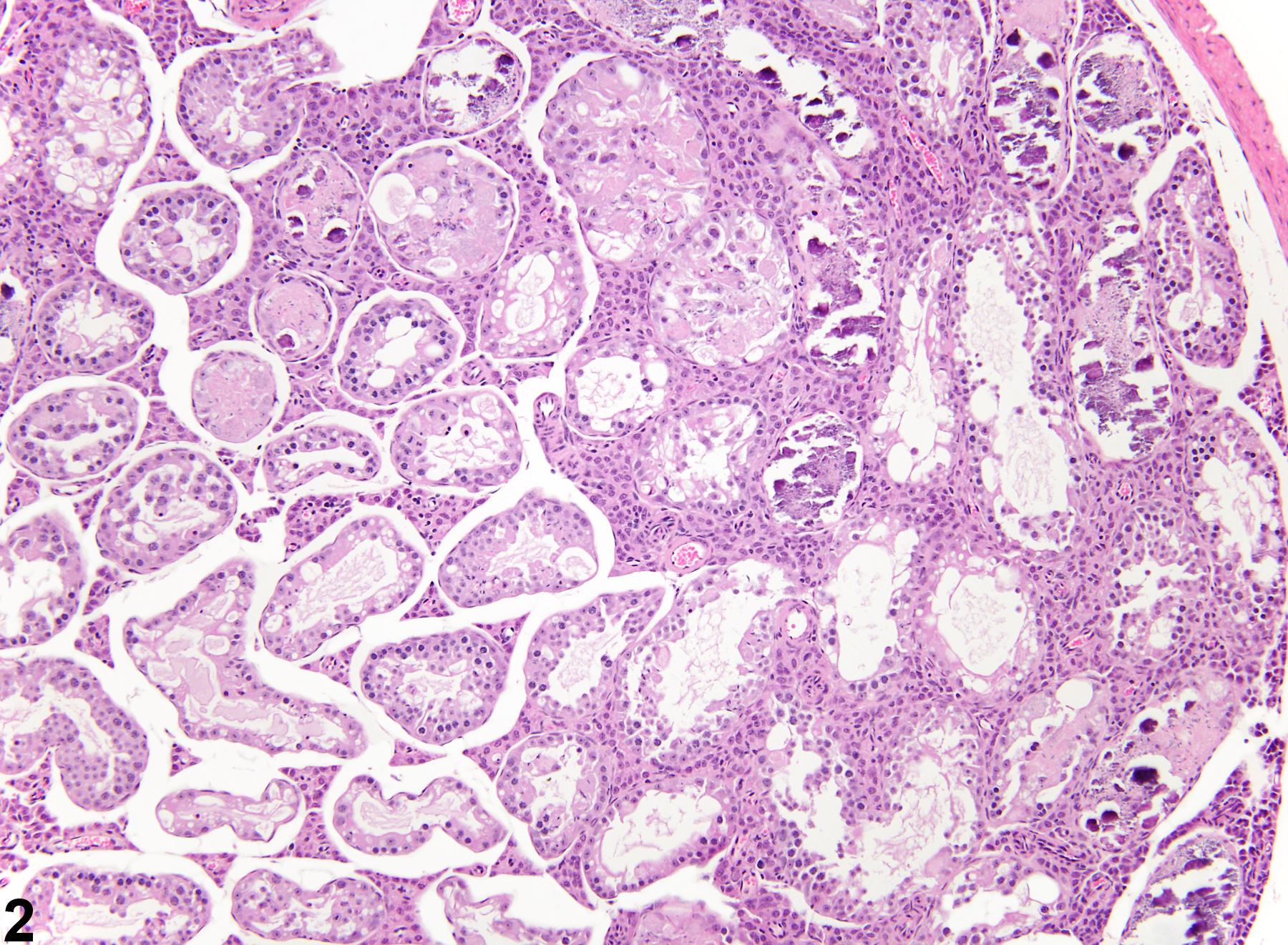
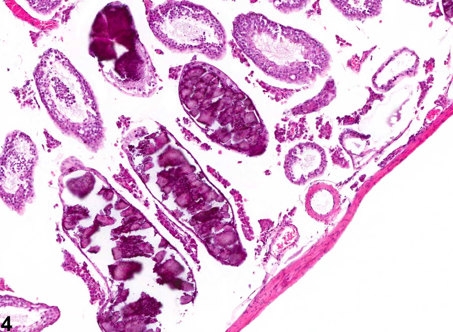
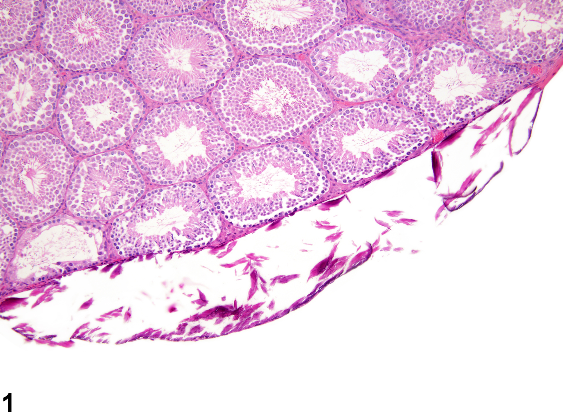
Dystrophic mineralization is a sequel of injury and may involve the capsule (Figure 1), blood vessels (Figure 3), or seminiferous tubules (Figure 2 and Figure 4). It is characterized by accumulation of basophilic, fine to coarsely granular to amorphous, laminated material, with or without distortion of the tissue architecture. It is often a sequel to sperm stasis within the seminiferous tubules. Mineralization is an age-related finding and can be seen unilaterally or bilaterally.
Recommendations:
Mineralization should be diagnosed, graded, and classified with respect to location through appropriate use of site modifiers (e.g., blood vessel, capsule, seminiferous tubule). If present in both testes, it should be diagnosed as bilateral. Associated lesions, such as degeneration, necrosis, or inflammation should be diagnosed separately if warranted by severity.
Testis - Mineralization in a male B6C3F1 mouse from a chronic study. Mineralization involving the testicular capsule.
All Images

Testis - Mineralization in a male B6C3F1 mouse from a chronic study. Mineralization involving the testicular capsule.

Testis - Mineralization in a male B6C3F1 mouse from a subchronic study. Mineralization involving seminiferous tubules.

Testis - Mineralization in a male F344/N rat from a chronic study. Mineralization involving blood vessels.

Testis - Mineralization in a male B6C3F1 mouse from a chronic study. Mineralized seminiferous tubules.