

Reproductive System, Male
Testis - Rete Hyperplasia
Narrative
Boorman GA, Chapin RE, Mitsumori K. 1990. Testis and epididymis. In: Pathology of the Fischer Rat: Reference and Atlas (Boorman GA, Eustis SL, Elwell MR, Montgomery CA, MacKenzie WF, eds). Academic Press, San Diego, 405-418.
Bullock BC, Newbold RR, McLachlan JA. 1988. Lesions of testis and epididymis associated with prenatal diethylstilbestrol exposure. Environ Health Perspect 77:29-31.
Abstract: http://www.ncbi.nlm.nih.gov/pubmed/3289905Creasy DM. 2012. Reproduction of the rat, primate, dog and pig. In: Background Lesions in Laboratory Animals: A Colour Atlas (McKinnes E, ed). Saunders Elselvier, Edinburgh. 101-122.
Abstract: http://www.sciencedirect.com/science/book/9780702035197Frith CH, Ward JM. 1988. Color Atlas of Neoplastic and Non-neoplastic Lesions in Aging Mice. Elsevier, Amsterdam.
Abstract: http://www.informatics.jax.org/frithbook/Gordon LR, Majka JA, Boorman GA. 1996. Spontaneous nonneoplastic and neoplastic lesions and experimentally induced neoplasms of the testes and accessory sex glands. In: Pathobiology of the Aging Mouse, Vol 1 (Mohr U, Dungworth DL, Capen CC, Carlton WW, Sundberg JP, Ward JM, eds). ILSI Press, Washington, DC, 421-441.
Abstract: http://catalog.hathitrust.org/Record/008994685Maekawa A, Hayashi Y. 1987. Adenomatous hyperplasia, rete testis, rat. In: Monographs on Pathology of Laboratory Animals: Genital System (Jones TC, Mohr U, Hunt RD, eds). Springer, Berlin, 234-236.
Abstract: http://www.springer.com/medicine/pathology/book/978-3-642-72552-4Mitsumori K, Elwell MR 1988. Proliferative lesions in the male reproductive system of F344 rats and B6C3F1 mice: Incidence and classification. Environ Health Perspect 77:11-21.
Abstract: http://www.ncbi.nlm.nih.gov/pubmed/3289903Rehm S, Harlemann J, Cary M, Creasy D, Ettlin R, Eustis S, Foley G, LeNet J, Maekawa A, Mitsumori K, McConnell RF, Reznik G. 2001. Male genital system. In: International Classification of Rodent Tumors: The Mouse (Mohr U, ed). Springer, Berlin, 163-210.
Abstract: http://www.springer.com/medicine/pathology/book/978-3-642-08422-5Yoshitomi K, Morii S. 1984. Benign and malignant epithelial tumors of the rete testis in mice. Vet Pathol 21:300-303.
Abstract: http://www.ncbi.nlm.nih.gov/pubmed/6730219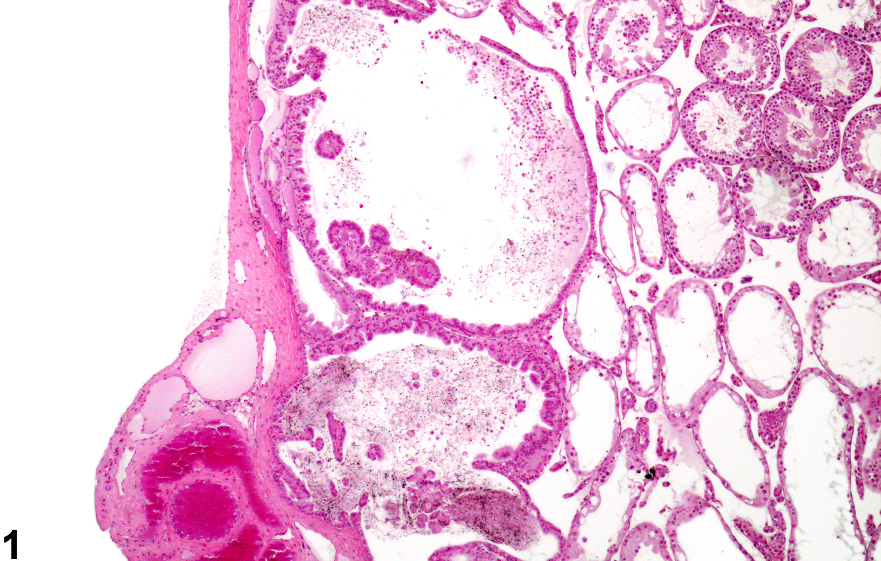
Testis, Rete testis - Hyperplasia in a male CD1 mouse. Proliferation of lining epithelium is present in the dilated rete testis. (Photograph courtesy of Dr. D. Creasy.)
All Images

Testis, Rete testis - Hyperplasia in a male CD1 mouse. Proliferation of lining epithelium is present in the dilated rete testis. (Photograph courtesy of Dr. D. Creasy.)

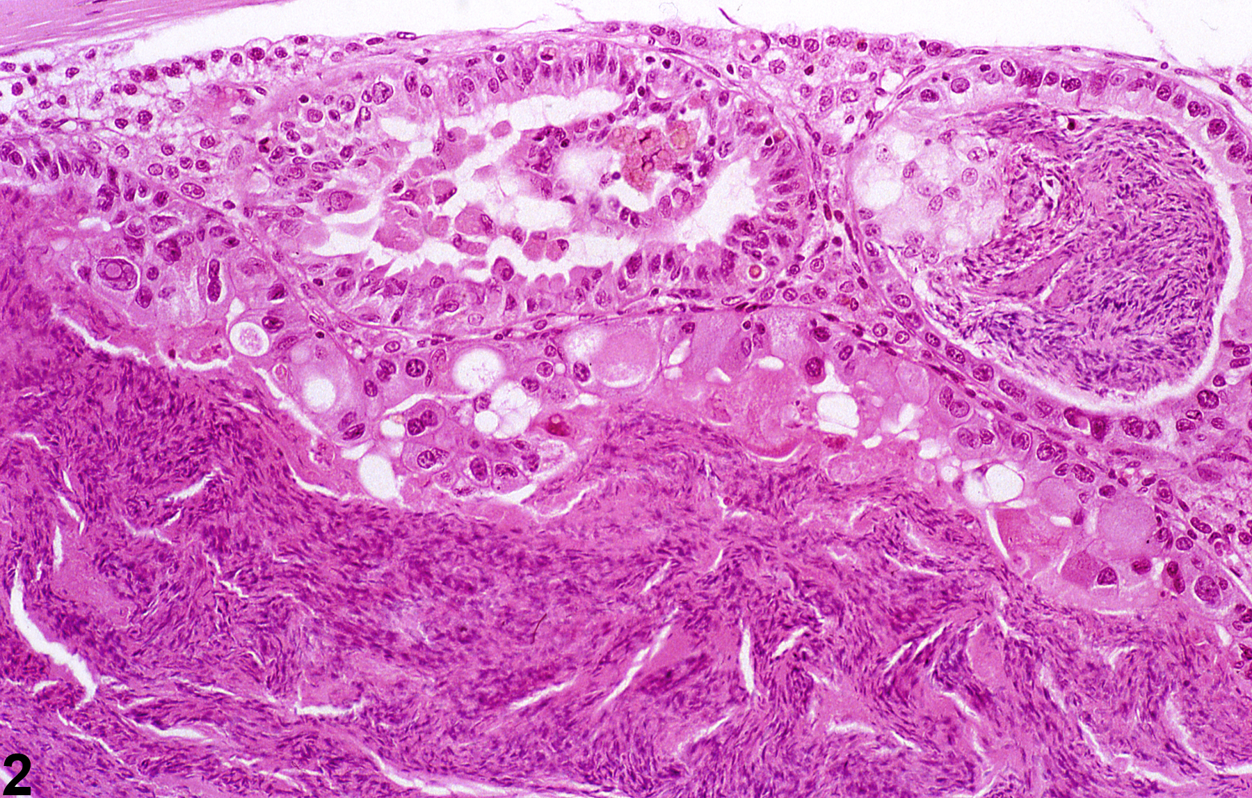
Testis, Rete testis - Hyperplasia in a male CD1 mouse. Hyperplasia of the lining epithelium of the rete testis. (Photograph courtesy of Dr. D. Creasy.)