U.S. flag

An official website of the United States government

Dot gov

The .gov means it's official.
Federal government websites often end in .gov or .mil. Before sharing sensitive information, make sure you're on a federal government site.

Https

The site is secure.
The https:// ensures that you are connecting to the official website and that any information you provide is encrypted and transmitted securely.

Skip to Main Navigation
Skip to Page Content
Skip to Atlas Navigation

Testis - Spermatocele

Image of spermatocele in the testis from a male B6C3F1 mouse in a chronic study
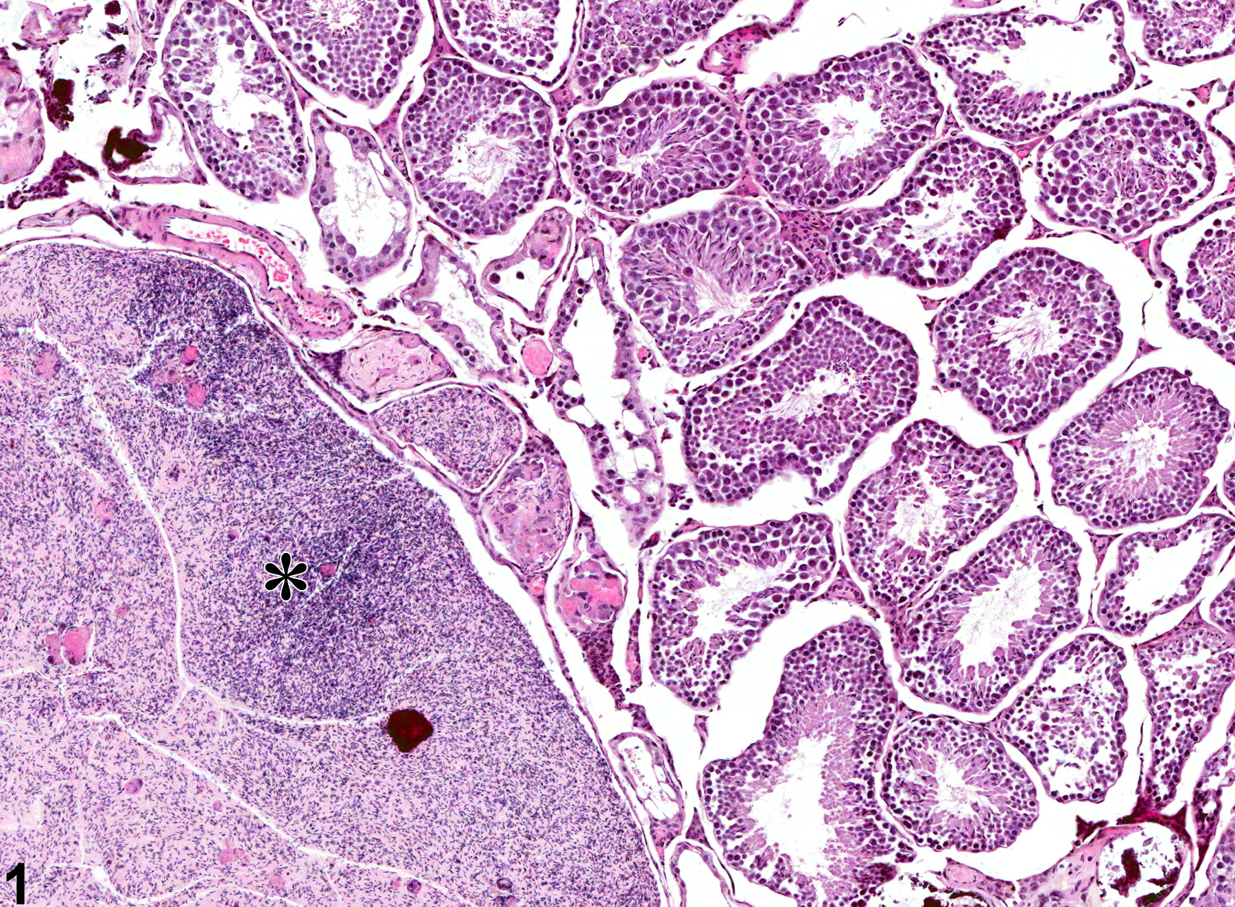
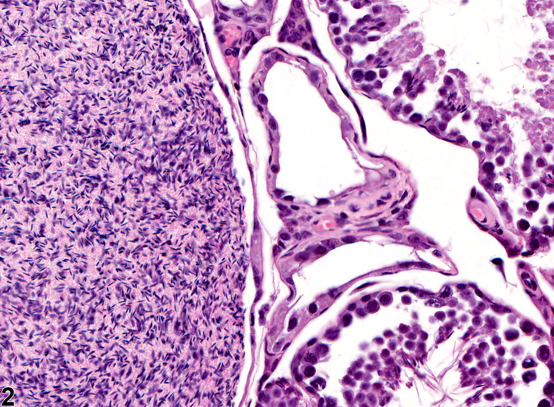
Testis - Spermatocele in a male B6C3F1 mouse from a chronic study. There is a large spermatocele in the testis (asterisk).
Figure 1 of 2
Image of spermatocele in the testis from a male B6C3F1 mouse in a chronic study
Testis - Spermatocele in a male B6C3F1 mouse from a chronic study. Higher magnification of the spermatocele in Figure 1.
Figure 2 of 2
next arrow

comment:

A spermatocele refers to the cystic accumulation or impaction of spermatozoa in a seminiferous tubule or duct causing it to increase to more than twice its normal diameter. If the diameter is less than this, the term “sperm stasis” should be used (see Testis - Sperm Stasis). If the accumulation is accompanied by an inflammatory reaction, the term “sperm granuloma” should be used (see Testis - Sperm Granuloma). Spermatoceles occur more commonly in the epididymis, but they are also sometimes seen within the testis, and particularly within the subcapsular rete testis of the mouse ( Figure 1image opens in a pop-up window , asterisk and Figure 2image opens in a pop-up window ). Spermatoceles can occur in association with reduced fluid absorption in the efferent ducts leading to sperm impaction and obstruction within the rete testis and adjacent seminiferous tubules. This can be a chemically induced finding, but in general spermatoceles are incidental, age-related findings. An association between age-related germ cell degeneration and spermatocele formation has been described.

recommendation:

Spermatocele should be diagnosed when present, but need not be graded. They should be discussed in the pathology narrative only if they are considered treatment related. Bilateral involvement should be recorded when present.

references:

Creasy D, Bube A, de Rijk E, Kandori H, Kuwahara M, Masson R, Nolte T, Reams R, Regan K, Rehm S, Rogerson P, Whitney K. (2012). Proliferative and nonproliferative lesions of the rat and mouse male reproductive system. Toxicol Pathol 40:40S-121S.
Abstract: https://doi.org/10.1177/0192623312454337

Gordon LR, Majka JA, Boorman GA. 1996. Spontaneous nonneoplastic and neoplastic lesions and experimentally induced neoplasms of the testes and accessory sex glands. In: Pathobiology of the Aging Mouse, Vol 1 (Mohr U, Dungworth DL, Capen CC, Carlton WW, Sundberg JP, Ward JM, eds). ILSI Press, Washington, DC, 421-441.
Abstract: http://catalog.hathitrust.org/Record/008994685

Itoh M, Li XQ, Miyamoto K, Takeuchi Y. 1999. Degeneration of the seminiferous epithelium with aging is a cause of spermatoceles. Int J Androl 22:91-96.
Abstract: http://www.ncbi.nlm.nih.gov/pubmed/10194640