

Musculoskeletal System
Bone - Callus
Narrative
Brighton CT, Hunt RM. 1991. Early histologic and ultrastructural changes in medullary fracture callus. J Bone Joint Surg 73:832-847.
Abstract: http://www.ncbi.nlm.nih.gov/pubmed/2071617Leininger JR, Riley MGI. 1990. Bones, joints, and synovia. In: Pathology of the Fischer Rat: Reference and Atlas (Boorman G, Eustis SL, Elwell MR, Montgomery CA, MacKenzie WF, eds). Academic Press, San Diego, 209-226.
Long PH, Leininger JR. 1999. Bones, joints, and synovia. In: Pathology of the Mouse (Maronpot R, Boorman G, Gaul BW, eds). Cache River Press, St Louis, 645-678.
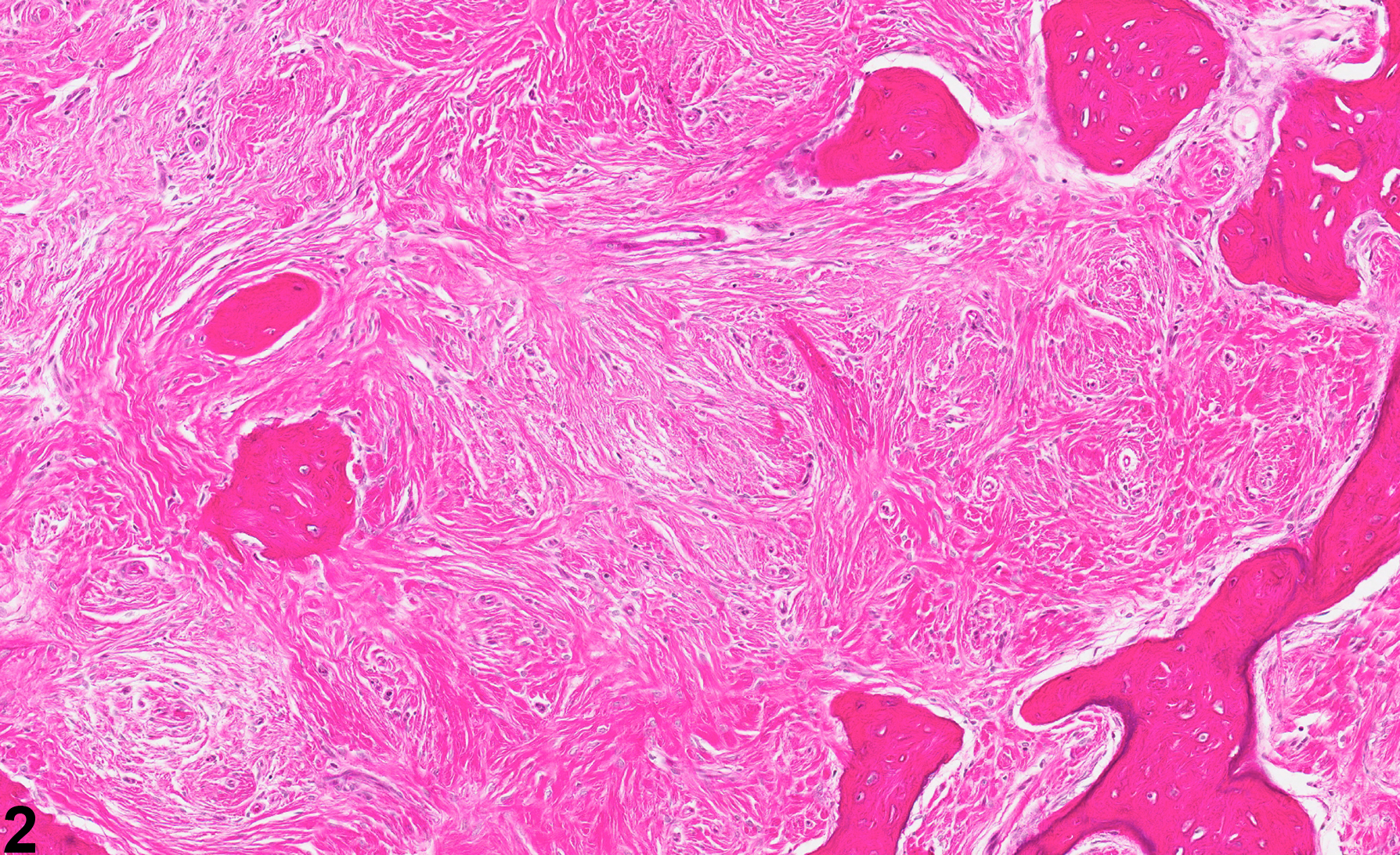
Bone - Callus in a male Wistar rat from a chronic study. There is callus formation surrounding a rib, characterized by a marked proliferative fibrous connective tissue response and bony remodeling.
All Images

Bone - Callus in a male Wistar rat from a chronic study. There is callus formation surrounding a rib, characterized by a marked proliferative fibrous connective tissue response and bony remodeling.

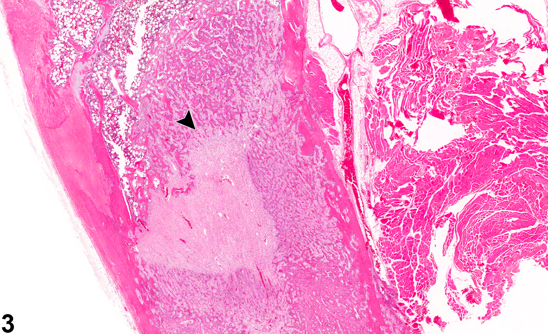
Bone - callus in a male Wistar rat from a chronic study (higher magnification of figure 1). Section of a bone callus, characterized by a marked proliferative fibrous connective tissue response and bony remodeling.

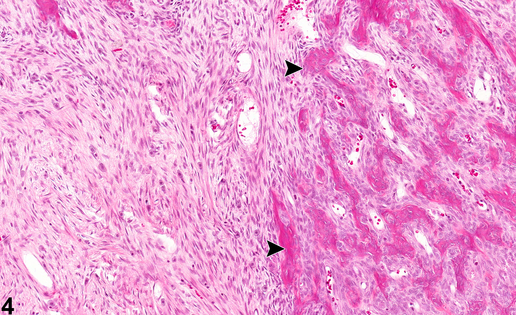
Bone - callus in a male F344/N rat from a chronic study. This section of long bone with a callus is characterized by a focal fibroblastic response (arrowhead).

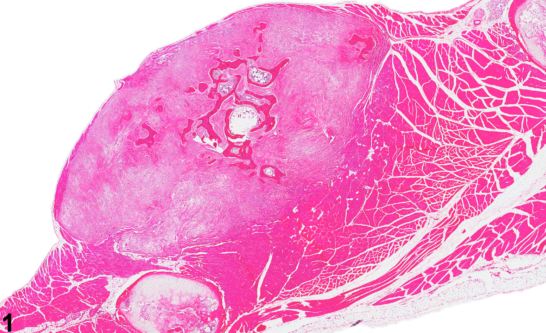
Bone - callus in a male F344/N rat from a chronic study (higher magnification of figure 3). This bone callus has a focal fibroblastic response and associated osteoid deposition and mineralization (arrowheads).