

Musculoskeletal System
Bone - Necrosis
Narrative
Leininger JR, Riley MGI. 1990. Bones, joints, and synovia. In: Pathology of the Fischer Rat: Reference and Atlas (Boorman G, Eustis SL, Elwell MR, Montgomery CA, MacKenzie WF, eds). Academic Press, San Diego, 209-226.
Long PH, Leininger JR. 1999. Bones, joints, and synovia. In: Pathology of the Mouse (Maronpot R, Boorman G, Gaul BW, eds). Cache River Press, St Louis, 645-678.
Yamasaki K. 1993. Morphological studies on the bone and cartilage of laboratory animals. Exp Anim 42:11-21.
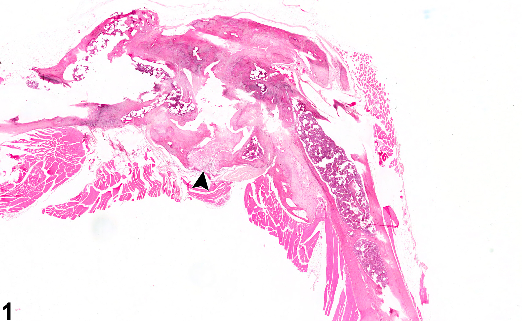
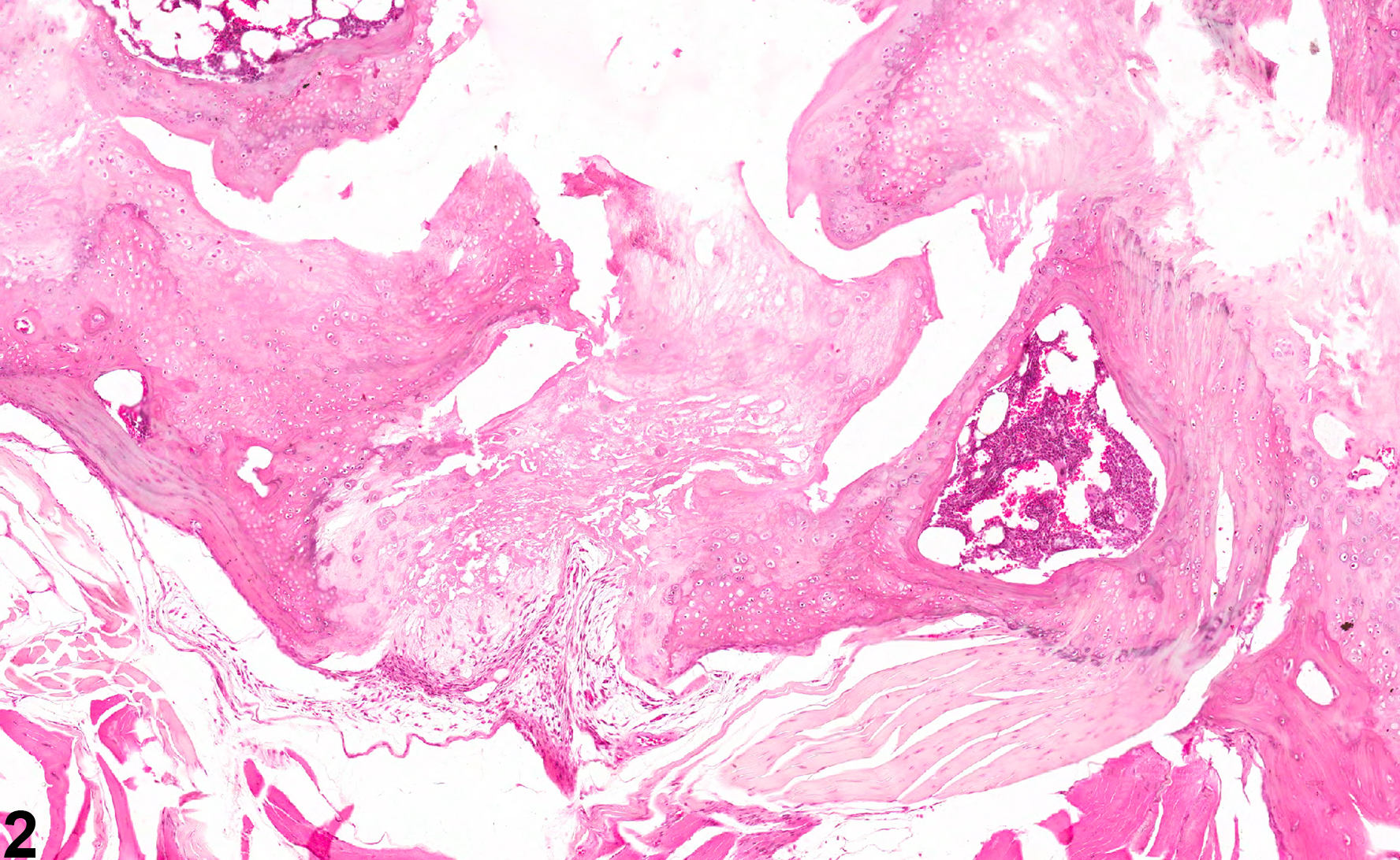
Bone, Femur - Necrosis in a male B6C3F1/N mouse from a chronic study. This section of a femur demonstrates focal necrosis (arrowhead) associated with marked joint degeneration.
All Images

Bone, Femur - Necrosis in a male B6C3F1/N mouse from a chronic study. This section of a femur demonstrates focal necrosis (arrowhead) associated with marked joint degeneration.

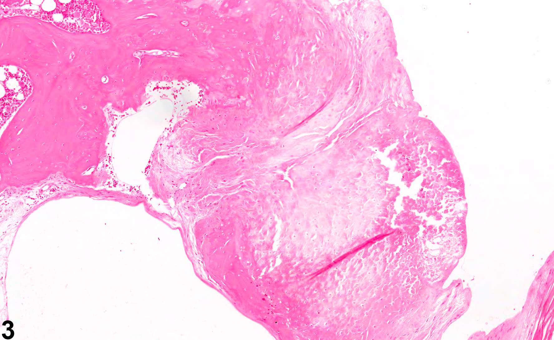
Bone, Femur - Necrosis in a male B6C3F1/N mouse from a chronic study (higher magnification of Figure 1). This section of femur illustrates focal necrosis associated with marked joint degeneration.

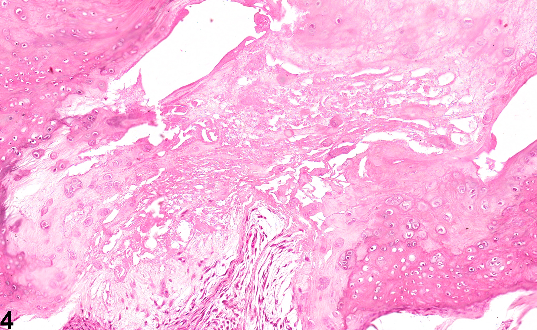
Bone, Femur - Necrosis in a male F344/N rat from a chronic study. Focal necrosis is associated with marked joint degeneration in a section of femur.

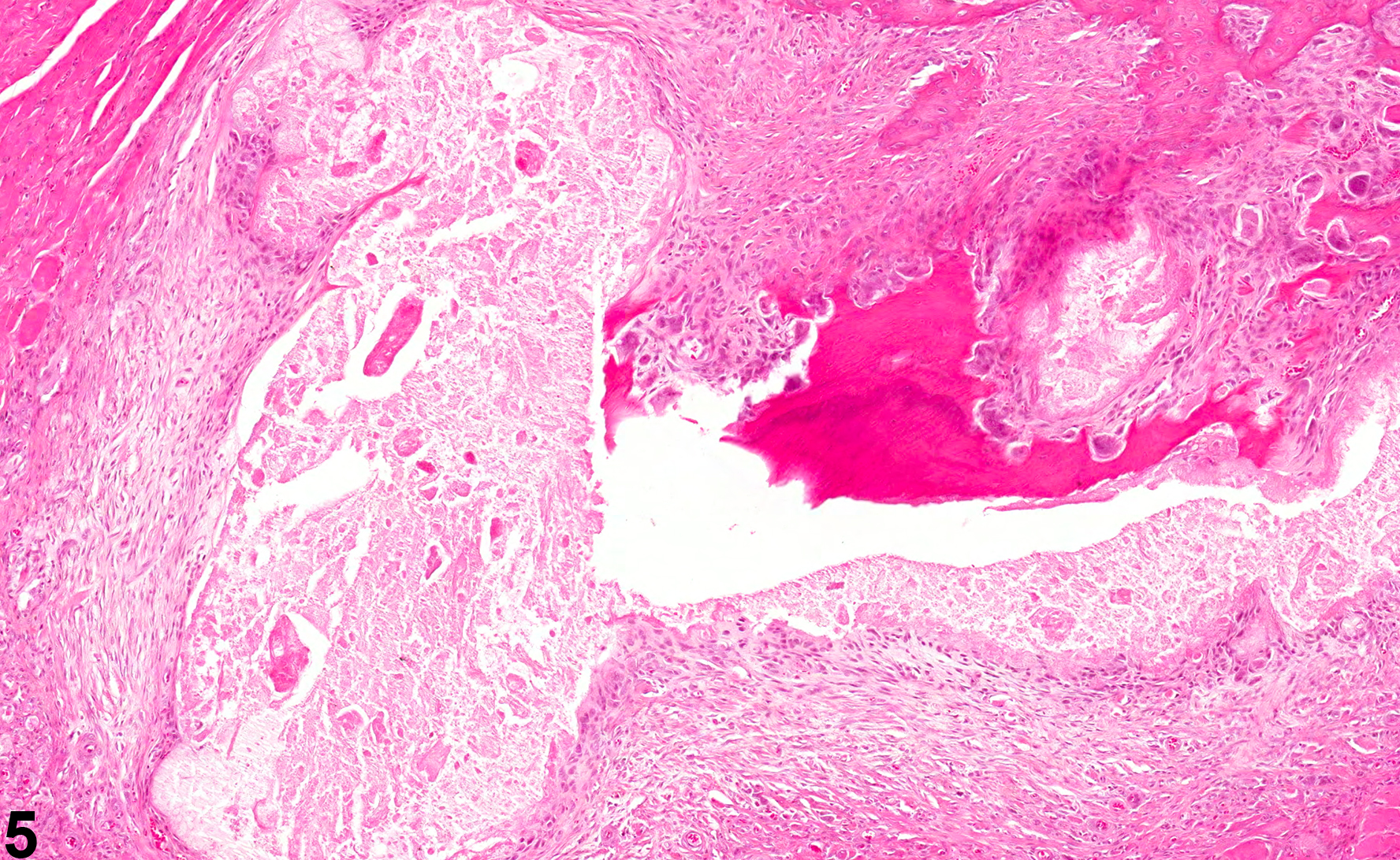
Bone, Femur - Necrosis in a male B6C3F1/N mouse from a chronic study. In this section of femur, focal necrosis is associated with marked joint degeneration.

Bone, Vertebra - Necrosis in a male F344/N rat from a chronic study. This section of a vertebra demonstrates marked necrosis characterized by loss of bony architecture, fragmentation, and debris associated with significant fibroplasia and bony remodeling.