

Musculoskeletal System
Skeletal Muscle - Edema
Narrative
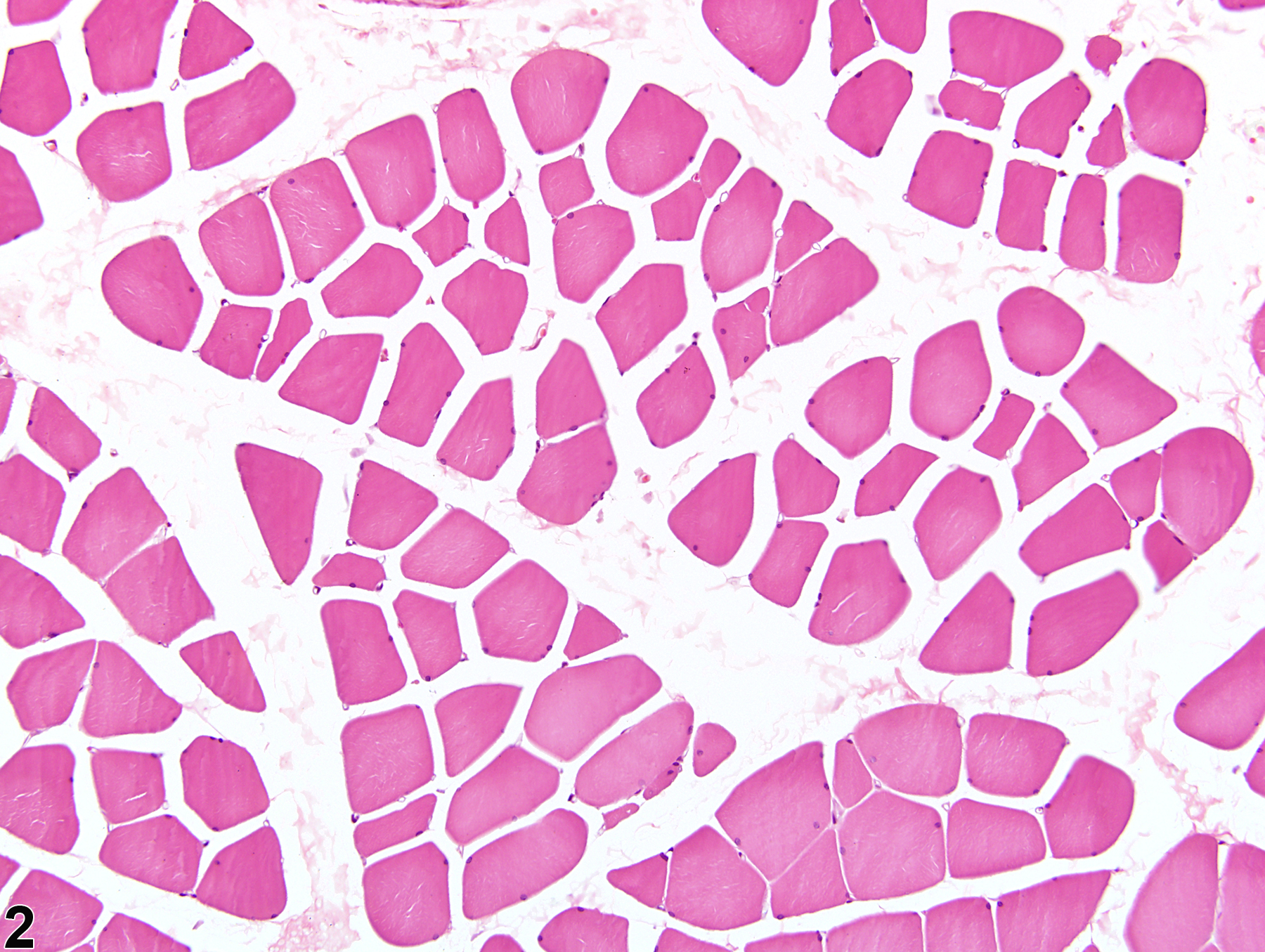
While edema can occur as a primary lesion (Figure 1, Figure 2, Figure 3, and Figure 4), it most commonly occurs secondary to necrosis and inflammation; regions of hemorrhage are often accompanied by edema. Intramuscular edema can be seen in association with autoimmune conditions, such as polymyositis and dermatomyositis; mild injuries; infectious myositis; subacute denervation; compartment syndrome; and rhabdomyolysis; it can also be seen as a transient, physiologic finding during and briefly following muscle exercise.
Berridge BR, Van Vleet JF, Herman E. 2013. Cardiac, vascular, and skeletal muscle systems. In: Haschek and Rousseaux’s Handbook of Toxicologic Pathology, 3rd ed (Haschek WM, Rousseaux CG, Wallig MA, Bolon B, Ochoa R, Mahler MW, eds). Elsevier, Amsterdam, 1635-1665.
Greaves P, Chouinard L, Ernst H, Mecklenburg L, Pruimboom-Brees IM, Rinke M, Rittinghausen S, Thibault S, von Erichsen J, Yoshida T. 2013. Proliferative and non-proliferative lesions of the rat and mouse soft tissue, skeletal muscle, and mesothelium. J Toxicol Pathol 26(3 suppl):1S-26S.
Abstract: http://www.ncbi.nlm.nih.gov/pubmed/25035576Mosier DA. 2007. Vascular disorders and thrombosis. In: Pathologic Basis of Veterinary Disease (McGavin MD, Zachary JF, eds). Mosby Elsevier, St Louis, 63-100.
Shimazaki C, Ochiai N, Uchida R, Fuchida S-I, Okano A, Ashihara E, Inaba T, Fujita N, Nakagawa M. 2003. Intramuscular edema as a complication of treatment with imatinib. Leukemia 17:804-805.
Full Text: http://www.nature.com/leu/journal/v17/n4/full/2402868a.htmlVahle JL, Leininger JR, Long PH, Hall DG, Ernst H. 2013. Bone, muscle, and tooth. In: Toxicologic Pathology: Nonclinical Safety Assessment (Sahota PS, Popp JA, Hardisty JF, Gopinath C, eds). CRC Press, Boca Raton, FL, 561-587.
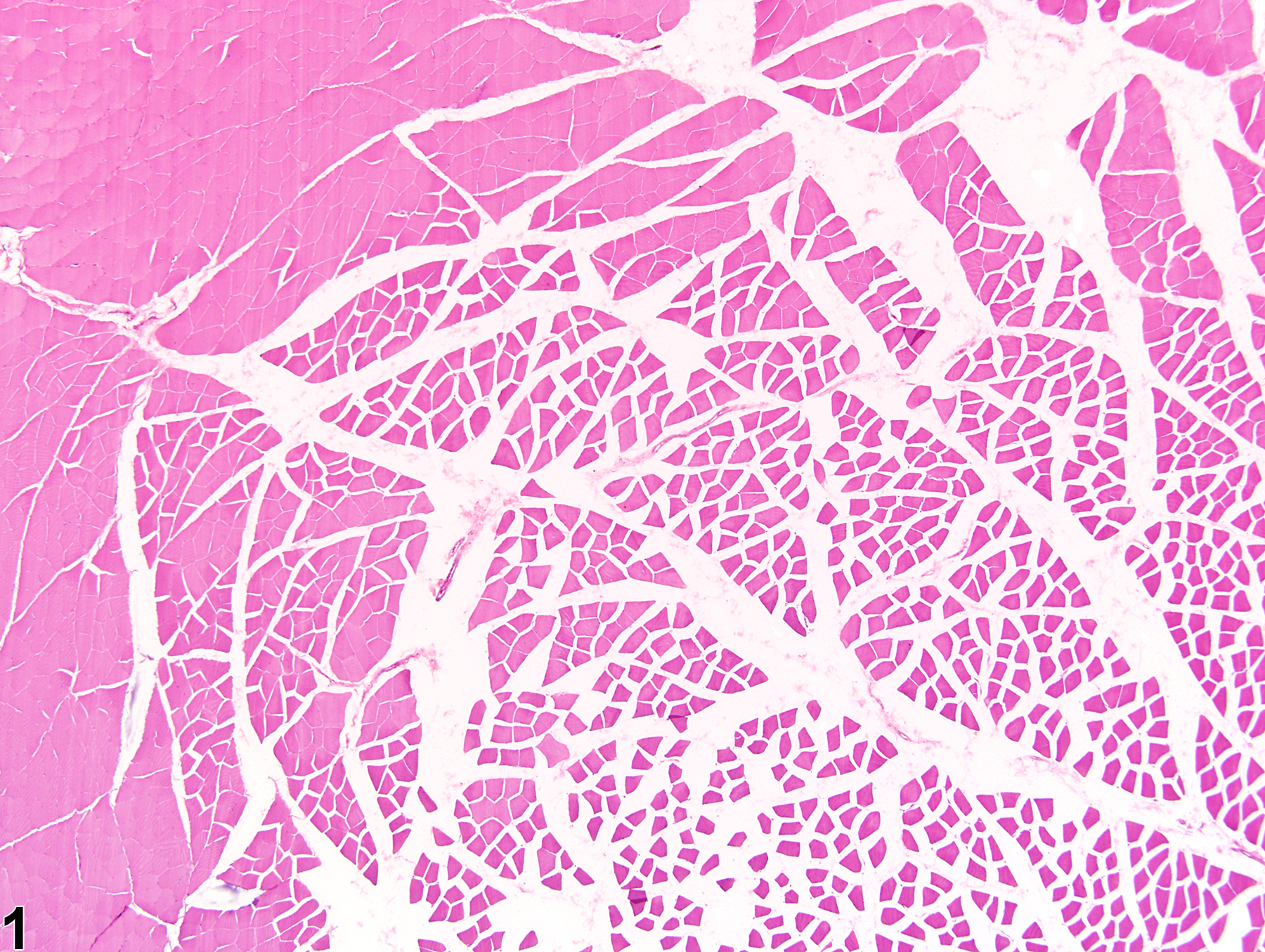
Skeletal muscle - Edema in a male F344/N rat from a chronic study. Muscle fibers and muscle bundles are separated by expanded interstitial spaces filled with pale pink material.
All Images

Skeletal muscle - Edema in a male F344/N rat from a chronic study. Muscle fibers and muscle bundles are separated by expanded interstitial spaces filled with pale pink material.

Skeletal muscle - Edema in a male F344/N rat from a chronic study (higher magnification of Figure 1). There are separated muscle fibers and muscle bundles due to an accumulation of pale pink interstitial material.

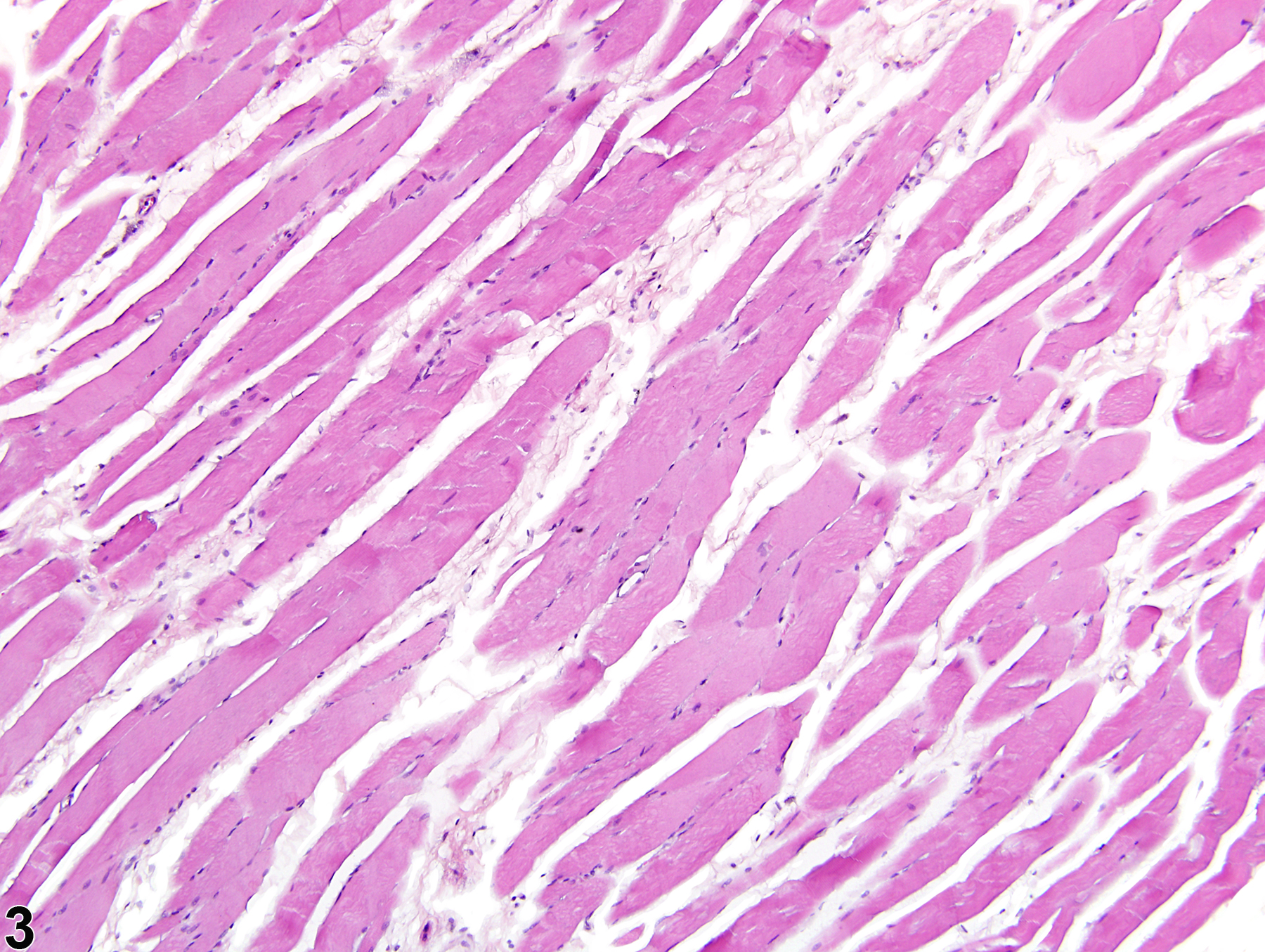
Skeletal muscle - Edema in a male F344/N rat from a chronic study. A longitudinal section of skeletal muscle shows expansion of the interstitial spaces due to an accumulation of poorly stained edema fluid.

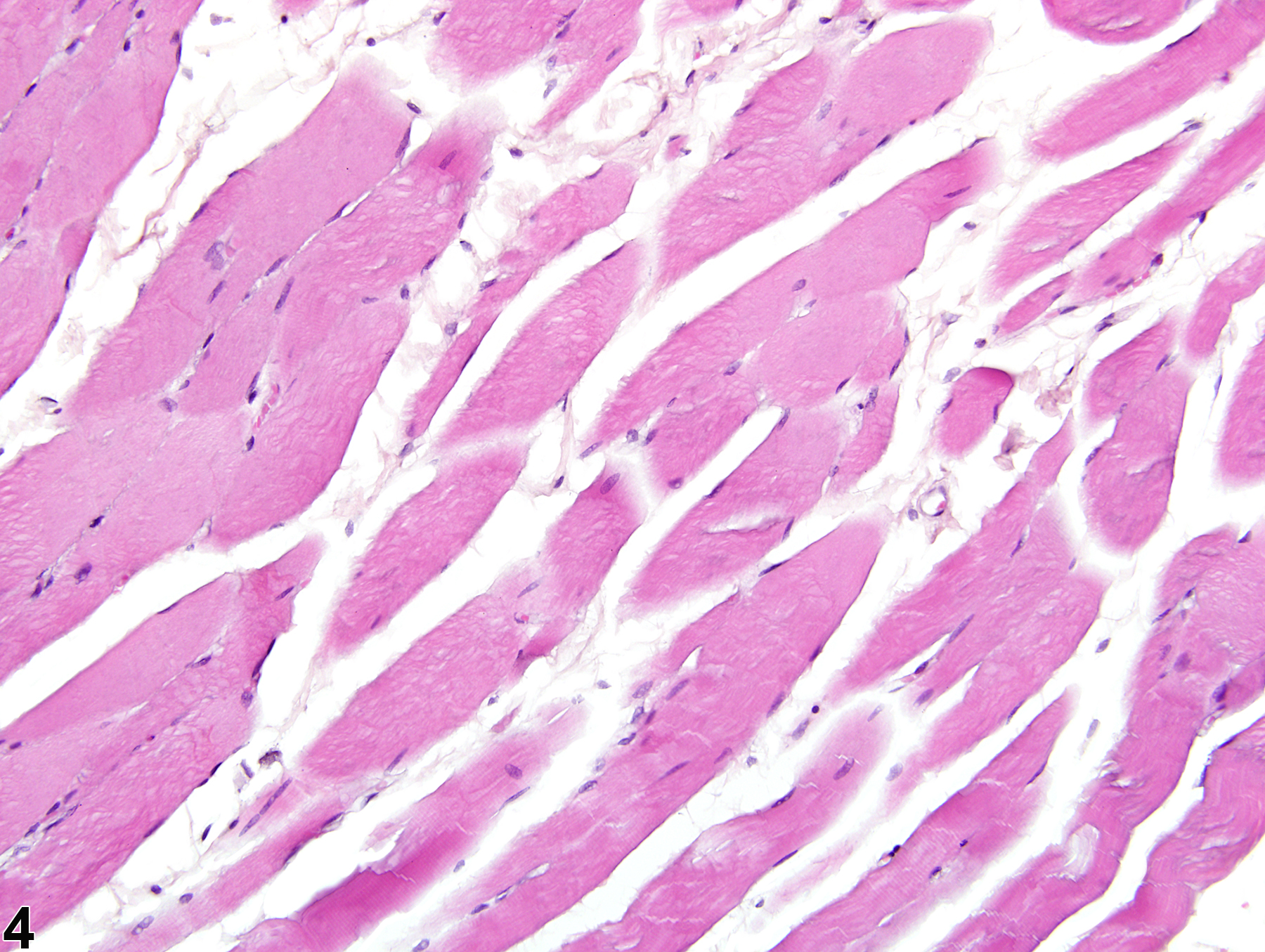
Skeletal muscle - Edema in a male F344/N rat from a chronic study (higher magnification of Figure 3). Interstitial spaces are expanded by poorly stained edema fluid.

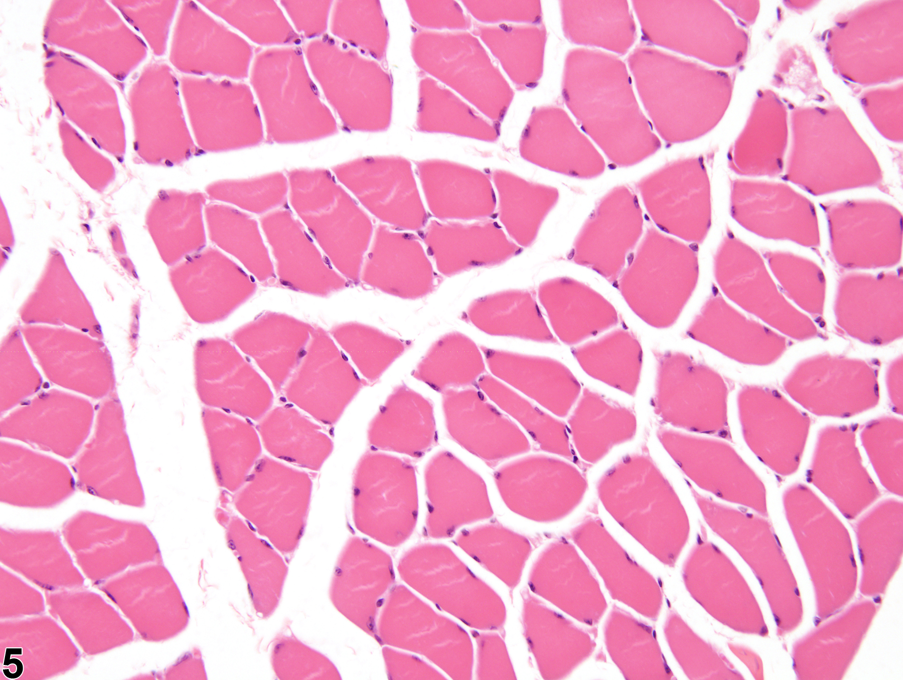
Skeletal muscle - Normal in a male B6C3F1/N mouse from a subchronic study. Expanded interstitial space caused by fixation and/or sectioning artifact.