U.S. flag

An official website of the United States government

Dot gov

The .gov means it's official.
Federal government websites often end in .gov or .mil. Before sharing sensitive information, make sure you're on a federal government site.

Https

The site is secure.
The https:// ensures that you are connecting to the official website and that any information you provide is encrypted and transmitted securely.

Skip to Main Navigation
Skip to Page Content
Skip to Atlas Navigation

Brain - Dysplasia

Image of dysplasia in the brain from a female F344/N rat in a 2 year study
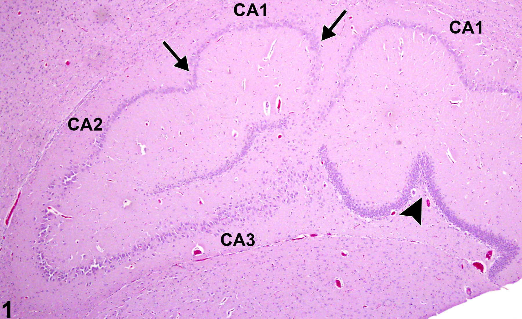
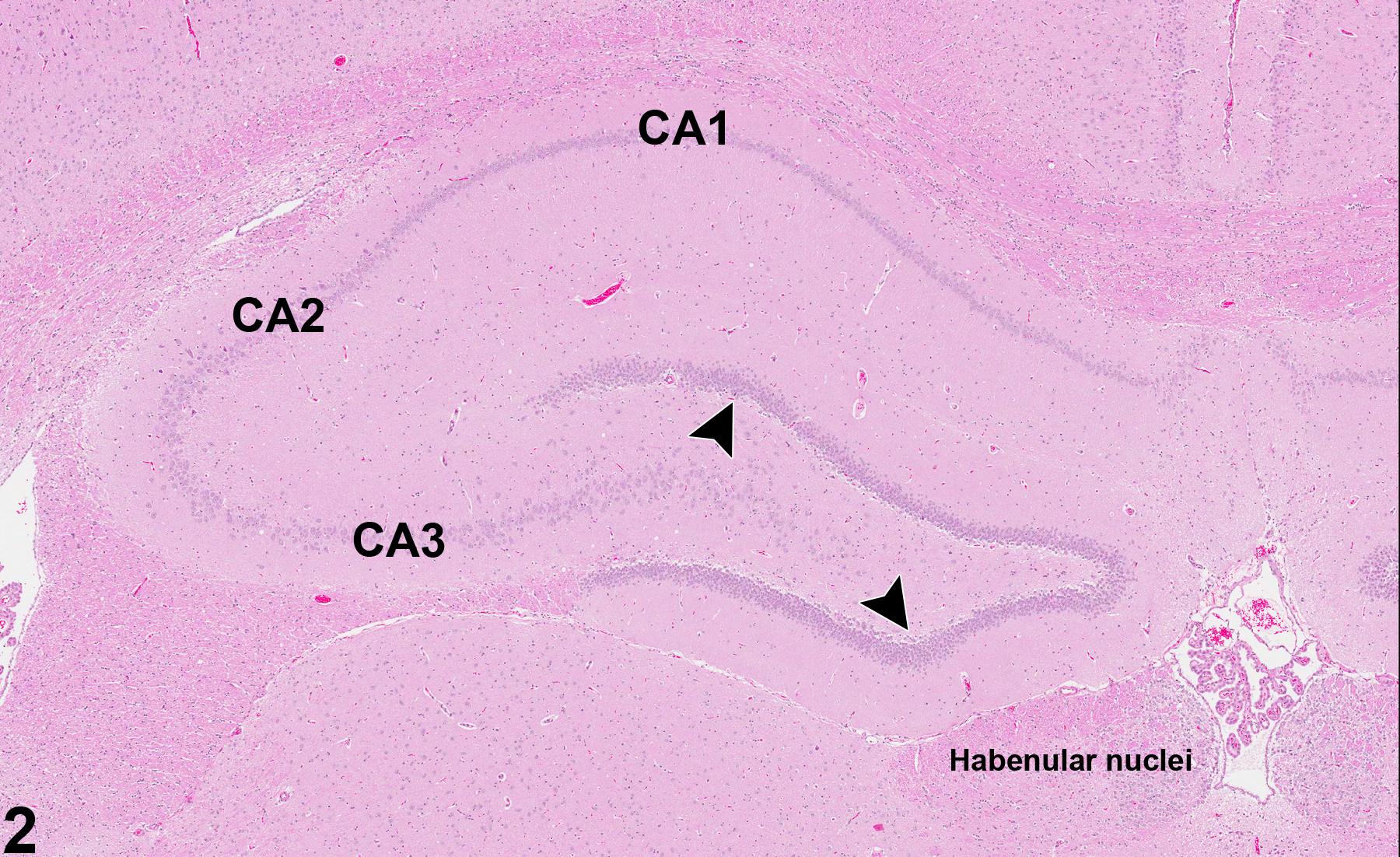
Incidental hippocampal dysplasia in a female F344/N rat from a 2-year study. Note the abnormal undulation of the CA1 region (arrows) and the distortion of the dentate gyrus (arrowhead) compared with that in Figure 2.
Figure 1 of 2
Image of dysplasia (normal comparison) in the brain from a rat
Normal hippocampus in a rat. Dentate gyrus is indicated by the arrowheads. Image provided courtesy Dr. D. Rao.
Figure 2 of 2
next arrow

comment:

With the increasing use of generational toxicity studies, recognizing neural dysplasia is becoming increasingly important. Dysplasias include developmental abnormalities in size, organization, and location of various elements of the neural structures. Figure 1image opens in a pop-up window shows, at low magnification, the appearance of uncommonly recognized incidental rodent hippocampal dysplasia. In this dramatic example, note the abnormal undulating CA1 region (arrows) and malformed dentate gyrus (arrowhead). CA3 appears relatively normal. Figure 2image opens in a pop-up window , labeled for comparison, shows normal rat hippocampal structure. Dysplastic neuronal lesions are usually encountered in the cortex, hippocampus, and cerebellum, but any part of the brain can be affected. In most cases, effects, if any, are not detected clinically but could account for abnormal clinical neurologic signs such as seizures in individual animals. Models of neural dysplasia in rats include treatment with methylazoxymethanol acetate followed by induction of epileptiform seizures by pilocarpine.

recommendation:

Whenever present in NTP studies, this lesion should be diagnosed and the subsite included in the diagnosis. Severity grading is not required.

references:

Colciaghi F, Finardi A, Frasca A, Balosso S, Nobili P, Carriero G, Locatelli D, Vezzani A, Battaglia G. 2011. Status epilepticus-induced pathologic plasticity in a rat model of focal cortical dysplasia. Brain 134:2828-2843.
Abstract: http://www.ncbi.nlm.nih.gov/pubmed/21482549

Kaufmann W, Groters S. 2006. Developmental neuropathology in DNT studies—a sensitive tool for the detection and characterization of developmental neurotoxicants. Reprod Toxicol 22:196-213.
Abstract: http://www.ncbi.nlm.nih.gov/pubmed/16781841

Park K, Chu K, Jung K, Kim J, Kang K, Lee S, Park H, Kim M, Lee S, Roh J. 2010. Role of cortical dysplasia in epileptogenesis following prolonged febrile seizure. Epilepsia 51:1809-1819.
Abstract: http://www.ncbi.nlm.nih.gov/pubmed/20738387