

Nervous System
Brain - Epidermoid Cyst
Narrative
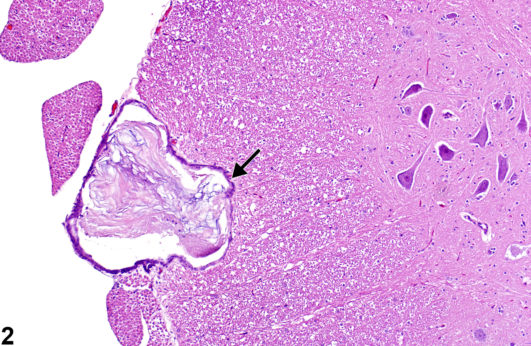
Figure 2 depicts a more advanced development of a sub-leptomeningeal epidermoid cyst located in the lumbar ventral spinal funiculus. Adjacent laterally are spinal nerve roots, and adjacent internally is the ventral spinal gray matter column. The extensive compression of the funicular tissue is apparent, but no clinical signs were detected in the animal.
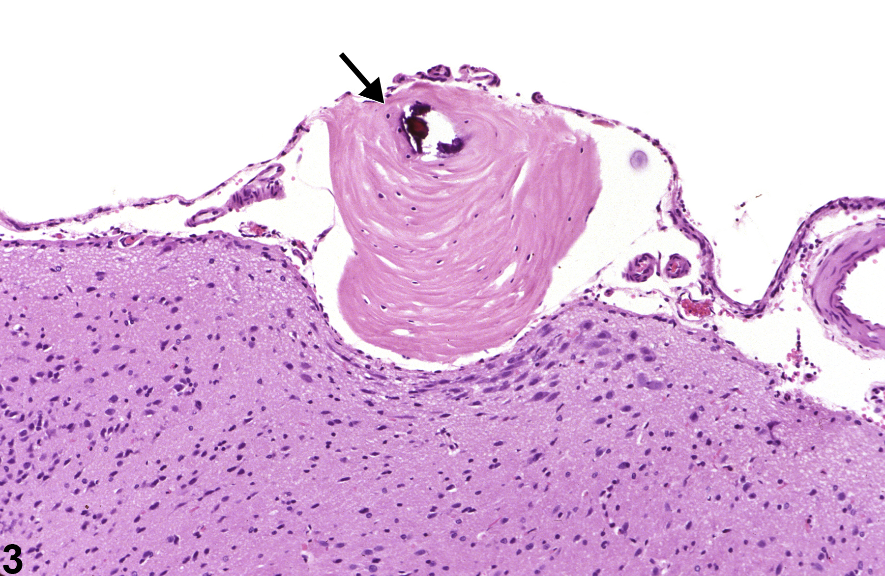
Figure 3 shows the location of an incidental cortical sub-leptomeningeal epidermoid cyst underlaid by the pia mater. There is compression of the underlying cortex and loss of neurons in the adjacent nucleus. Dystrophic mineralization of the central portion of the keratin core is present.
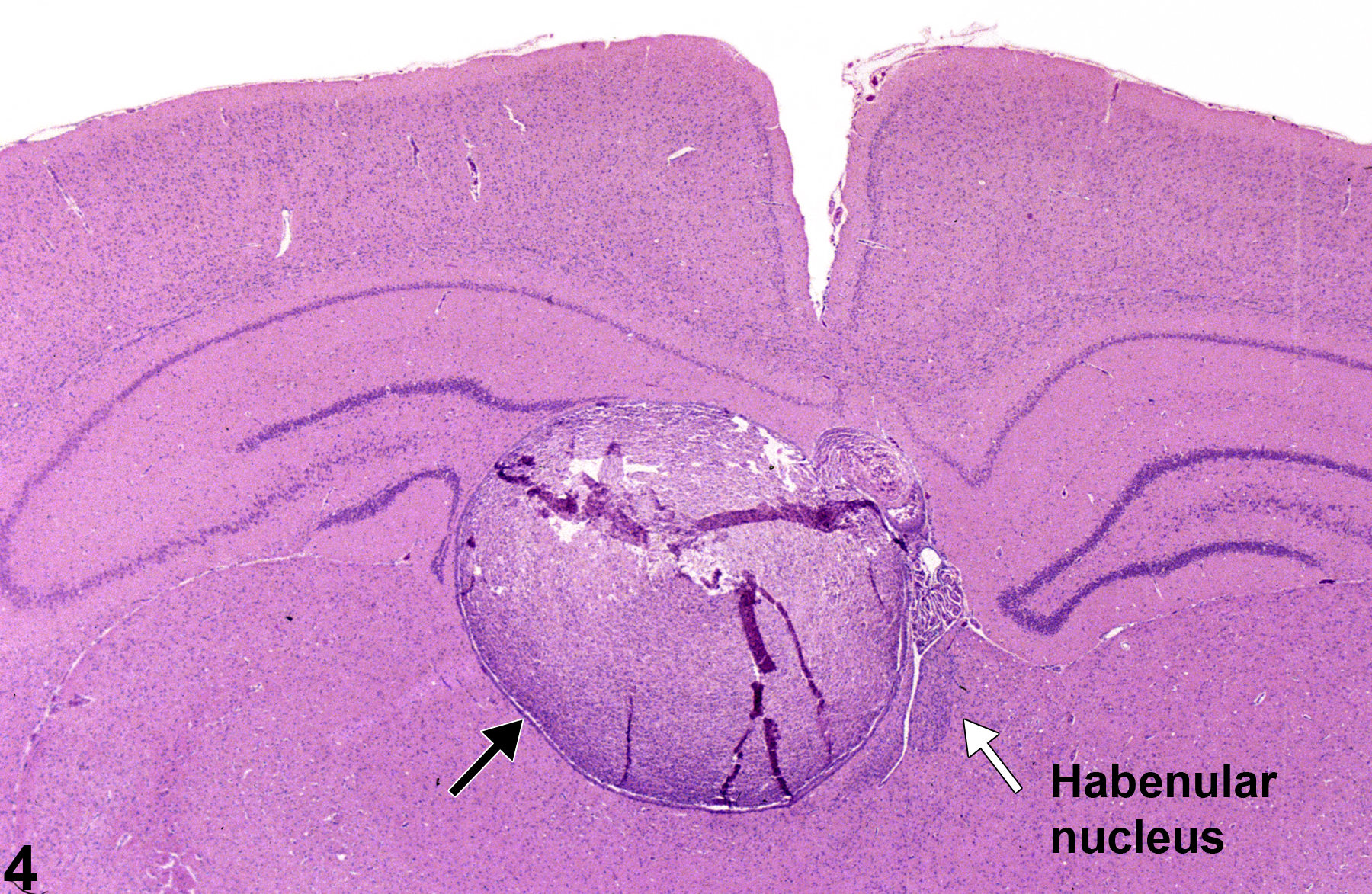
In Figure 4, this large incidental epidermoid cyst has a midline location above the diencephalon and compresses the adjacent hippocampal dentate gyrus and habenular nuclear structures. Note that the cyst is cellular and relatively poorly keratinized.
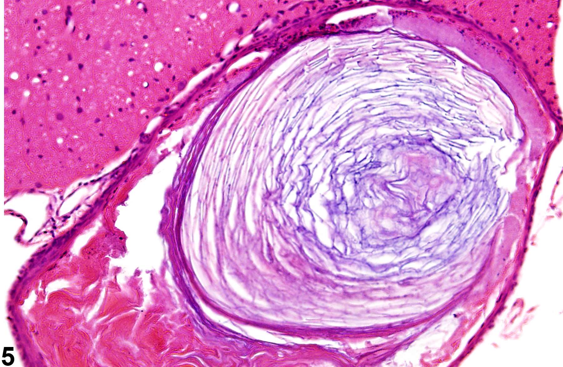
In Figure 5, the epidermoid cyst confines itself to a spinal radicle (nerve root), where its progressive enlargement has totally effaced the nerve fibers, leaving only the epineurium and perineurium intact.
Iaconetta G, Carvalho GA, Vorkapic P, Samii M. 2001. Intracerebral epidermoid tumor: A case report and review of the literature. Surg Neurol 55:218-222.
Abstract: http://www.ncbi.nlm.nih.gov/pubmed/11358593Krinke GJ, Kaufmann W, Mahrous AT, Schaetti P. 2000. Morphologic characterization of spontaneous nervous system tumors in mice and rats. Toxicol Pathol 28:178-192.
Abstract: http://www.ncbi.nlm.nih.gov/pubmed/10669006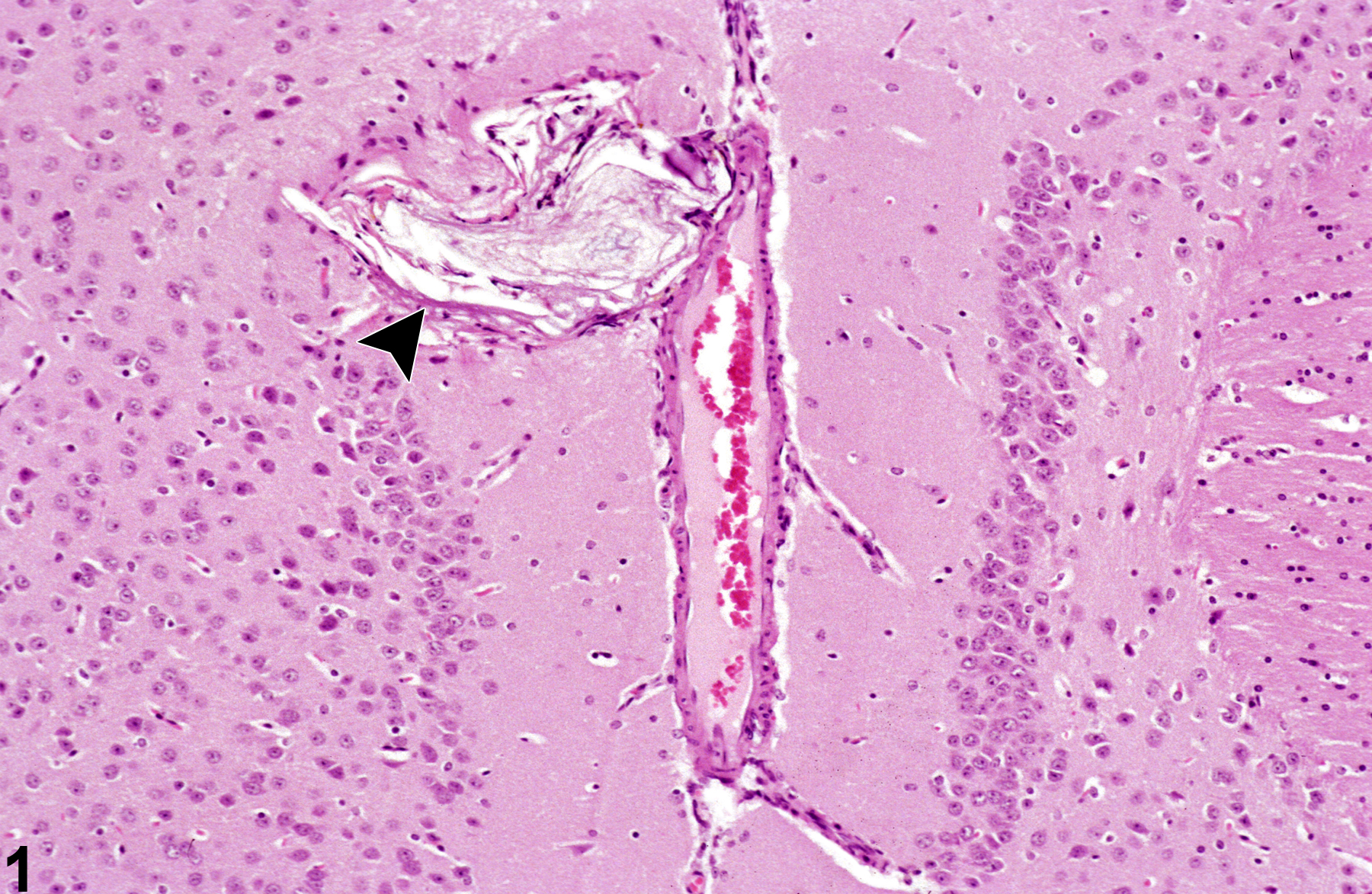
Brain, cingulate cortex-incidental epidermoid cyst (arrowhead) in a male B6C3F1 mouse from a chronic study.
All Images

Brain, cingulate cortex-incidental epidermoid cyst (arrowhead) in a male B6C3F1 mouse from a chronic study.


Cortex, sub-leptomeningeal-incidental epidermoid cyst (arrow) in a male F344/N rat from a chronic study.

Brain, hippocampus-incidental epidermoid cyst (arrow) in a male F344/N rat from a chronic study.

Spinal radicle-incidental epidermoid cyst in a female F344 rat.