

Nervous System
Brain, Neuron - Cell Loss
Narrative
Ozaki HS, Murakami TH, Shimada M. 1983. Learning deficits on avoidance task and hippocampal lesions in area CA3 following intraperitoneal administration of 3-acetylpyridine. J Neurosci Res 10(4):425-435.
Abstract: http://www.ncbi.nlm.nih.gov/pubmed/6686615Purpura DP, Gonzalez-Monteagudo O. 1960. Acute effects of methoxypyridoxine on hippocampal end-blade neurons; an experimental study of “special pathoclisis” in the cerebral cortex. J Neuropathol Exp Neurol 19:421-432.
Abstract: http://www.ncbi.nlm.nih.gov/pubmed/14435353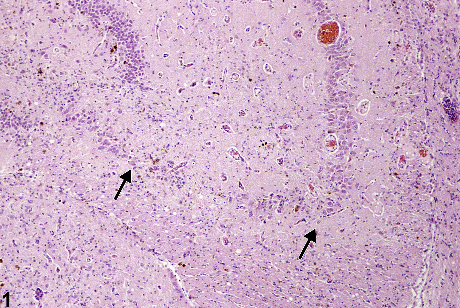
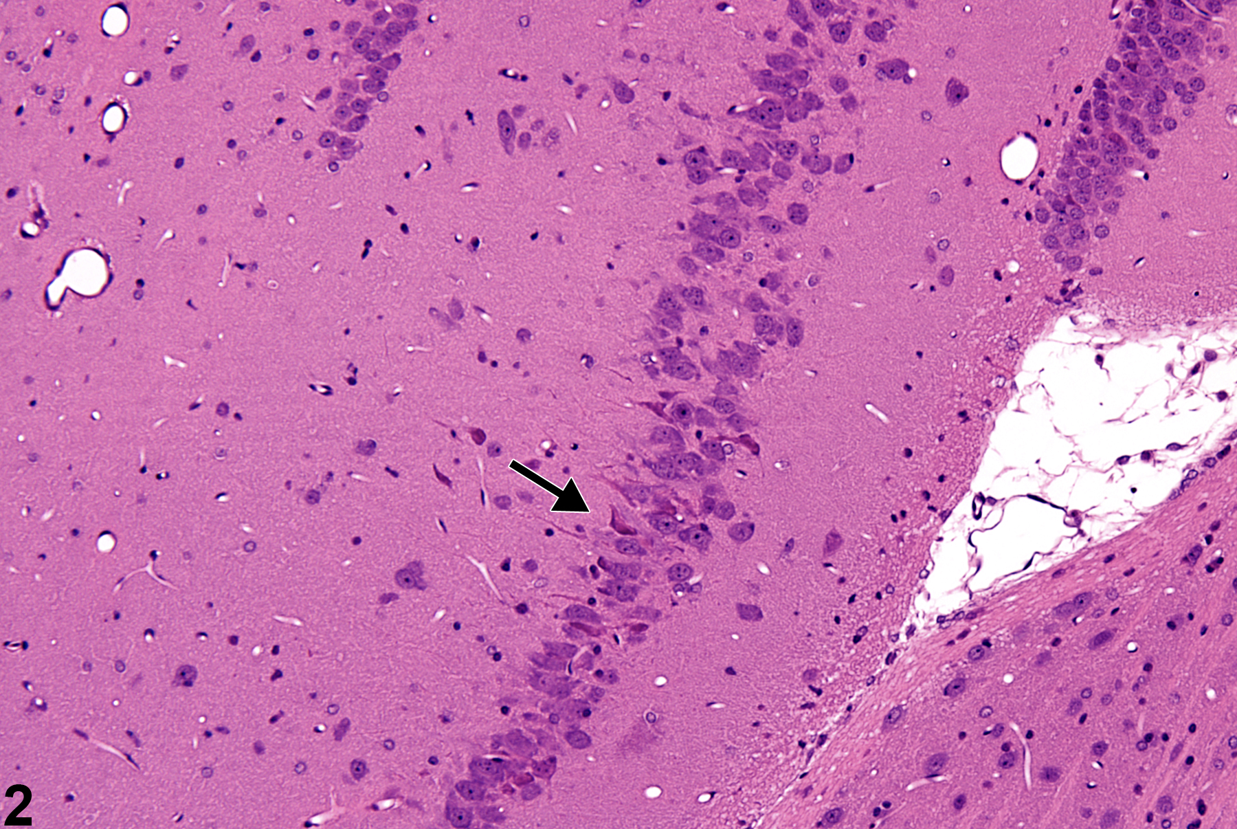
Neuronal cell loss in a female F344/N from a 2-year study. Note the loss of neurons in CA3 region of the hippocampus (arrows).
All Images

Neuronal cell loss in a female F344/N from a 2-year study. Note the loss of neurons in CA3 region of the hippocampus (arrows).

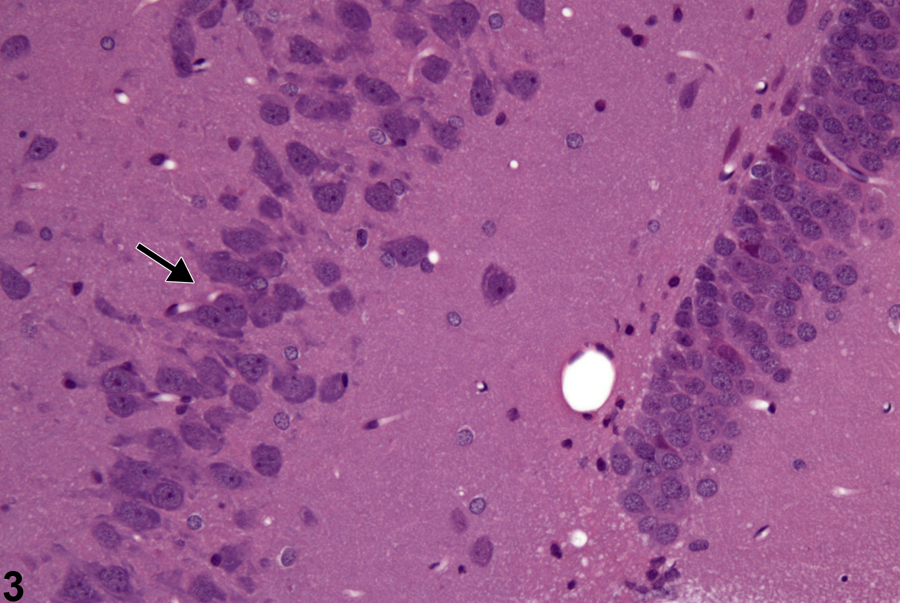
Normal number and morphology of CA3 neurons (arrow) in a control male rat from a single-dose acute gavage study.

Normal number and morphology of CA3 neurons (arrow) in a control male rat from a single-dose acute gavage study.