

Nervous System
Brain - Pigment
Narrative
Comment:
Various forms of pigment, such as yellow-golden ceroid/lipofuscin and dark brown hemosiderin, are deposited in the brain sometimes as part of the aging process or metabolic abnormalities. Ceroid/lipofuscin autofluoresces at 365 nm wavelength, and periodic acid Schiff stain highlights the deposits. The normal iron content of hemosiderin can be detected as blue to purple particles using Perls Prussian blue stain. The presence of hemosiderin-laden macrophages indicates prior neural injury with associated hemorrhage resulting in macrophage phagocytic degradation of extravasated red blood cells and their hemoglobin content. Figure 1 (arrow) shows multiple brown-pigmented hemosiderin-laden macrophages present in the corpus callosum suggesting previous hemorrhage.
Recommendations:
The presence of pigments (type of pigment need not be specified in the diagnosis), their severity grading, and anatomic subsite location should be documented in NTP studies. Definitive pigment identification is often difficult in histological sections, even with a battery of special stains. Therefore, it is recommended that a diagnosis of pigment (as opposed to diagnosing the type of pigment, e.g., hemosiderin or lipofuscin) is most appropriate. The pathology narrative should describe the morphological features of the pigmentation. Not all pigments have to be diagnosed, as some are ubiquitous in aging animals or related to some other disease process and are not toxicologically meaningful. The pathologist should use his or her judgment in deciding whether or not secondary deposits of pigment are prominent enough to warrant a separate diagnosis.
References:
Culling CFA, Allison RT, Barr WT. 1985. Cellular Pathology Technique, 4th ed. Butterworth, London.
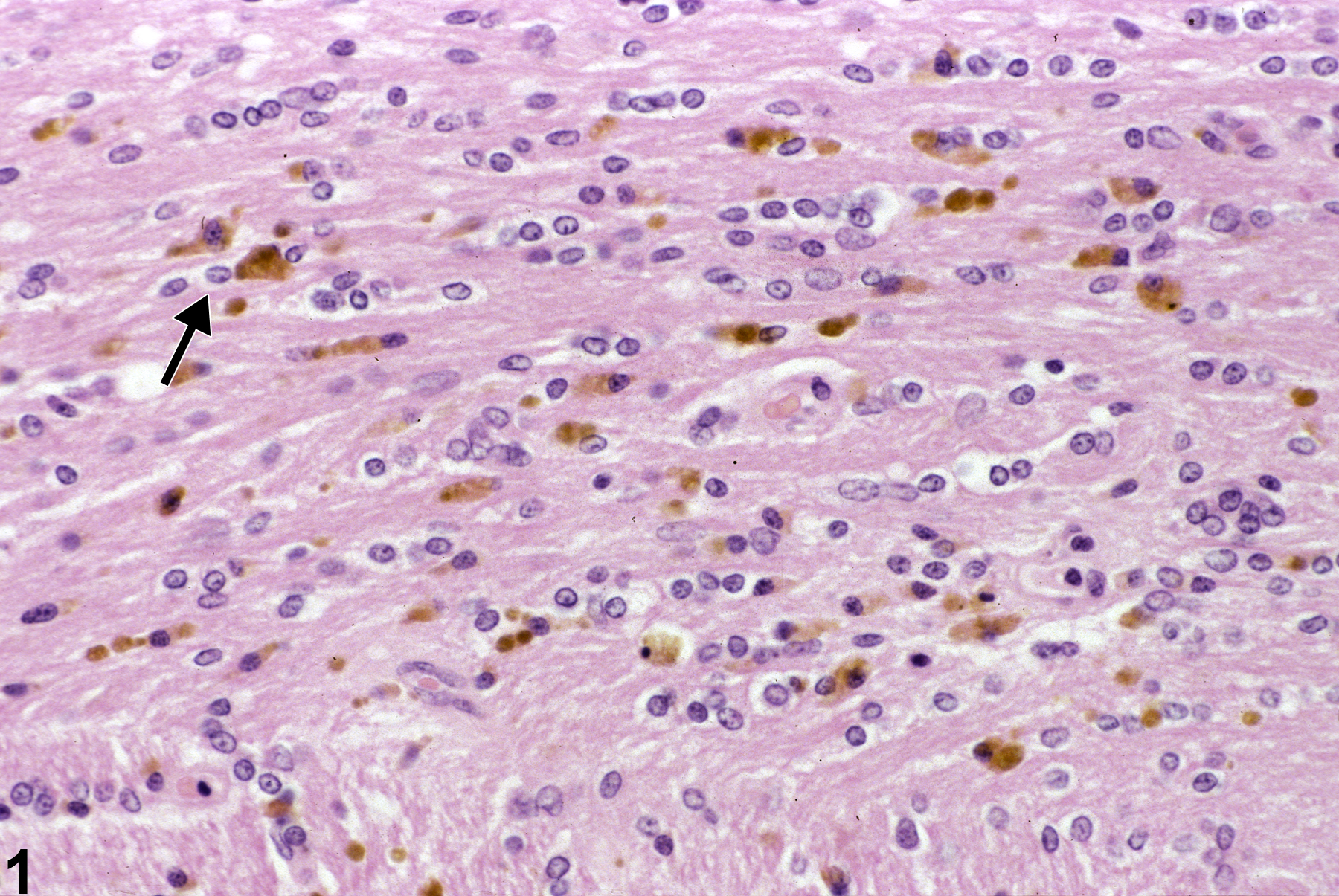
Pigment. A site of former hemorrhage (arrow) in the corpus callosum in a female F344/N rat from a chronic study. Multiple brown-pigmented hemosiderin-laden macrophages are present.
All Images

Pigment. A site of former hemorrhage (arrow) in the corpus callosum in a female F344/N rat from a chronic study. Multiple brown-pigmented hemosiderin-laden macrophages are present.