U.S. flag

An official website of the United States government

Dot gov

The .gov means it's official.
Federal government websites often end in .gov or .mil. Before sharing sensitive information, make sure you're on a federal government site.

Https

The site is secure.
The https:// ensures that you are connecting to the official website and that any information you provide is encrypted and transmitted securely.

Skip to Main Navigation
Skip to Page Content
Skip to Atlas Navigation

Lung - Amyloid

Image of amyloid in the lung from a female B6C3F1/N mouse in a chronic study
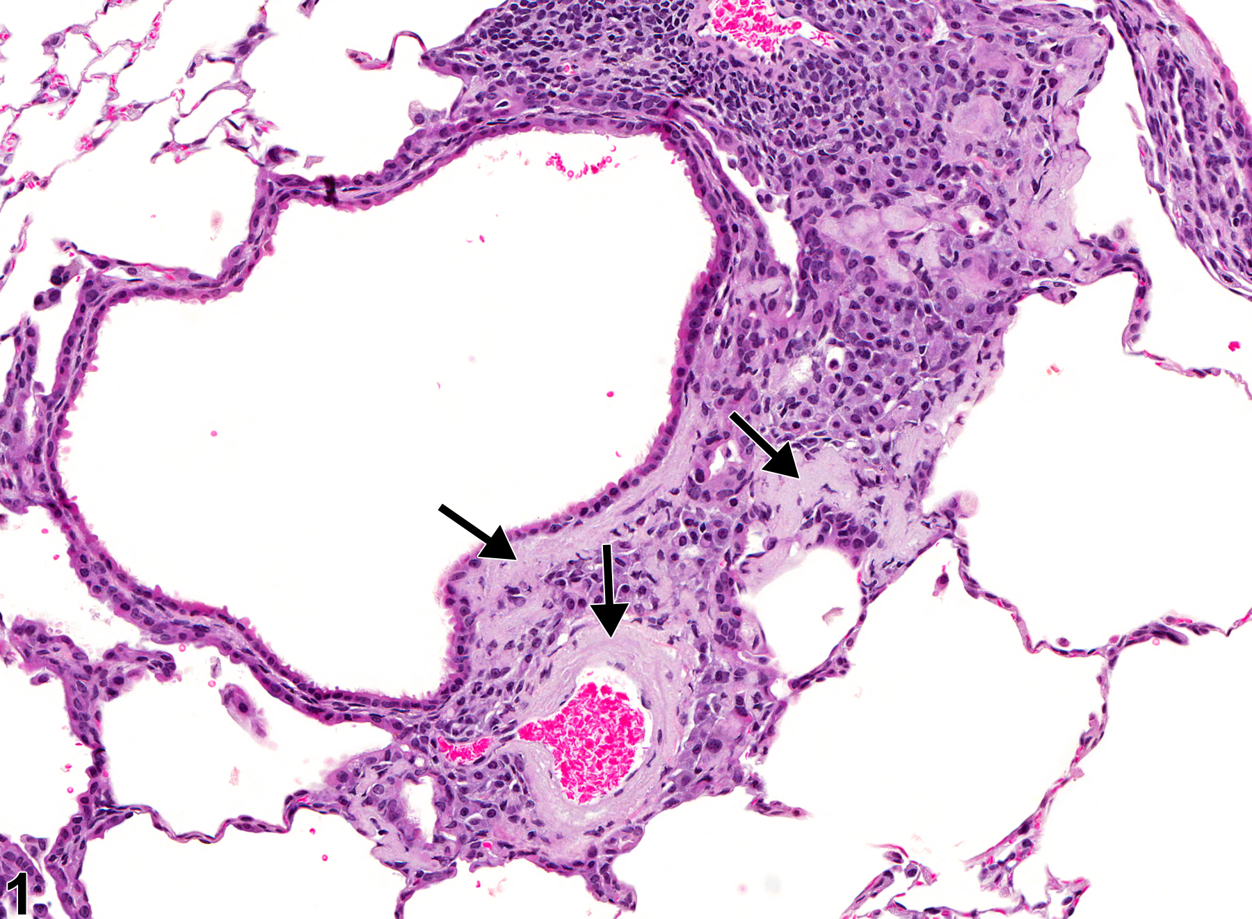
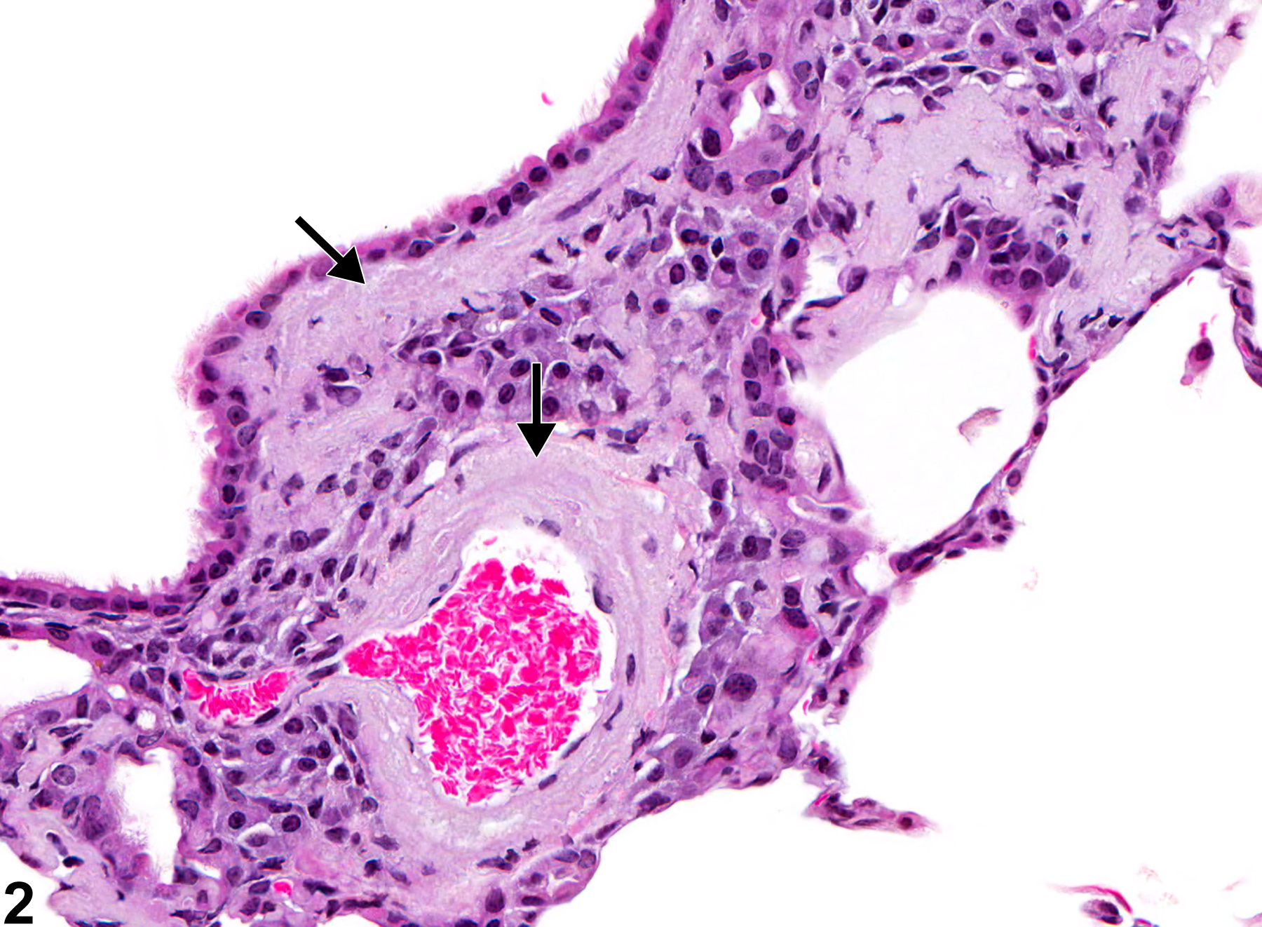
Lung - Amyloid in a female B6C3F1/N mouse from a chronic study. There is perivascular and peribronchiolar amyloid deposition (arrows).
Figure 1 of 4
Image of amyloid in the lung from a female B6C3F1/N mouse in a chronic study
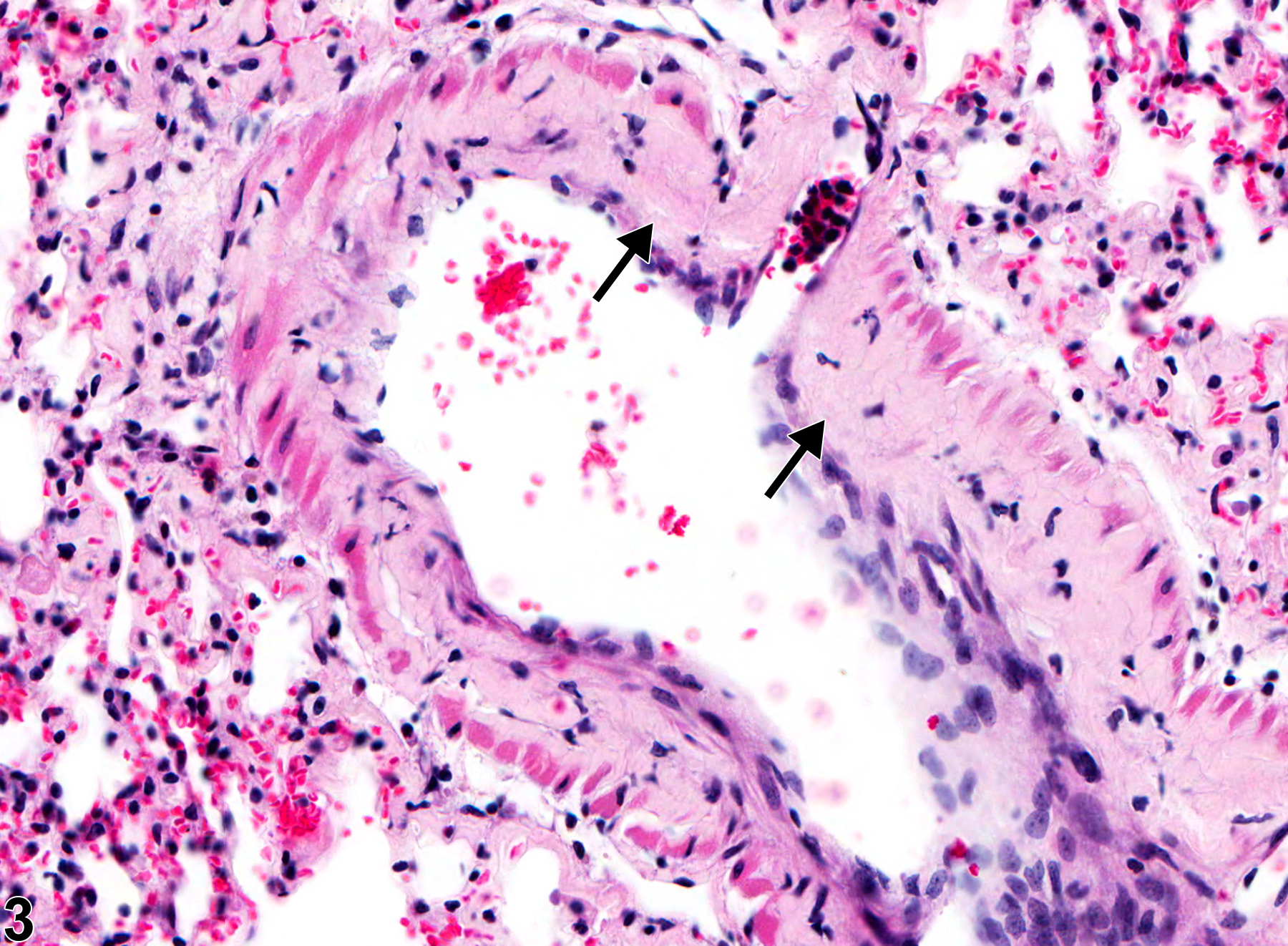
Lung - Amyloid in a female B6C3F1/N mouse from a chronic study (higher magnification of Figure 1). There is perivascular and peribronchiolar, acellular, eosinophilic material (arrows).
Figure 2 of 4
Image of amyloid in the lung from a male B6C3F1/N mouse in a chronic study
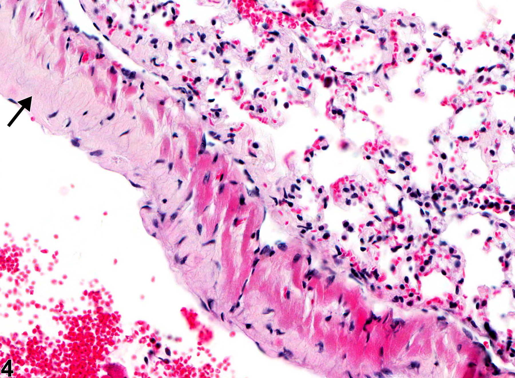
Lung - Amyloid in a male B6C3F1/N mouse from a chronic study. There is deposition of amyloid in the vascular wall (arrows).
Figure 3 of 4
Image of amyloid in the lung from a male B6C3F1/N mouse in a chronic study
Lung - Amyloid in a male B6C3F1/N mouse from a chronic study. Acellular, eosinophilic material (amyloid) is present in the vessel wall (arrow).
Figure 4 of 4
next arrow

comment:

Amyloid ( Figure 1image opens in a pop-up window , Figure 2image opens in a pop-up window , Figure 3image opens in a pop-up window , and Figure 4image opens in a pop-up window ) refers to insoluble, extracellular and/or intracellular proteins that appear as amorphous, eosinophilic, acellular material histologically. It stains positively with Congo red stain and has a characteristic apple green birefringence under polarized light. Deposition of amyloid can be focal, multifocal, or diffuse. Involvement of vascular walls is not uncommon, but it may also be seen in peribronchial areas. In severe cases, the pulmonary parenchyma may be effaced. Approximately 90% of the deposits consist of amyloid fibrils formed by the aggregation of misfolded proteins, and 10% of the deposits consist of glycosaminoglycans, apolipoprotein E, and serum amyloid P. Amyloidosis is found as a background change in some strains, such as CD-1 mice. Pulmonary amyloidosis is a spontaneous lesion in aging rodents and may be localized to the lung, or it may be a component of systemic amyloidosis involving spleen, kidney, liver, and other organs. It may also be associated with chronic inflammation. Since an association between amyloid deposition in the lung and xenobiotic administration has not been reported, amyloid deposition is generally considered a background finding. It may, however, be the cause of morbidity or mortality in some animals and can affect the outcome of a study.

recommendation:

Because this lesion is biologically important, Lung - Amyloid should be diagnosed and assigned a severity grade whenever present. The location of the amyloid deposits should be indicated in the diagnosis by using a site modifier. Any secondary lesions, such as necrosis or degeneration, should not be diagnosed separately unless warranted by severity but should be described in the pathology narrative.

references:

Buxbaum JN. 2004. The systemic amyloidoses. Curr Opin Rheumatol 16:67-75.
Abstract: https://www.ncbi.nlm.nih.gov/pubmed/14673392

Frith CH, Manik C. 1991. Incidence, distribution, and morphology of amyloidosis in Charles Rivers CD-1 mice. Toxicol Pathol 19:123-127.
Abstract: https://www.ncbi.nlm.nih.gov/pubmed/1771365

Higuchi K, Matsumura A, Honma A, Takeshita S, Hashimoto K, Hosokawa M, Yasuhira K, Takeda T. 1983. Systemic senile amyloid in senescence-accelerated mice. A unique fibril protein demonstrated in tissues from various organs by the unlabeled immunoperoxidase method. Lab Invest 48:231-240.
Abstract: https://www.ncbi.nlm.nih.gov/pubmed/6337302

Homburger F, Russfield AB, Weisburger JH, Lim S, Chak SP, Weisburger EK. 1975. Aging changes in CD-1 HaM/ICR mice reared under standard laboratory conditions. J Natl Cancer Inst 55:37-45.
Abstract: https://www.ncbi.nlm.nih.gov/pubmed/169366

Takeshita S, Hosokawa M, Irino M, Higuchi K, Shimizu K, Yasuhira K, Takeda T. 1982. Spontaneous age-associated amyloidosis in senescence-accelerated mouse (SAM). Mech Ageing Dev 20:13-23.
Abstract: https://www.ncbi.nlm.nih.gov/pubmed/7176700

Westermark P, Benson MD, Buxbaum JN, Cohen AS, Frangione B, Ikeda S, Masters CL, Merlini G, Saraiva MJ, Sipe JD. 2007. A primer of amyloid nomenclature. Amyloid 14:179-183.
Abstract: https://www.ncbi.nlm.nih.gov/pubmed/17701465