U.S. flag

An official website of the United States government

Dot gov

The .gov means it's official.
Federal government websites often end in .gov or .mil. Before sharing sensitive information, make sure you're on a federal government site.

Https

The site is secure.
The https:// ensures that you are connecting to the official website and that any information you provide is encrypted and transmitted securely.

Skip to Main Navigation
Skip to Page Content
Skip to Atlas Navigation

Lung - Bronchiectasis

Image of bronchiectasis in the lung from a male F344/N rat in a chronic study
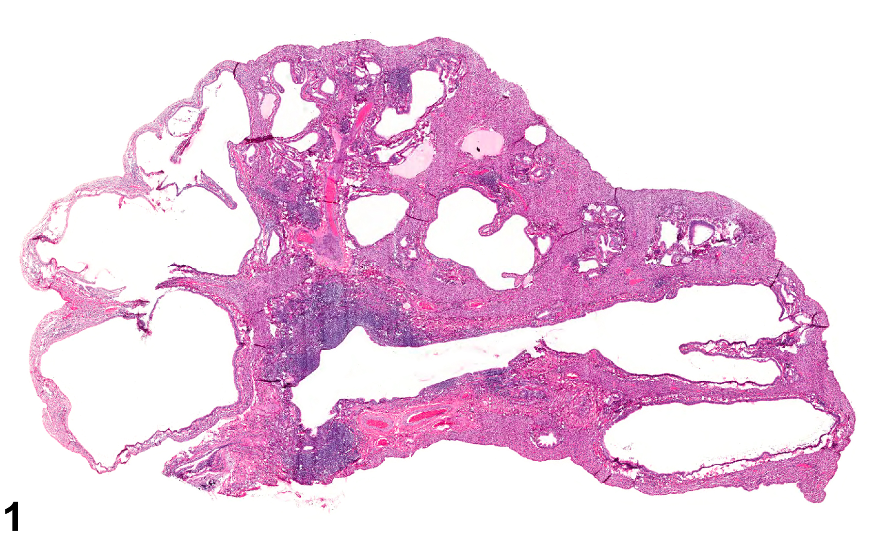
Lung - Bronchiectasis in an F344/N rat. The airways are markedly dilated.
Figure 1 of 1
next arrow

comment:

Bronchiectasis ( Figure 1image opens in a pop-up window ), permanent dilation of the airways, is very uncommon in NTP studies. The affected airways are dilated and often are inflamed. There may be damage to the mucosa, including the submucosal glands, and there may be regeneration, hyperplasia, or metaplasia of the epithelium. With chronicity, there may be fibrosis of the affected airways. Bronchiectasis may need to be differentiated from emphysema. While bronchiectasis is a lesion of the airways, emphysema is an enlarged airspace in the alveolar parenchyma caused by breakdown of the septal walls (see Lung - Emphysema). Bronchiectasis is usually secondary to bronchitis, such as that caused by pulmonary infection, aspiration, or, less commonly, test-article-associated effects. In rats, it has often been caused by infection with Mycoplasma pulmonis, although this is rare due to the implementation of modern husbandry techniques. Severe bronchiectasis can lead to atelectasis of the dependent lung tissue ( Figure 1image opens in a pop-up window ).

recommendation:

Bronchiectasis should be diagnosed whenever present and given a severity grade. A site modifier should be included in the diagnosis to indicate which airways are affected. If bronchi and bronchioles are both affected, the site modifier may be omitted and the location of the lesion described in the pathology narrative. It should be noted in the pathology narrative whether or not the airway dilation is treatment related, or secondary to an infectious disease.

related links:

Lung - Emphysema

references:

Boorman GA, Eustis SL. 1990. Lung. In: Pathology of the Fischer Rat: Reference and Atlas (Boorman GA, Eustis SL, Elwell MR, Montgomery CA, MacKenzie WF, eds). Academic Press, San Diego, CA, 339-367.

Costa DL, Lehmann JR, Slatkin DN, Popenoe EA, Drew RT. 1983. Chronic airway obstruction and bronchiectasis in the rat after intratracheal bleomycin. Lung 161:287-300.

Dungworth DL. 1993. The respiratory system. In: Pathology of Domestic Animals, Vol 2, 4th ed (Jubb KVF, Kennedy PC, Palmer N, eds). Academic Press, San Diego, CA, 539-699.

Kohn DF. 1971. Bronchiectasis in rats infected with Mycoplasma pulmonis: An electron microscopy study. Lab Anim Sci 21:856-861.