U.S. flag

An official website of the United States government

Dot gov

The .gov means it's official.
Federal government websites often end in .gov or .mil. Before sharing sensitive information, make sure you're on a federal government site.

Https

The site is secure.
The https:// ensures that you are connecting to the official website and that any information you provide is encrypted and transmitted securely.

Skip to Main Navigation
Skip to Page Content
Skip to Atlas Navigation

Lung, Epithelium, Alveolus - Hyperplasia

Image of alveolar epithelial hyperplasia in the lung from a male B6C3F1/N mouse in a chronic study
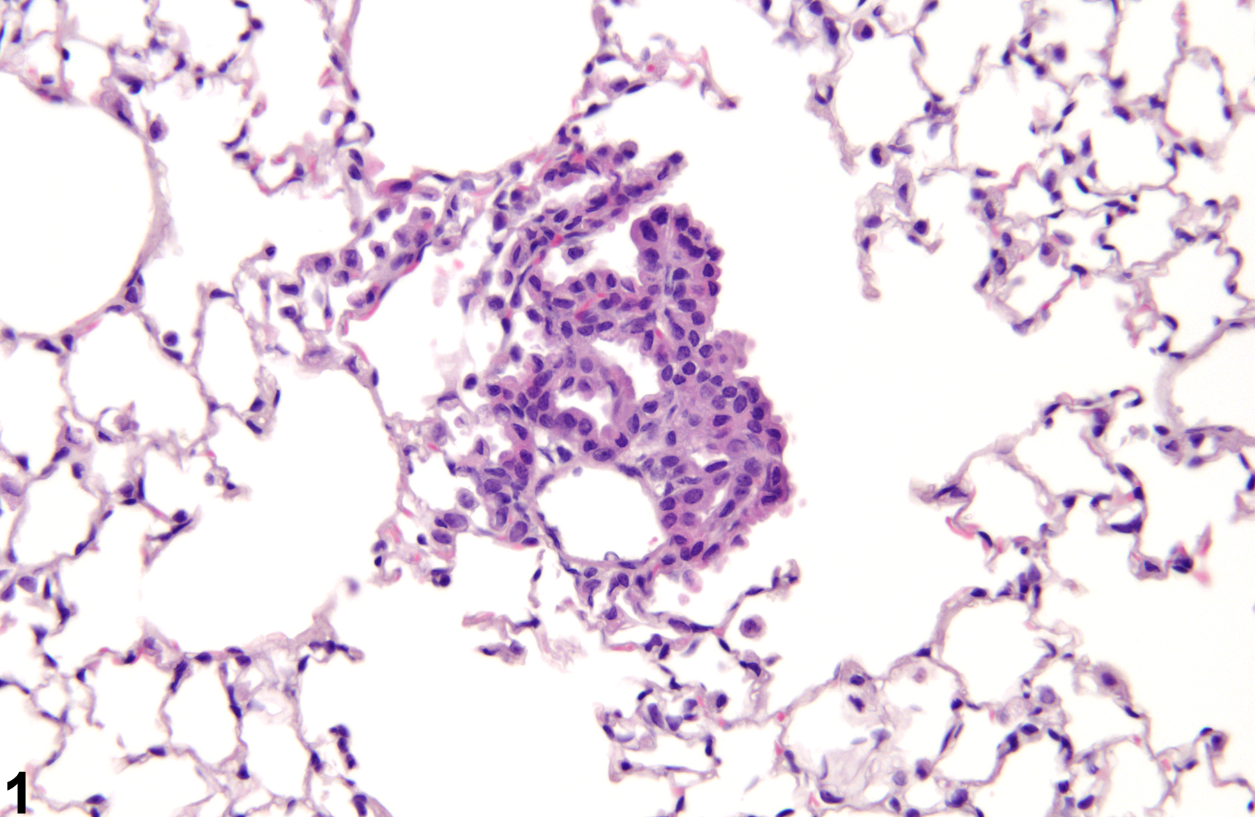
Lung, Epithelium, Alveolus - Hyperplasia in a male B6C3F1/N mouse from a chronic study. There is a small proliferation of alveolar epithelial cells with no inflammation.
Figure 1 of 6
Image of alveolar epithelial hyperplasia in the lung from a male F344/N rat in a chronic study
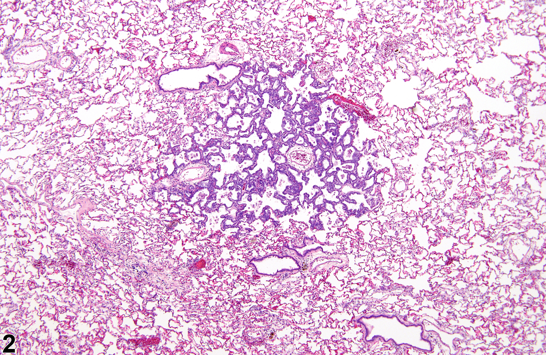
Lung, Epithelium, Alveolus - Hyperplasia in a male F344/N rat from a chronic study. There is no change in the normal alveolar architecture in this focus of alveolar epithelial hyperplasia.
Figure 2 of 6
Image of alveolar epithelial hyperplasia in the lung from a male F344/N rat in a chronic study
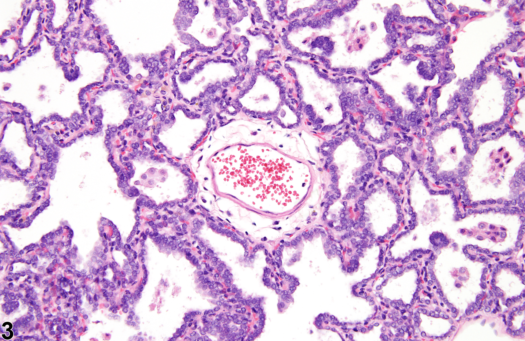
Lung, Epithelium, Alveolus - Hyperplasia in a male F344/N rat from a chronic study (higher magnification of Figure 2). The hyperplastic epithelial cells are cuboidal.
Figure 3 of 6
Image of alveolar epithelial hyperplasia in the lung from a male B6C3F1/N mouse in a chronic study
Lung, Epithelium, Alveolus - Hyperplasia in a male B6C3F1/N mouse from a chronic study. The hyperplastic epithelial cells line slightly thickened alveolar septa.
Figure 4 of 6
Image of alveolar epithelial hyperplasia in the lung from a male F344/N rat in a subchronic study
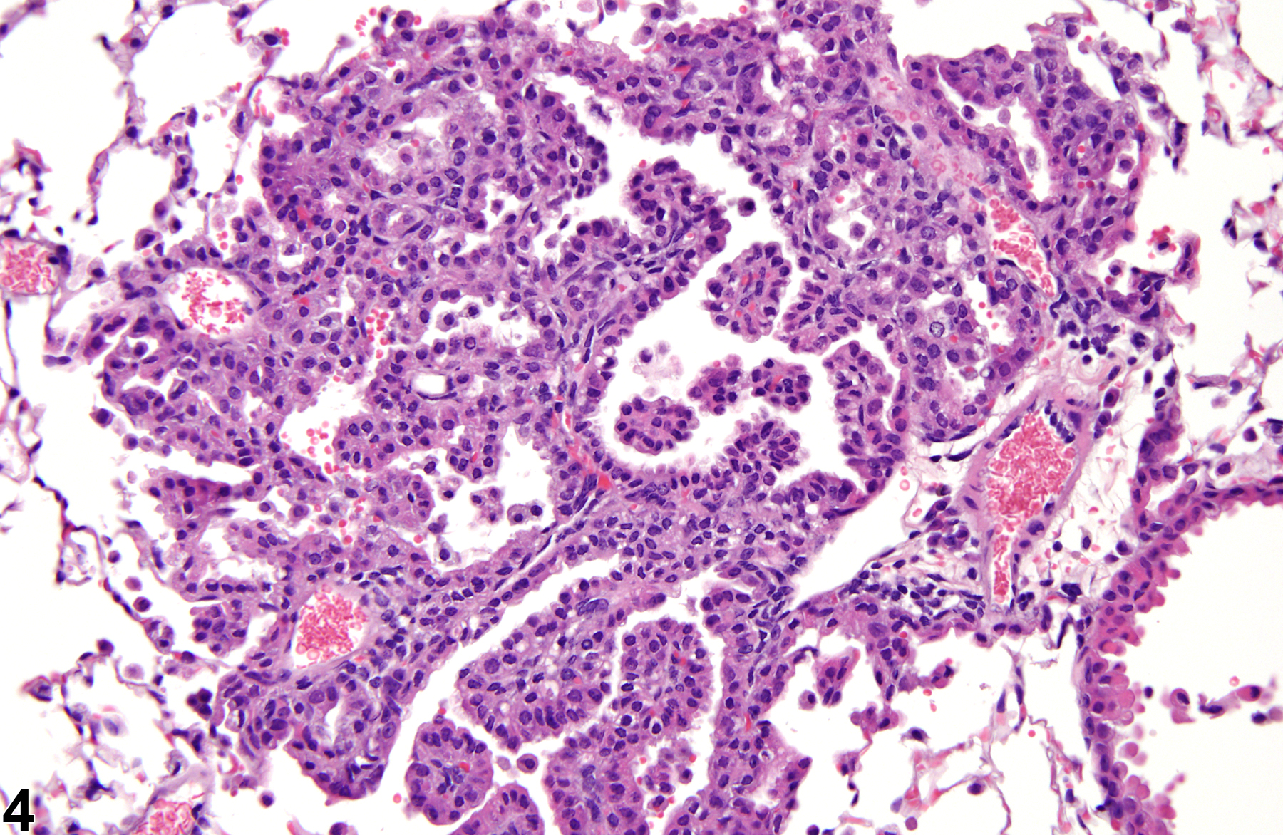
Lung, Epithelium, Alveolus - Hyperplasia in a male F344/N rat from a subchronic study. Alveolar epithelial hyperplasia secondary to inflammation is associated with numerous alveolar macrophages and minimal hemorrhage.
Figure 5 of 6
Image of alveolar epithelial hyperplasia in the lung from a male F344/N rat in a chronic study
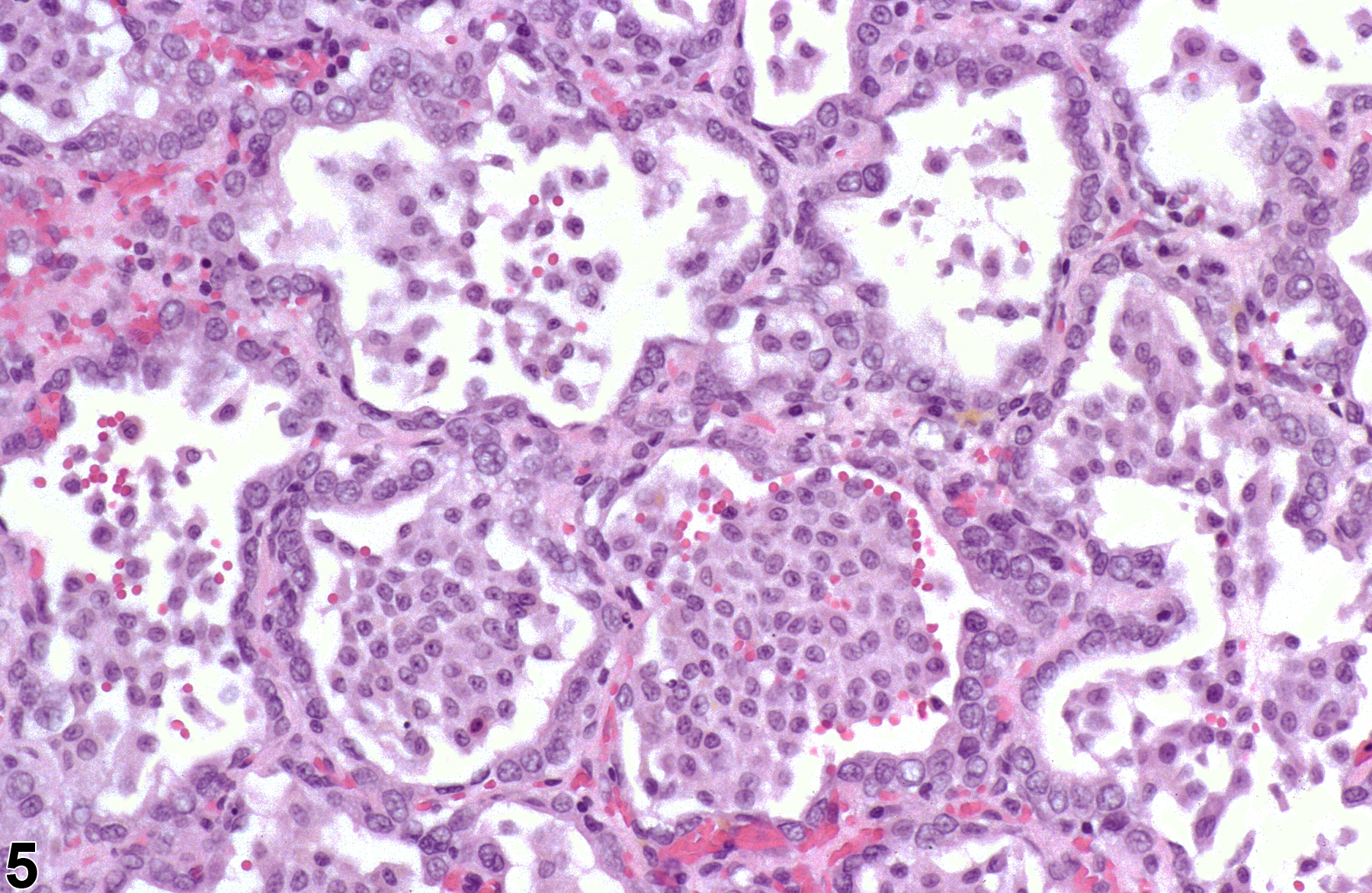
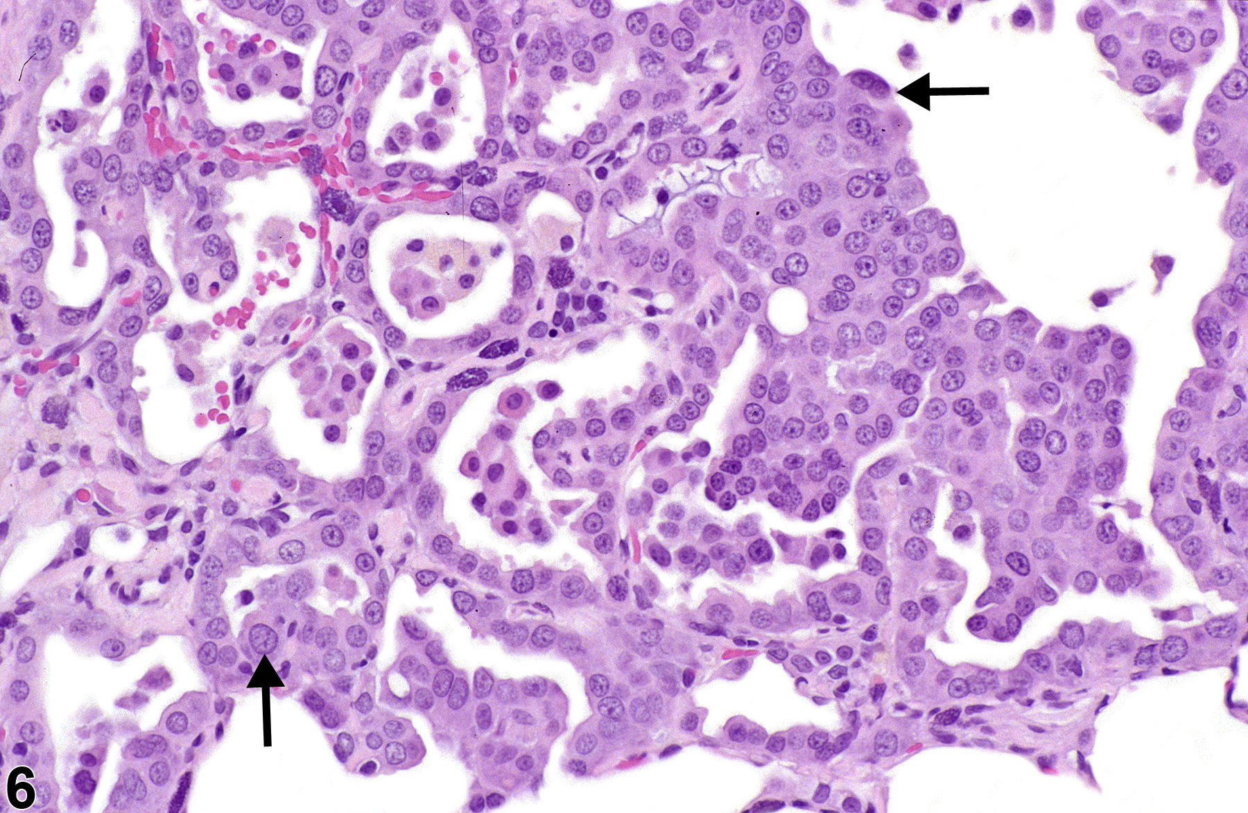
Lung, Epithelium, Alveolus - Hyperplasia, Atypical from a male F344/N rat in a chronic study. Atypical cells (arrows) are enlarged with enlarged nuclei.
Figure 6 of 6
next arrow

comment:

Alveolar epithelial hyperplasia is a proliferation of type II pneumocytes and may be primary ( Figure 1image opens in a pop-up window , Figure 2image opens in a pop-up window , Figure 3image opens in a pop-up window , and Figure 4image opens in a pop-up window ) or secondary to type I pneumocyte injury or inflammation ( Figure 5image opens in a pop-up window ). Type II pneumocytes are thought to be the progenitors of type I cells. Type I pneumocytes are especially vulnerable to oxidant injury, and proliferation of type II pneumocytes is often seen following injury and loss of type I cells. When alveolar epithelial hyperplasia is secondary to type I pneumocyte damage, it is usually associated with inflammation, as opposed to primary alveolar epithelial hyperplasia, which is typically not associated with inflammation. Foci of hyperplasia are characterized by an increase in closely spaced, cuboidal type II cells lining alveolar septa. The cells lack cellular atypia, seldom contain mitotic figures, and may or may not have prominent cytoplasmic vacuoles. The alveolar architecture is maintained, and the focus does not compress the surrounding parenchyma. These morphologic characteristics differentiate hyperplasia from adenoma, in which cellular atypia, mitotic figures, and compression of adjacent tissue may be present and the normal architecture is distorted. Accumulations of alveolar macrophages may be associated with primary hyperplasia (although the accumulations are most severe when associated with alveolar bronchiolar carcinomas). In some cases, some of the cells in a focus of alveolar epithelial hyperplasia (primary or secondary) may have features of atypia ( Figure 6image opens in a pop-up window ), such as enlarged or oddly shaped nuclei or binucleation. Primary alveolar epithelial hyperplasia is thought to be the precursor lesion to alveolar/bronchiolar adenomas and carcinomas, especially when there are features of cellular atypia.

recommendation:

Lung, Epithelium, Alveolus - Hyperplasia should be diagnosed and given a severity grade whenever present. When the alveolar epithelial hyperplasia is secondary to type I pneumocyte injury or inflammation, it need not be recorded separately unless the pathologist feels that its severity warrants a separate diagnosis. If it is not diagnosed separately, it should be described in the pathology narrative as a component of the inflammatory lesion. If both primary and secondary alveolar epithelial hyperplasia is present in a study, and the pathologist feels that both should be diagnosed, then they must be assigned different terms. This can be accomplished by adding an appropriate modifier, such as "focal." That is, primary alveolar epithelial hyperplasia can be recorded as Lung, Epithelium, Alveolus - Hyperplasia, Focal and secondary alveolar epithelial hyperplasia can be recorded as Lung, Epithelium, Alveolus - Hyperplasia. However, the distinction must be made in the pathology narrative. If the hyperplastic cells have significant features of atypia, the modifier "atypical" should be included in the diagnosis (i.e., Lung, Epithelium, Alveolus - Hyperplasia, Atypical).

related links:

Lung - Atypia, Cellular

references:

Boorman GA, Eustis SL. 1990. Lung. In: Pathology of the Fischer Rat: Reference and Atlas (Boorman GA, Eustis SL, Elwell MR, Montgomery CA, MacKenzie WF, eds). Academic Press, San Diego, CA, 339-367.

Dixon D, Herbert RA, Sills RC, Boorman GA. 1999. Lungs, pleura, and mediastinum. In: Pathology of the Mouse: Reference and Atlas (Maronpot RR, Boorman GA, Gaul BW, eds). Cache River Press, Vienna, IL, 293-332.

Renne, R, Brix A, Harkema J, Herbert R, Kittle B, Lewis D, March T, Nagano K, Pino M, Rittinghausen S, Rosenbruch M, Tellier P, Wohrmann T. 2009. Proliferative and nonproliferative lesions of the rat and mouse respiratory tract. Toxicol Pathol 37(suppl):5S-73S.
Abstract: https://www.ncbi.nlm.nih.gov/pubmed/20032296