U.S. flag

An official website of the United States government

Dot gov

The .gov means it's official.
Federal government websites often end in .gov or .mil. Before sharing sensitive information, make sure you're on a federal government site.

Https

The site is secure.
The https:// ensures that you are connecting to the official website and that any information you provide is encrypted and transmitted securely.

Skip to Main Navigation
Skip to Page Content
Skip to Atlas Navigation

Lung - Fibrosis

Image of fibrosis in the lung from a mouse
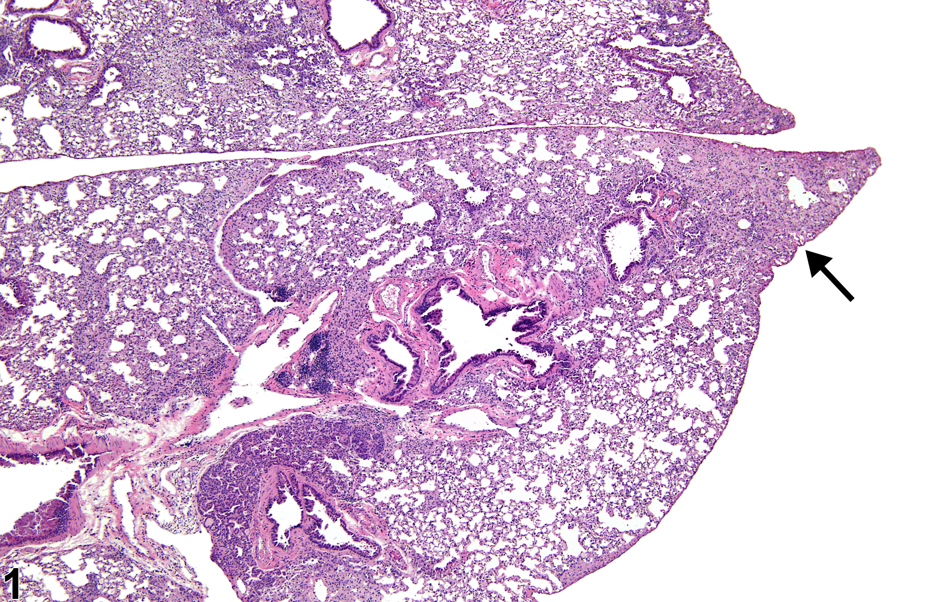
Lung - Fibrosis in a mouse. The airspaces have been obliterated at the tip of the lung lobe (arrow).
Figure 1 of 8
Image of fibrosis in the lung from a mouse
Lung - Fibrosis in a mouse (same animal as in Figure 1). The blue staining indicates interstitial fibrosis. Masson�s trichrome stain.
Figure 2 of 8
Image of fibrosis in the lung from a male F344/N rat in a chronic study
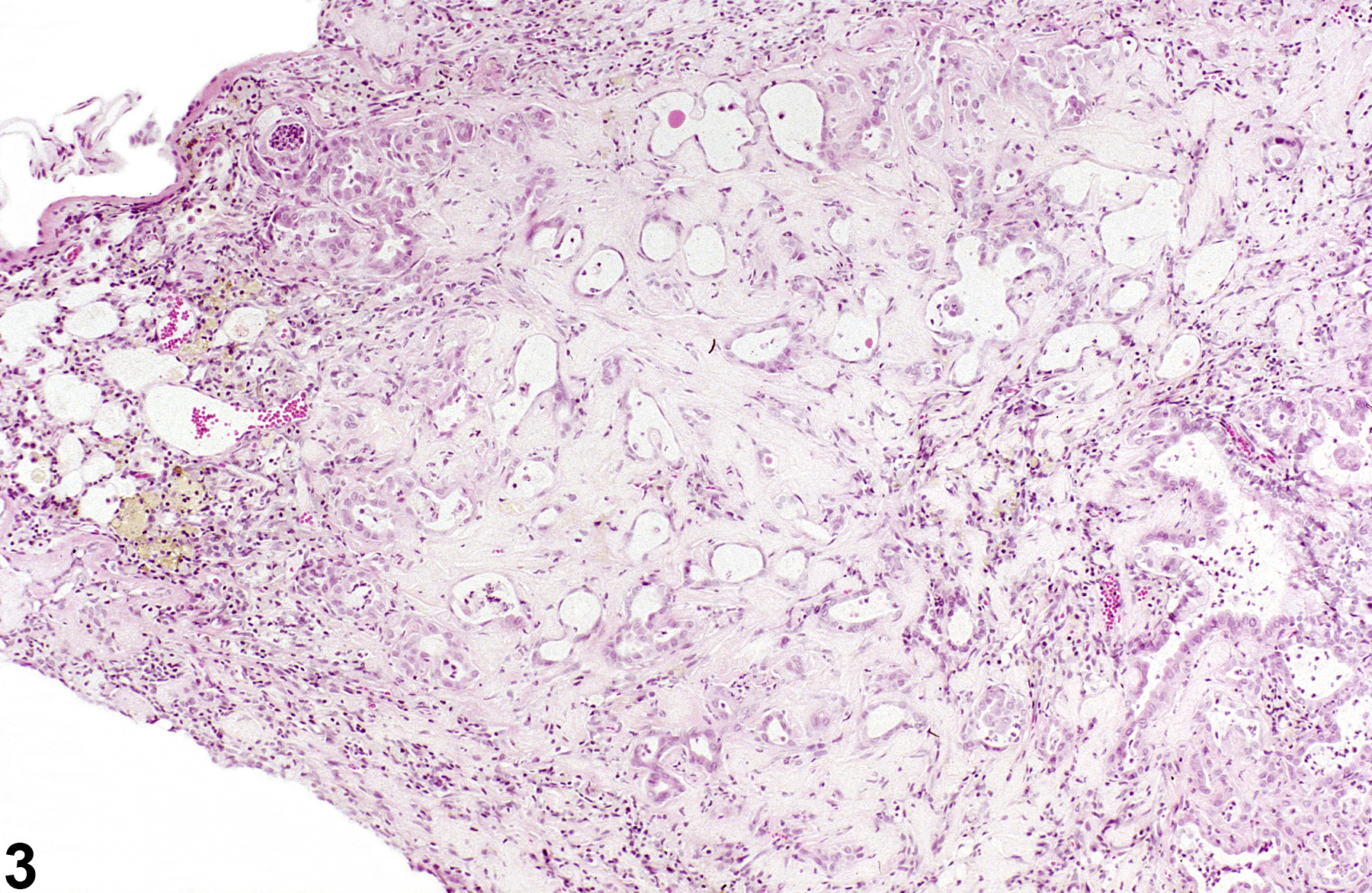
Lung - Fibrosis in a male F344/N rat from a chronic study. Severe interstitial fibrosis replaces the lung parenchyma.
Figure 3 of 8
Image of fibrosis in the lung from a female F344/N rat in a subchronic study
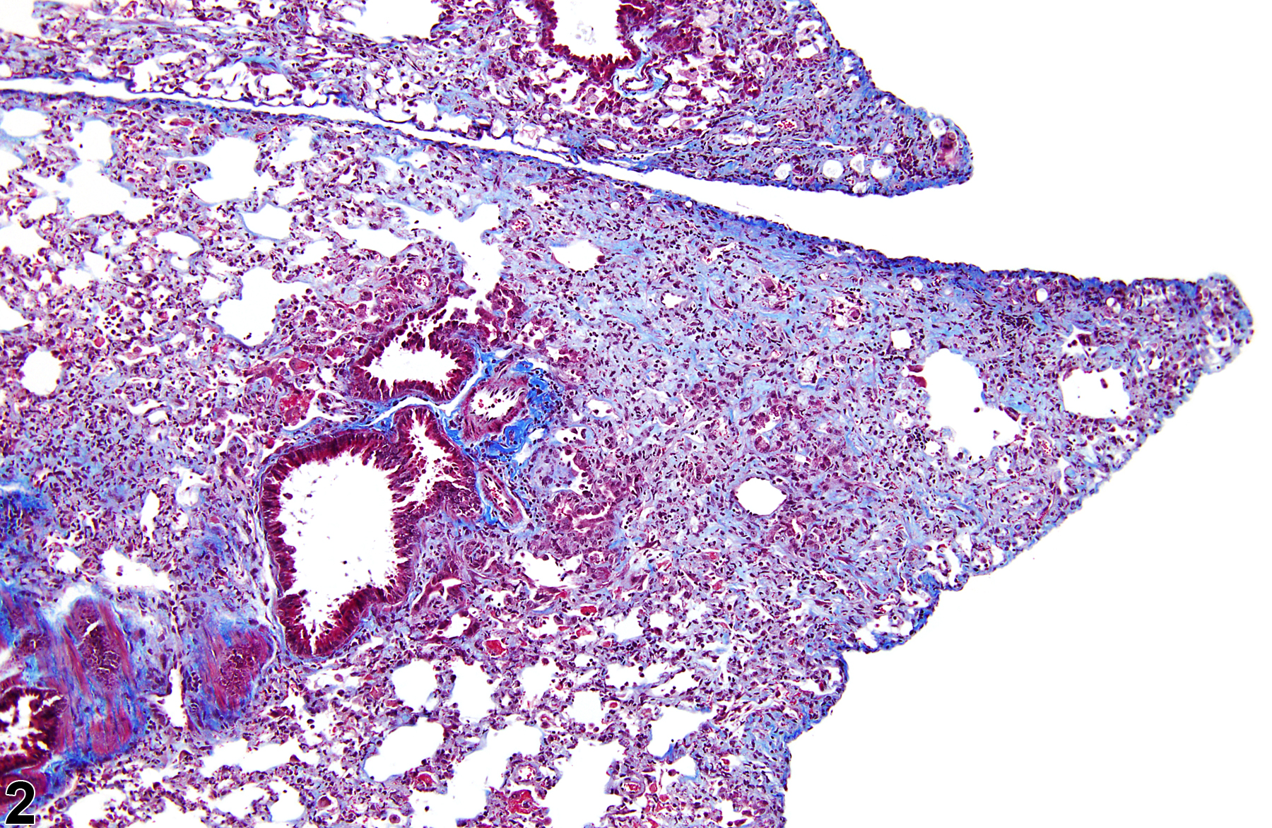
Lung, Bronchiole - Fibrosis in a female F344/N rat in a subchronic study. There is intraluminal and mural bronchiolar fibrosis.
Figure 4 of 8
Image of fibrosis in the lung from a male B6C3F1/N mouse in a chronic study
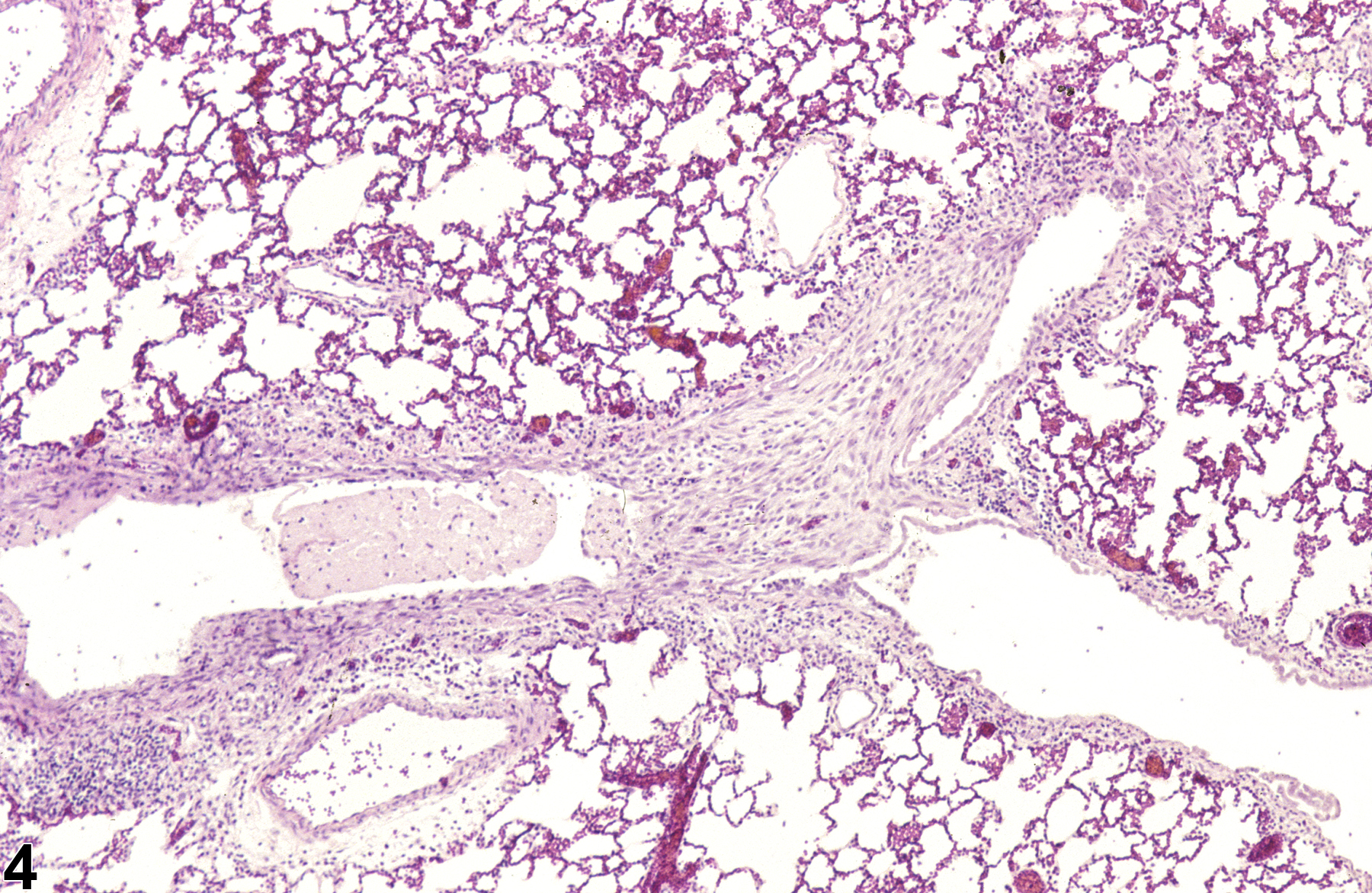
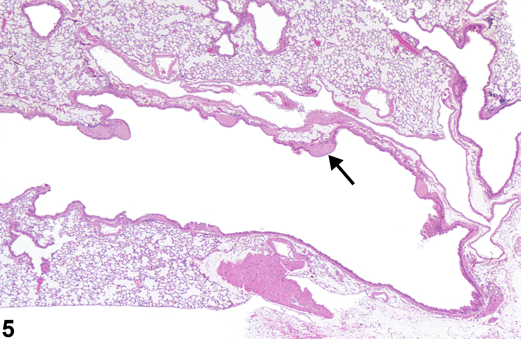
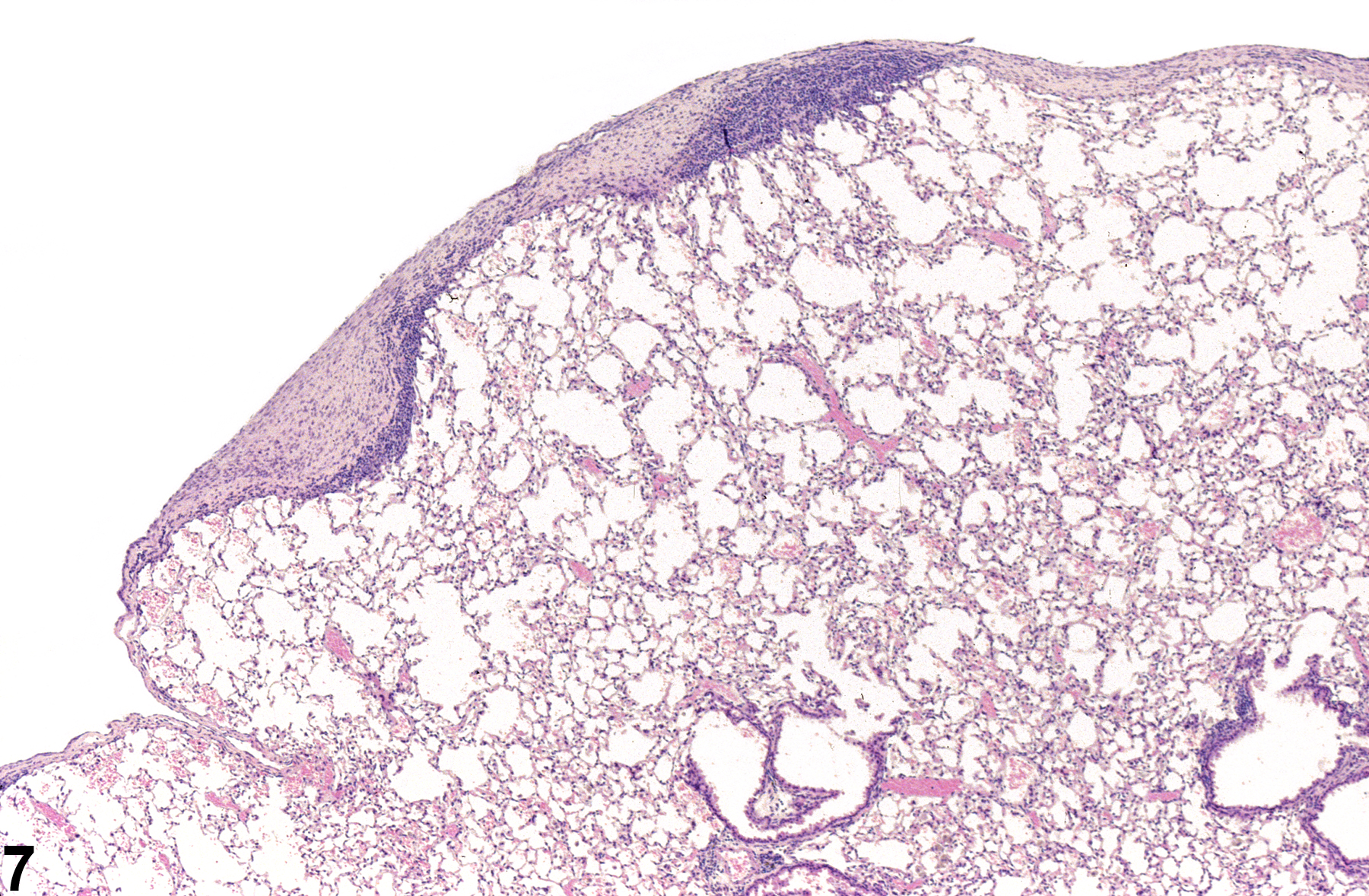
Lung, Bronchus - Fibrosis in a male B6C3F1/N mouse from a chronic study. There is multifocal subepithelial bronchial fibrosis (arrow).
Figure 5 of 8
Image of fibrosis in the lung from a male B6C3F1/N mouse in a chronic study
Lung, Bronchus - Fibrosis in a male B6C3F1/N mouse from a chronic study (higher magnification of Figure 5). There is a focus of mature subepithelial bronchial fibrosis with epithelial atrophy.
Figure 6 of 8
Image of pleural fibrosis in the lung from a female B6C3F1/N mouse in a chronic study
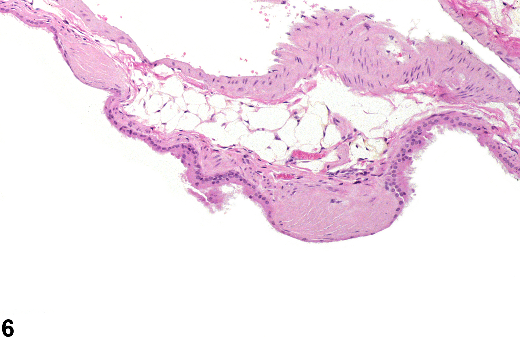
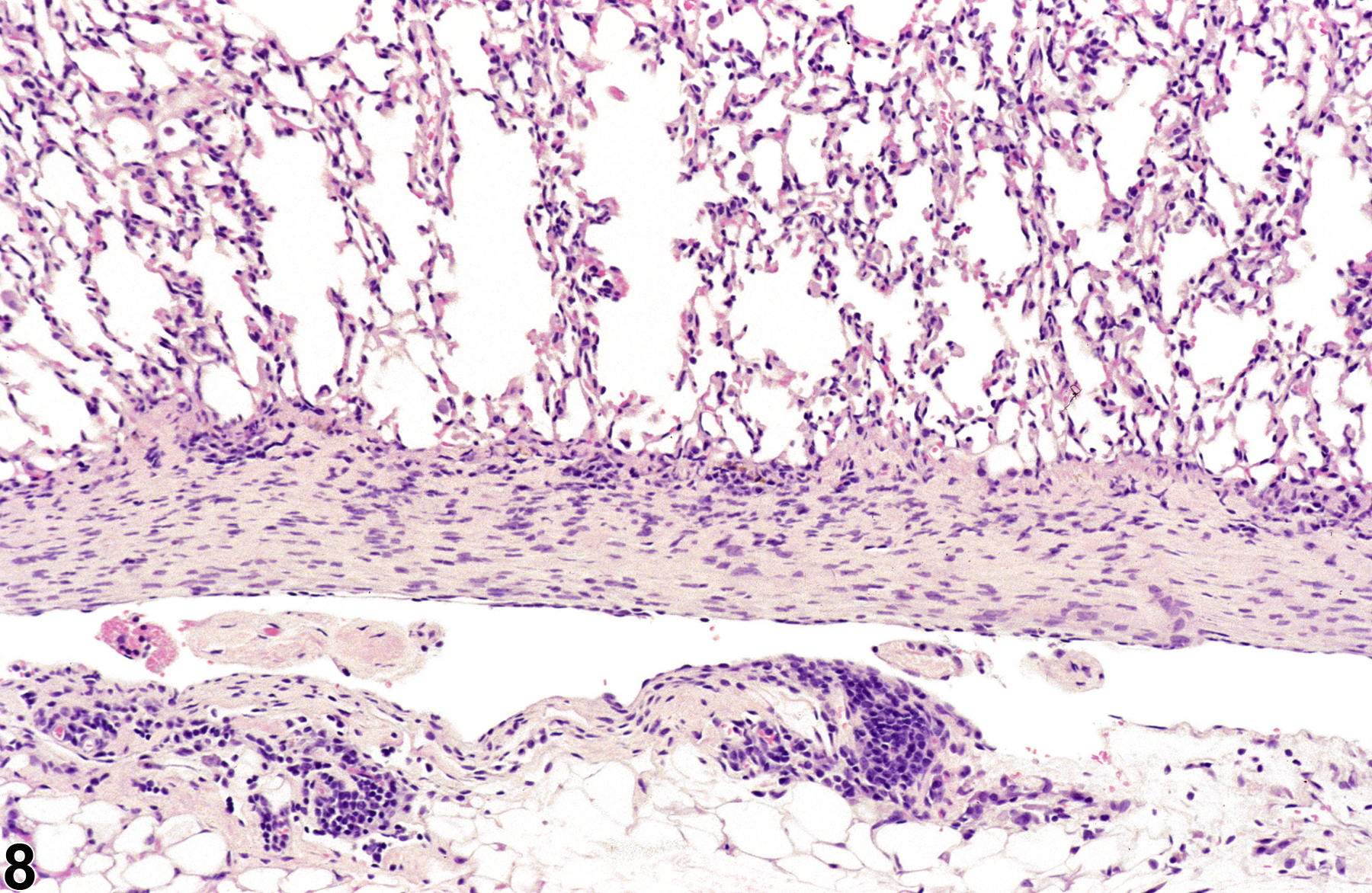
Lung, Pleura - Fibrosis in a female B6C3F1/N mouse from a chronic study. The pleura is thickened by fibrosis and inflammation.
Figure 7 of 8
Image of pleural fibrosis in the lung from a female B6C3F1/N mouse in a chronic study
Lung, Pleura - Fibrosis in a female B6C3F1/N mouse from a chronic study. The pleura is diffusely thickened by fibrosis and inflammation.
Figure 8 of 8
next arrow

comment:

Fibrosis appears microscopically as an increase in the amount of fibrous tissue that either replaces or expands normal structures. Fibrosis can occur anywhere in the lung, including alveolar septa or interstitium ( Figure 1image opens in a pop-up window , Figure 2image opens in a pop-up window , and Figure 3image opens in a pop-up window ), airways ( Figure 4image opens in a pop-up window , Figure 5image opens in a pop-up window , and Figure 6image opens in a pop-up window ), and pleura ( Figure 7image opens in a pop-up window and Figure 8image opens in a pop-up window ), depending on the site at which a particular agent produces damage. Fibrosis generally consists of mature, dense collagen, although areas of loose immature collagen may also be present. In airways, the fibrosis commonly occurs as focal projections of fibrous tissue into the airway lumen that may partially occlude the lumen (Figure 4) but may also occur as circumferential thickening of the airway wall ( Figure 5image opens in a pop-up window and Figure 6image opens in a pop-up window ). Because fibrosis is often a response to tissue damage (it is meant to replace damaged tissue when repair is not possible), it is often associated with inflammation. Fibrosis can be confirmed with special stains for collagen. With the Mason's trichrome staining technique, mature collagen stains blue ( Figure 2image opens in a pop-up window ).

recommendation:

Lung - Fibrosis should be diagnosed and graded whenever present. The location of the fibrosis (alveolus, bronchus/bronchiole, pleura) should be included in the diagnosis as a site modifier. The preferred terminology for fibrosis involving the interalveolar septa (alveolar walls) is Lung, Interstitium - Fibrosis. If fibrosis occurs in conjunction with inflammation, it should be diagnosed separately, though if the degree of fibrosis is very minimal or sporadic (occurring in only a few animals), it may be described in the pathology narrative in lieu of a separate diagnosis.

references:

Boorman GA, Eustis SL. 1990. Lung. In: Pathology of the Fischer Rat: Reference and Atlas (Boorman GA, Eustis SL, Elwell MR, Montgomery CA, MacKenzie WF, eds). Academic Press, San Diego, CA, 339-367.

Dixon D, Herbert RA, Sills RC, Boorman GA. 1999. Lungs, pleura, and mediastinum. In: Pathology of the Mouse: Reference and Atlas (Maronpot RR, Boorman GA, Gaul BW, eds). Cache River Press, Vienna, IL, 293-332.

Renne RA, Dungworth DL, Keenan CM, Morgan KT, Hahn FF, Schwartz LW. 2003. Non-proliferative lesions of the respiratory tract in rats. In: Guides for Toxicologic Pathology. STP/ARP/AFIP, Washington, DC.