U.S. flag

An official website of the United States government

Dot gov

The .gov means it's official.
Federal government websites often end in .gov or .mil. Before sharing sensitive information, make sure you're on a federal government site.

Https

The site is secure.
The https:// ensures that you are connecting to the official website and that any information you provide is encrypted and transmitted securely.

Skip to Main Navigation
Skip to Page Content
Skip to Atlas Navigation

Lung - Infiltration Cellular, Histiocyte

Image of infiltration cellular, histiocyte in the lung from a male Wistar Han rat in a subchronic study
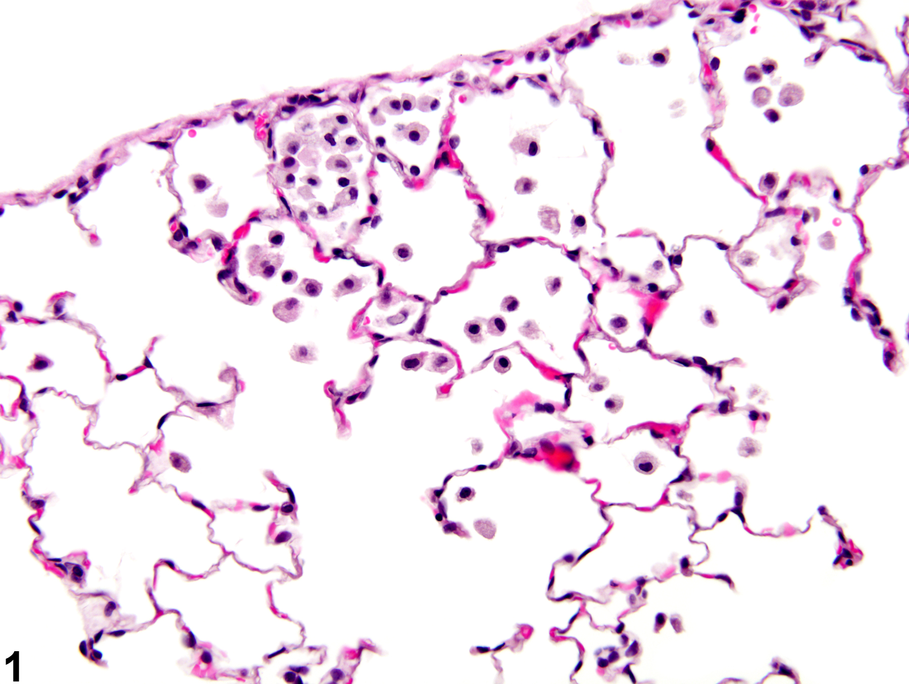
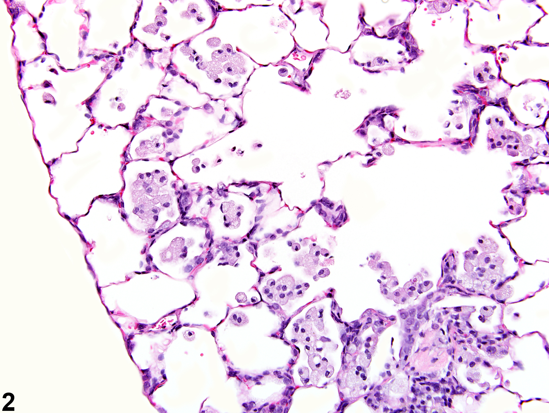
Lung - Infiltration cellular, Histiocyte in a male Wistar Han rat from a subchronic study. There is a small, subpleural accumulation of alveolar histiocytes.
Figure 1 of 6
Image of infiltration cellular, histiocyte in the lung from a female Harlan Sprague-Dawley rat in a chronic study
Lung - Infiltration cellular, Histiocyte in a female Harlan Sprague-Dawley rat from a chronic study. There is a small, subpleural accumulation of alveolar histiocytes with some thickening of associated alveolar septae.
Figure 2 of 6
Image of infiltration cellular, histiocyte in the lung from a male F344/NTac rat in a subchronic study
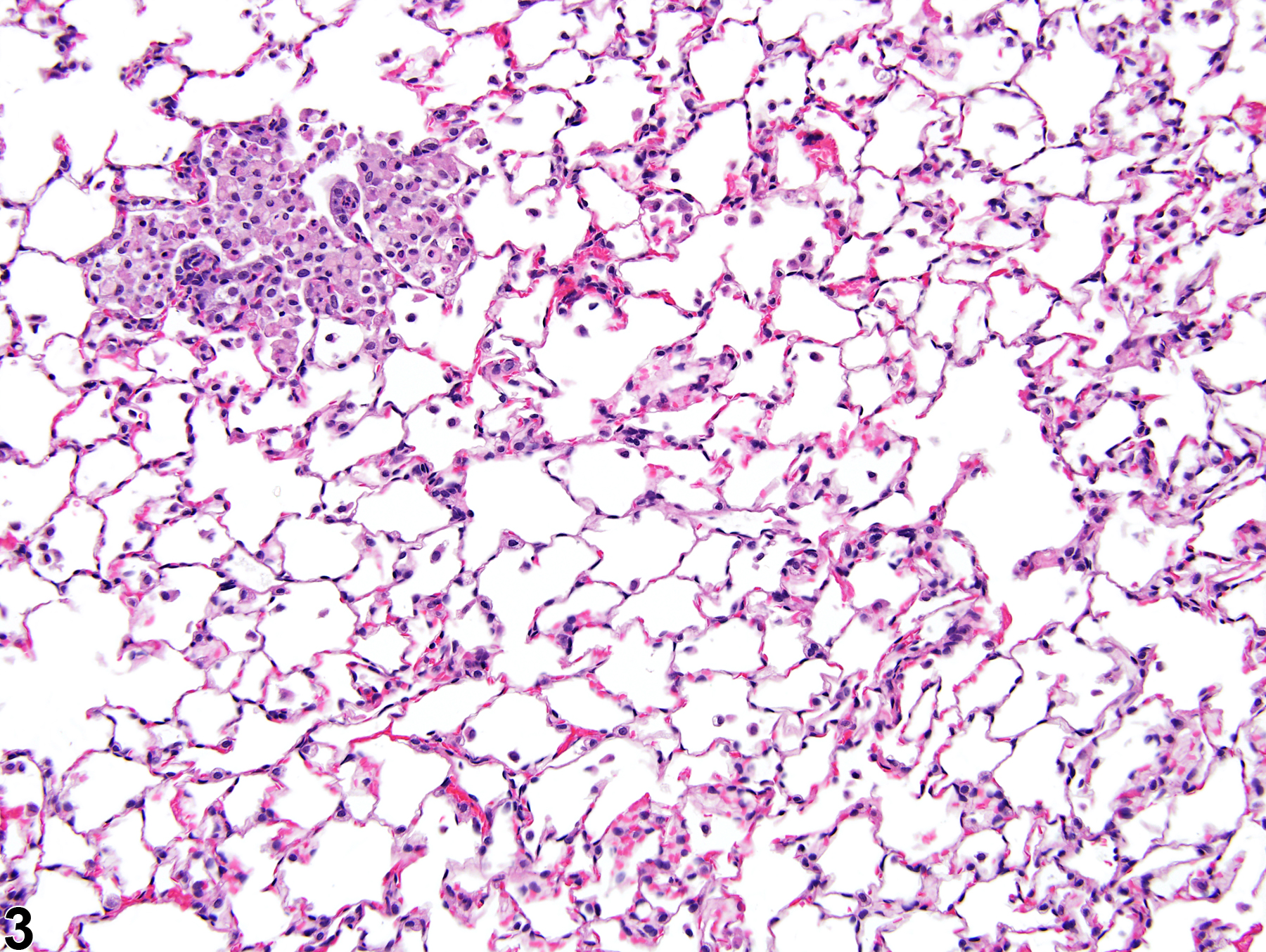
Lung - Infiltration cellular, Histiocyte in a control male F344/NTac rat from a subchronic study. There is a focal accumulation of histiocytes in several adjacent alveoli.
Figure 3 of 6
Image of infiltration cellular, histiocyte in the lung from a male F344/NTac rat in a subchronic study
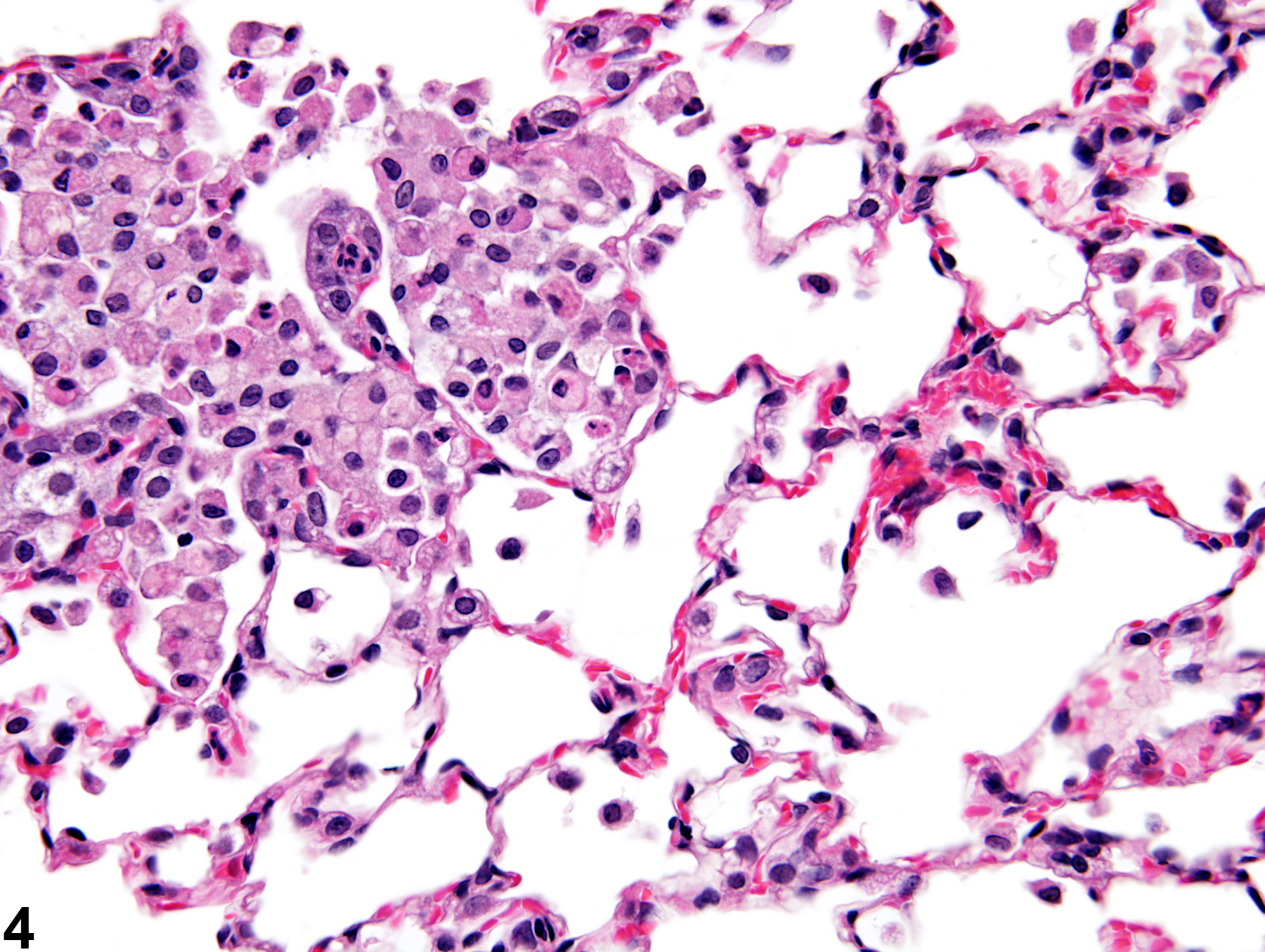
Lung - Infiltration cellular, Histiocyte in a control male F344/NTac rat form a subchronic study (higher magnification of Figure 3). Many of the alveolar macrophages in this focal accumulation contain variably sized vacuoles.
Figure 4 of 6
Image of infiltration cellular, histiocyte in the lung from a male Wistar Han rat in a subchronic study
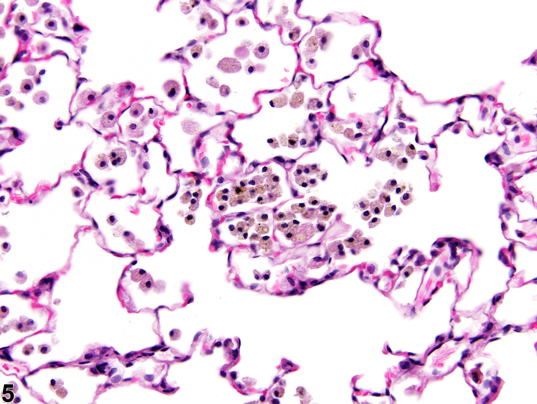
Lung - Infiltration cellular, Histiocyte in a male Wistar Han rat from a subchronic study. These histiocytes contain brown material, which is considered to be the test agent, and this animal would also be diagnosed with foreign material.
Figure 5 of 6
Image of infiltration cellular, histiocyte in the lung from a male Harlan Sprague-Dawley rat in a subchronic study
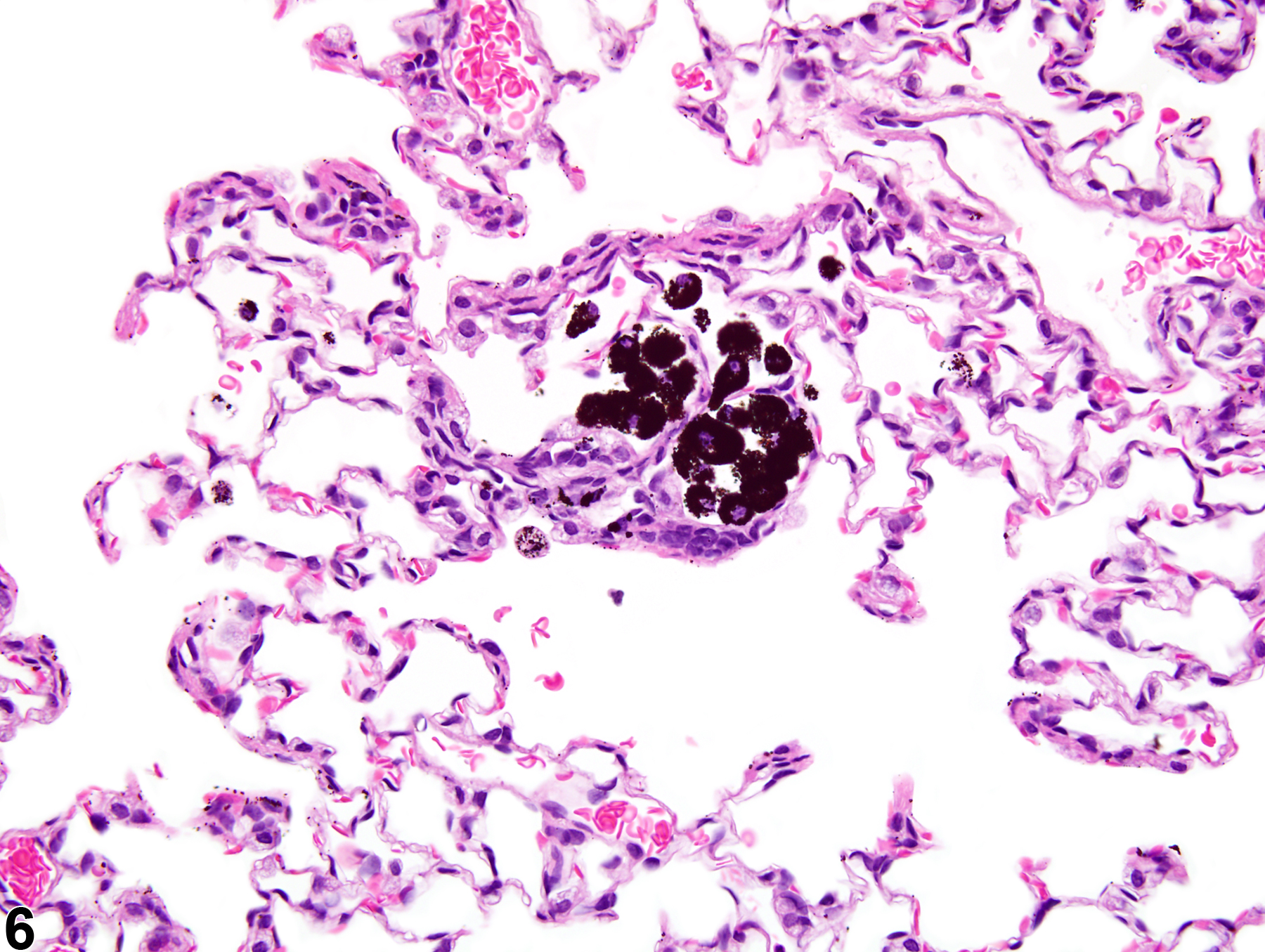
Lung - Infiltration, Cellular, Histiocyte in a male Harlan Sprague-Dawley rat from a subchronic study. These histiocytes contain black material, which is considered to be the test agent; this animal would also be diagnosed with foreign material.
Figure 6 of 6
next arrow

comment:

Responses to inhaled toxicants often include the accumulation of alveolar macrophages within alveoli. Accumulation of alveolar macrophages is also part of the response to necrosis and may be associated with pulmonary neoplasms or alveolar/bronchiolar hyperplasia. Focal histiocytic infiltrates may be observed spontaneously, most often in subpleural regions of older animals. Alveolar histiocytic infiltrates consist of intra-alveolar accumulations of macrophages. The macrophages can appear activated (enlarged with foamy cytoplasm) or quiescent. Though the lesion is often limited to the accumulation of alveolar macrophages ( Figure 1image opens in a pop-up window , Figure 3image opens in a pop-up window , Figure 4image opens in a pop-up window , Figure 5image opens in a pop-up window , and Figure 6image opens in a pop-up window ), associated findings may be present, such as a few other inflammatory cells (lymphocytes or neutrophils), minimal edema, hemorrhage, or thickening of alveolar septa ( Figure 2image opens in a pop-up window ). Also, the macrophages may contain pigment, which may represent hemosiderin, or foreign material consistent with the test article ( Figure 5image opens in a pop-up window and Figure 6image opens in a pop-up window ).

Histiocytic infiltration must be differentiated from inflammation, which can sometimes be difficult. In general, histiocytic infiltration is characterized by increased numbers of alveolar histiocytes with no (or minimal) other evidence of inflammation (e.g., the presence of other types of inflammatory cells, edema, hemorrhage, alveolar septal thickening).

recommendation:

Lung - Infiltration cellular, Histiocyte should be diagnosed and given a severity grade in most cases. If the histiocytes contain the test agent, it should be diagnosed and graded separately foreign material (see Lung - Foreign Material). If the alveolar macrophages are considered to be a component of an inflammatory response, then inflammation should be diagnosed in lieu of histiocytic infiltration. Accumulations of alveolar histiocytes that are clearly associated with a neoplasm should not be recorded but should be described in the narrative as being secondary to the neoplasm. Small, subpleural accumulations of alveolar macrophages are frequently seen as background lesions in rats. If histiocytic infiltration is a diffuse change related to test article administration, and focal, subpleural (background) aggregates might skew incidence statistics, then either the non-test-article-related subpleural foci in chronic studies should not be recorded (i.e., considered to be within normal limits), or they may be recorded separately using modifier (e.g., Infiltration cellular, Histiocyte, Focal or Infiltration cellular, Histiocyte, Diffuse). In some cases, it may be best to diagnose both lesions. For example, if there is a continuum from alveolar histiocyte infiltration (with no inflammatory changes) in the low-dose groups to alveolar histiocyte infiltration with areas of inflammation in the high-dose groups, then it may be necessary to diagnose both lesions to fully capture the effects of the test article. The pathologist will need to use his or her judgment in recording these lesions and must take care to maintain consistency throughout the study.

related links:

Lung - Foreign Material

references:

Boorman GA, Eustis SL. 1990. Lung. In: Pathology of the Fischer Rat: Reference and Atlas (Boorman GA, Eustis SL, Elwell MR, Montgomery CA, MacKenzie WF, eds). Academic Press, San Diego, CA, 339-367.

Dixon D, Herbert RA, Sills RC, Boorman GA. 1999. Lungs, pleura, and mediastinum. In: Pathology of the Mouse: Reference and Atlas (Maronpot RR, Boorman GA, Gaul BW, eds). Cache River Press, Vienna, IL, 293-332.

Renne, R, Brix A, Harkema J, Herbert R, Kittle B, Lewis D, March T, Nagano K, Pino M, Rittinghausen S, Rosenbruch M, Tellier P, Wohrmann T. 2009. Proliferative and nonproliferative lesions of the rat and mouse respiratory tract. Toxicol Pathol 37(suppl):5S-73S.
Abstract: https://www.ncbi.nlm.nih.gov/pubmed/20032296