

Respiratory System
Lung - Inflammation
Narrative
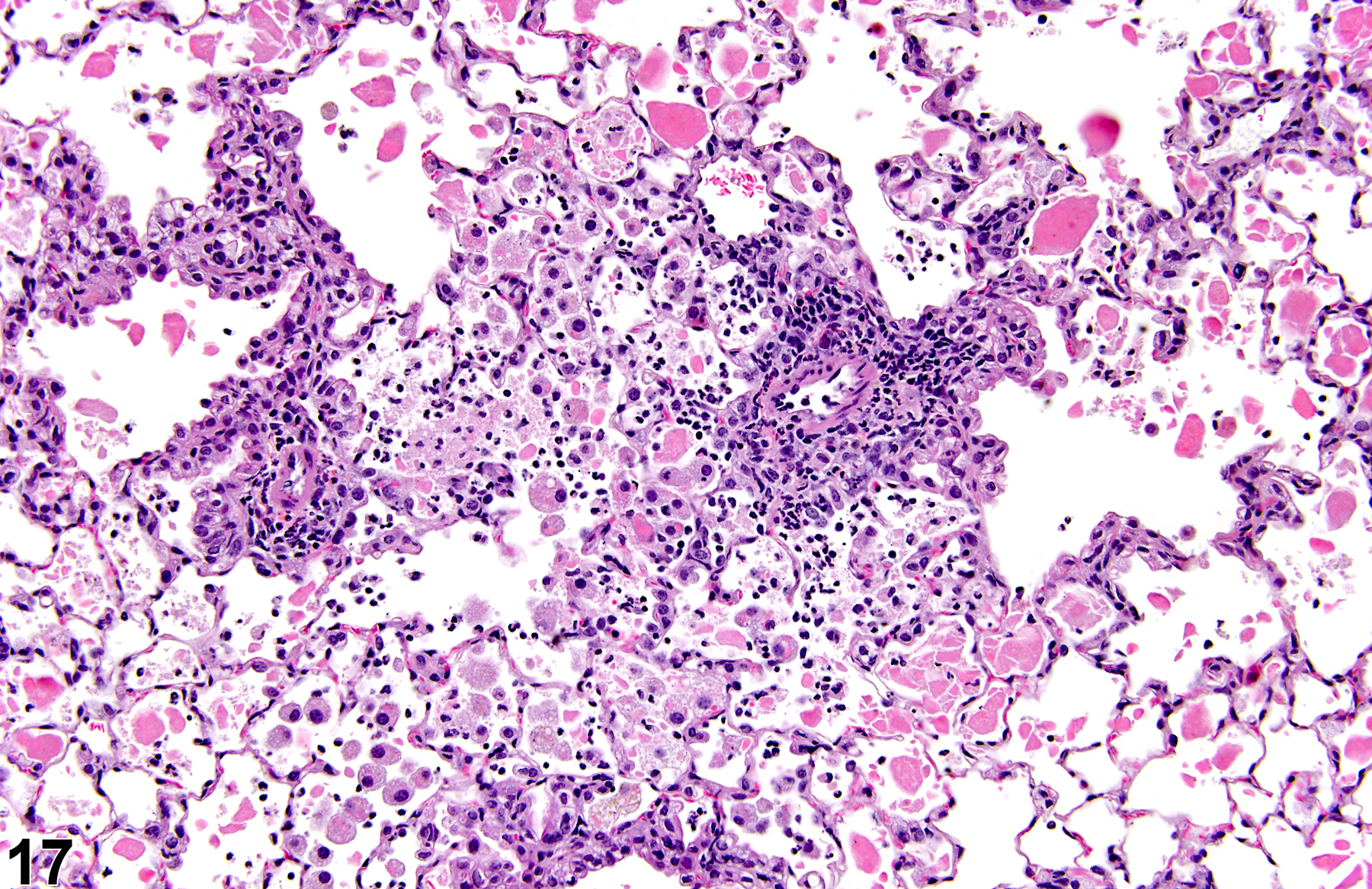
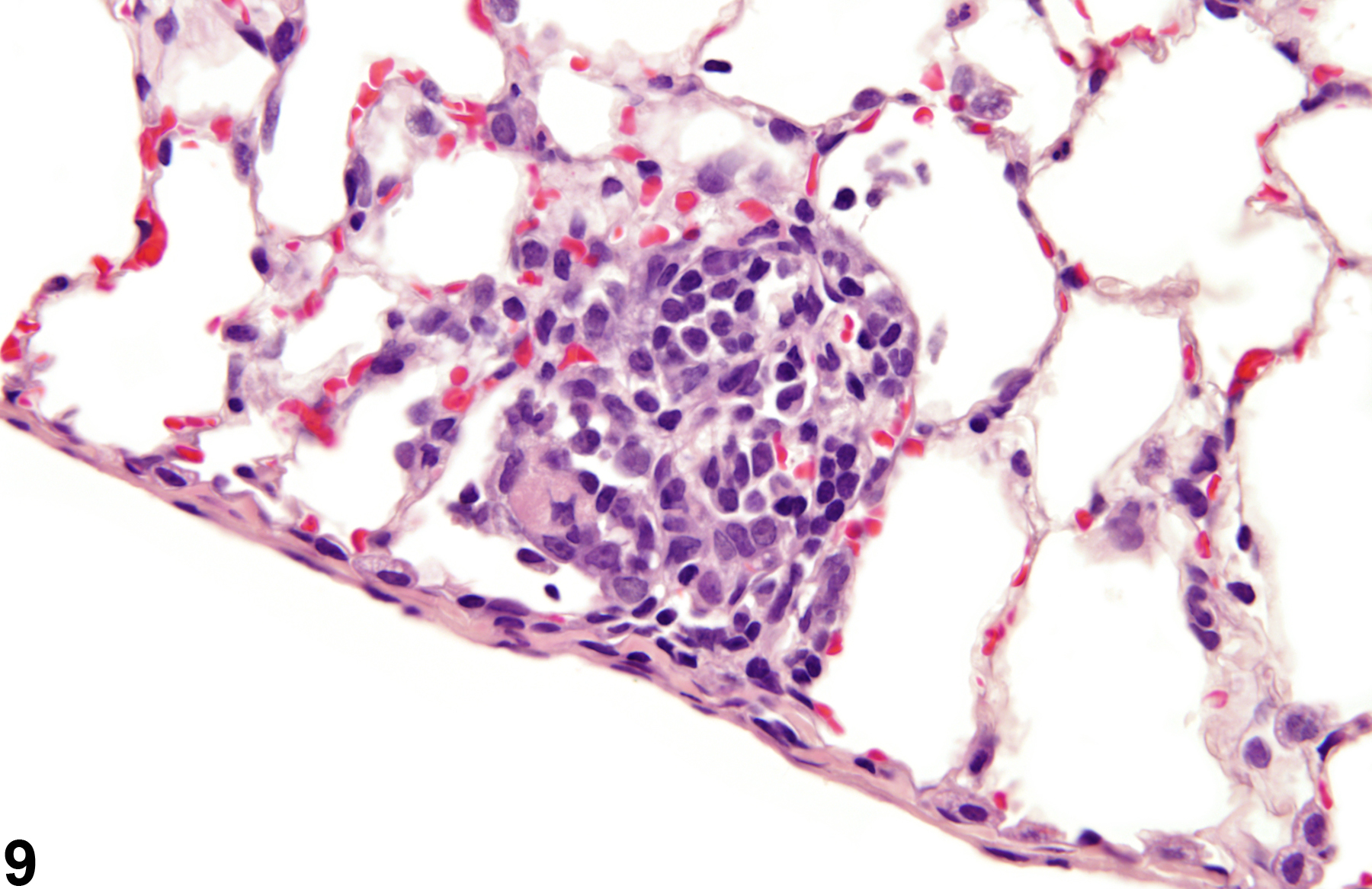
A test agent may stimulate epithelial cells and resident macrophages to secrete cytokines, thus inducing an inflammatory response. Alternatively, a test agent may cause tissue damage, which secondarily results in inflammation. Several compartments within the lung may be inflamed, including the airways (bronchi and bronchioles), the alveoli or alveolar septa (interstitium), the terminal bronchiole/alveolar duct region (acinar region), perivascular areas, and the pleura. Occasionally, focal, minimal inflammation in the lung is seen as a background lesion in mice and rats (Figure 9 and Figure 10), particularly in the subpleural region. Small aggregates of alveolar histiocytes can also be seen (see) as a background lesion and must be differentiated from inflammation. Inflammation can also be caused by a number of infectious agents, but these are rare under current husbandry practices. Systemic bacterial infections affecting the pulmonary interstitium via the bloodstream often produce a suppurative alveolitis, whereas viral infections tend to induce suppurative or mononuclear perivascular inflammation. Inflammation can also be associated with pulmonary neoplasms.
Boorman GA, Eustis SL. 1990. Lung. In: Pathology of the Fischer Rat: Reference and Atlas (Boorman GA, Eustis SL, Elwell MR, Montgomery CA, MacKenzie WF, eds). Academic Press, San Diego, CA, 339-367.
Dixon D, Herbert RA, Sills RC, Boorman GA. 1999. Lungs, pleura, and mediastinum. In: Pathology of the Mouse: Reference and Atlas (Maronpot RR, Boorman GA, Gaul BW, eds). Cache River Press, Vienna, IL, 293-332.
Dungworth DL, Ernst H, Nolte T, Mohr U. 1992. Nonneoplastic lesions in the lungs. In: Pathobiology of the Aging Rat (Mohr U, Dungworth DL, Capen CC, eds). ILSI Press, Washington, DC, 143-160.
Plopper CG, Dungworth DL. 197. Structure, function, cell injury and cell renewal of bronchiolar and alveolar epithelium. In: Lung Carcinomas (McDowell EM, ed). Churchill Livingstone, Edinburgh, 94-128.
Renne, R, Brix A, Harkema J, Herbert R, Kittle B, Lewis D, March T, Nagano K, Pino M, Rittinghausen S, Rosenbruch M, Tellier P, Wohrmann T. 2009. Proliferative and nonproliferative lesions of the rat and mouse respiratory tract. Toxicol Pathol 37(suppl):5S-73S.
Abstract: https://www.ncbi.nlm.nih.gov/pubmed/20032296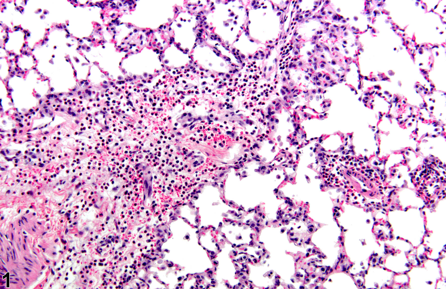
Lung - Inflammation, Acute in a male Wistar Han rat from a subchronic study. The majority of the inflammatory cells are neutrophils; there is also a small amount of hemorrhage.
All Images

Lung - Inflammation, Acute in a male Wistar Han rat from a subchronic study. The majority of the inflammatory cells are neutrophils; there is also a small amount of hemorrhage.

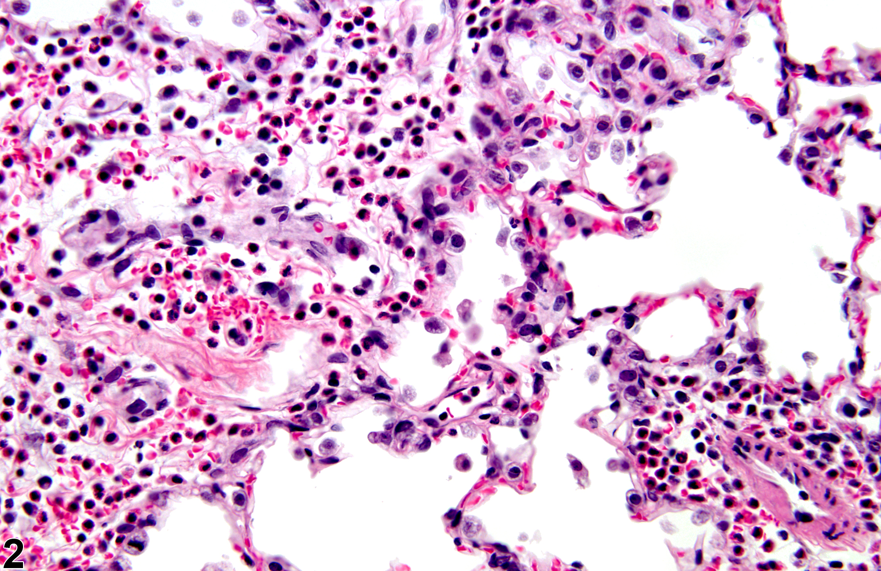
Lung - Inflammation, Acute in a male Wistar Han rat from a subchronic study (higher magnification of Figure 1). The majority of the inflammatory cells are neutrophils, but there are also mononuclear cells, including alveolar macrophages.
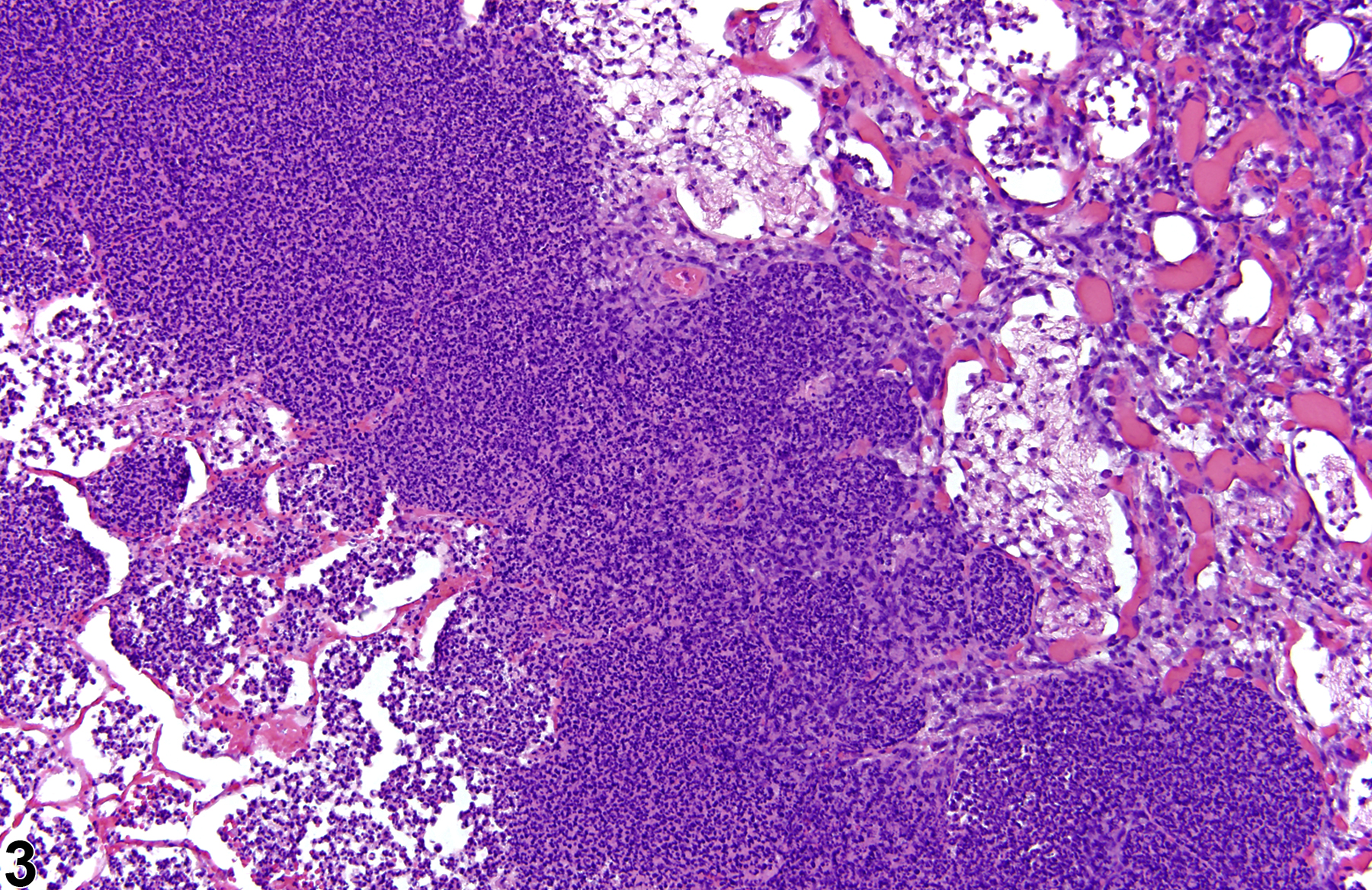
Lung - Inflammation, Suppurative in a male Wistar Han rat from a chronic study. Large numbers of degenerate neutrophils fill and replace alveoli.

Lung - Inflammation, Suppurative in a male Wistar Han rat from a chronic study (higher magnification of Figure 3). Abundant necrotic debris is admixed with the degenerate neutrophils.

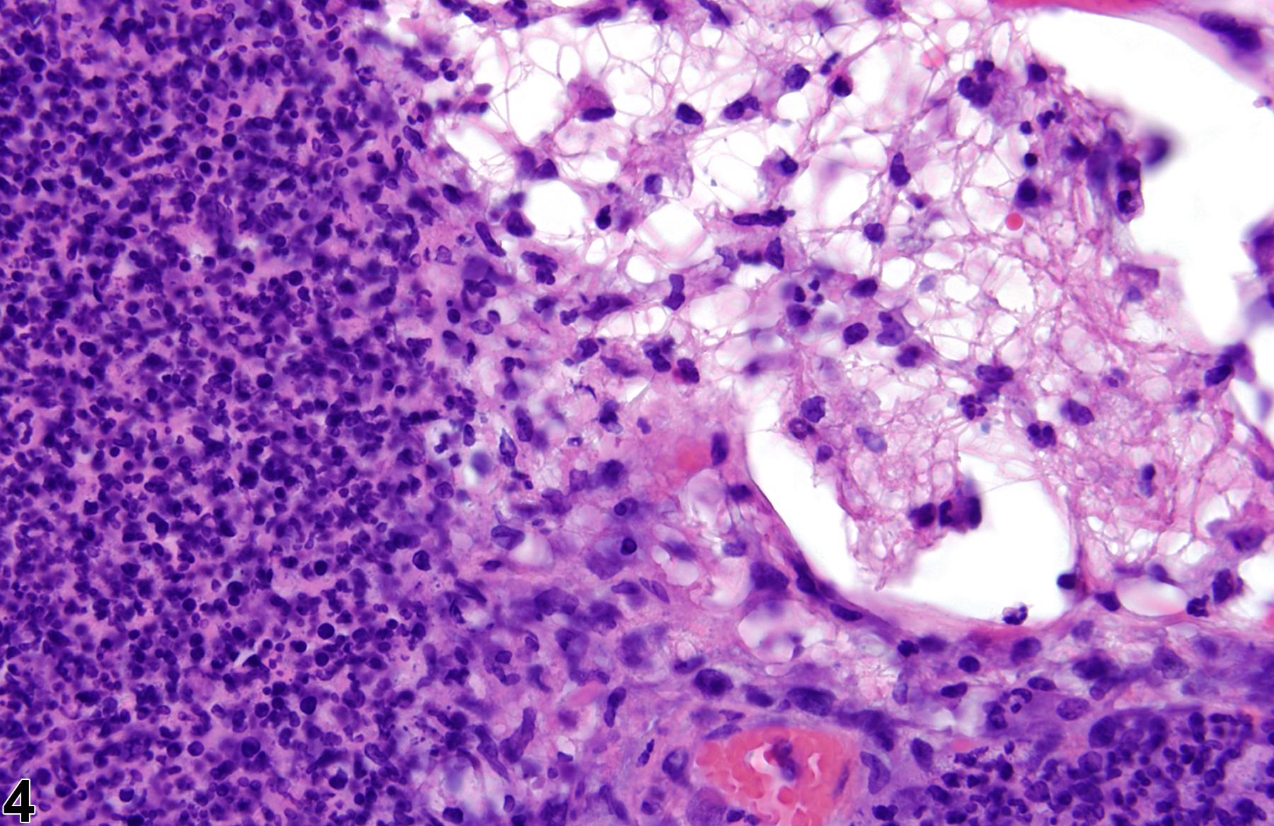
Lung, Bronchiole - Inflammation, Suppurative in a male B6C3F1/N mouse from a chronic study. Degenerate neutrophils fill the bronchiole.

Lung - Inflammation, Suppurative in a female B6C3F1/N mouse from a chronic study. There are numerous large, foamy, activated alveolar macrophages amid the degenerate neutrophils.
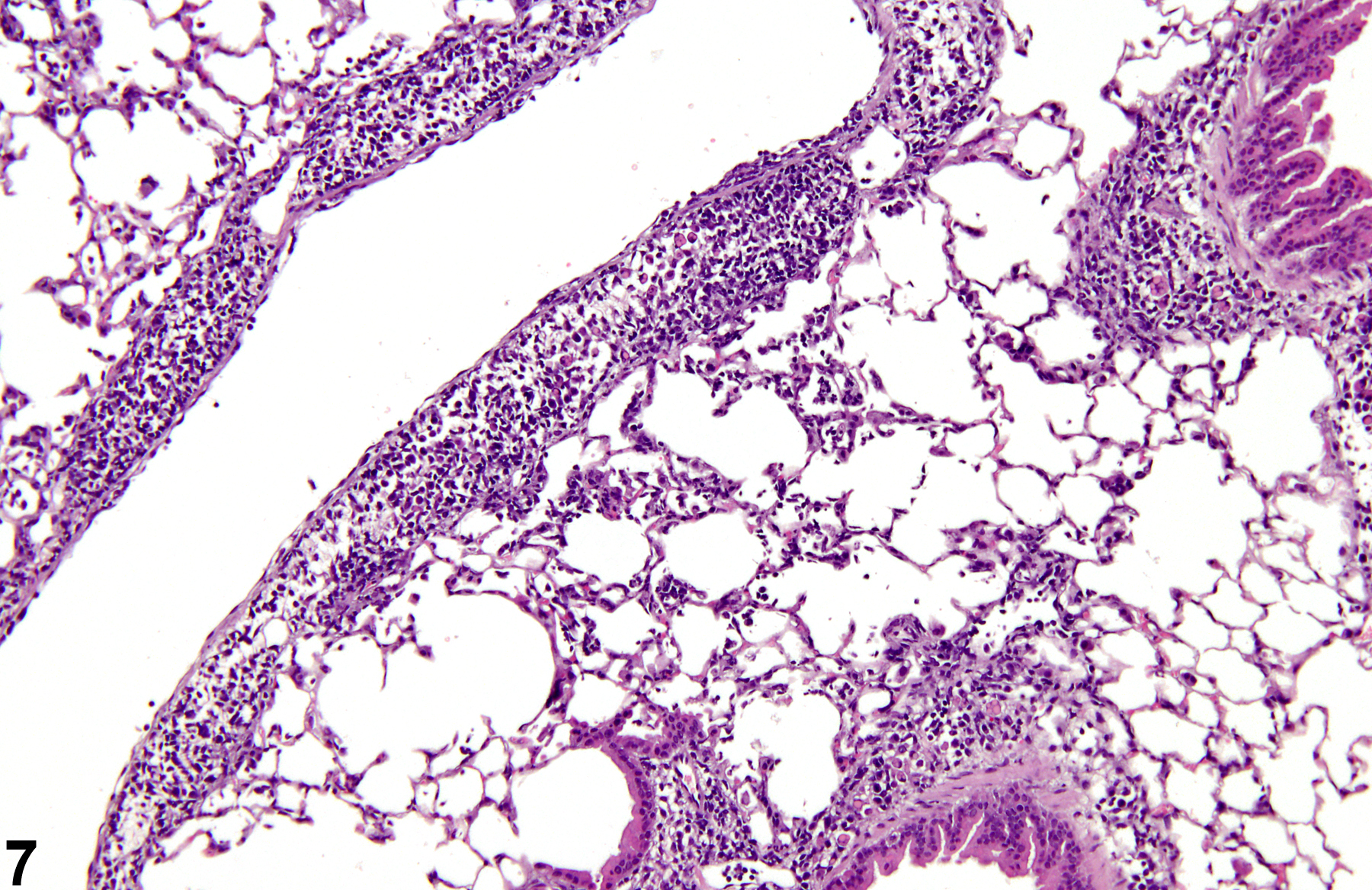
Lung - Inflammation, Chronic in a female B6C3F1/N mouse from a chronic study. The mononuclear inflammatory cells are largely perivascular, peribronchiolar, and subpleural.

Lung - Inflammation, Chronic in a female B6C3F1/N mouse from a chronic study (higher magnification of Figure 7). Mott cells are visible amid the lymphocytes and plasma cells in the subpleural region.

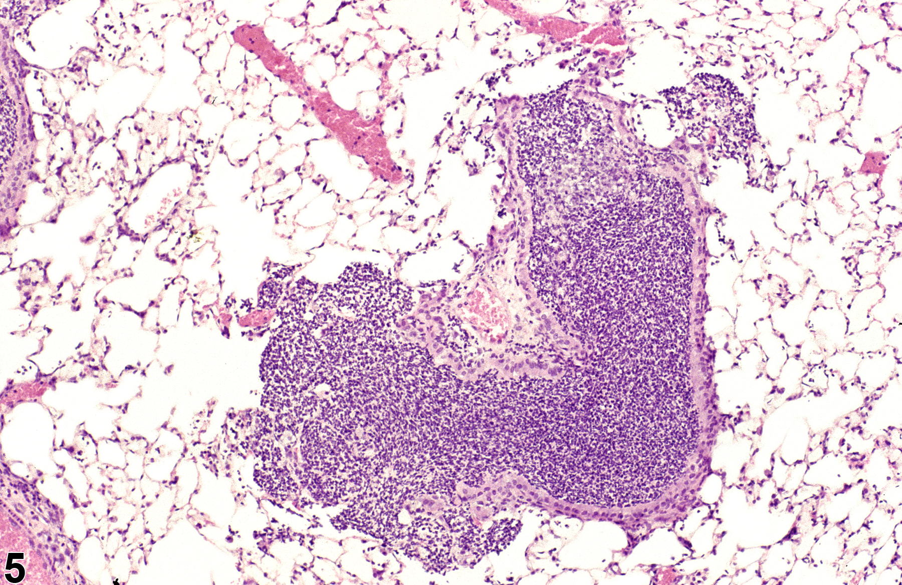
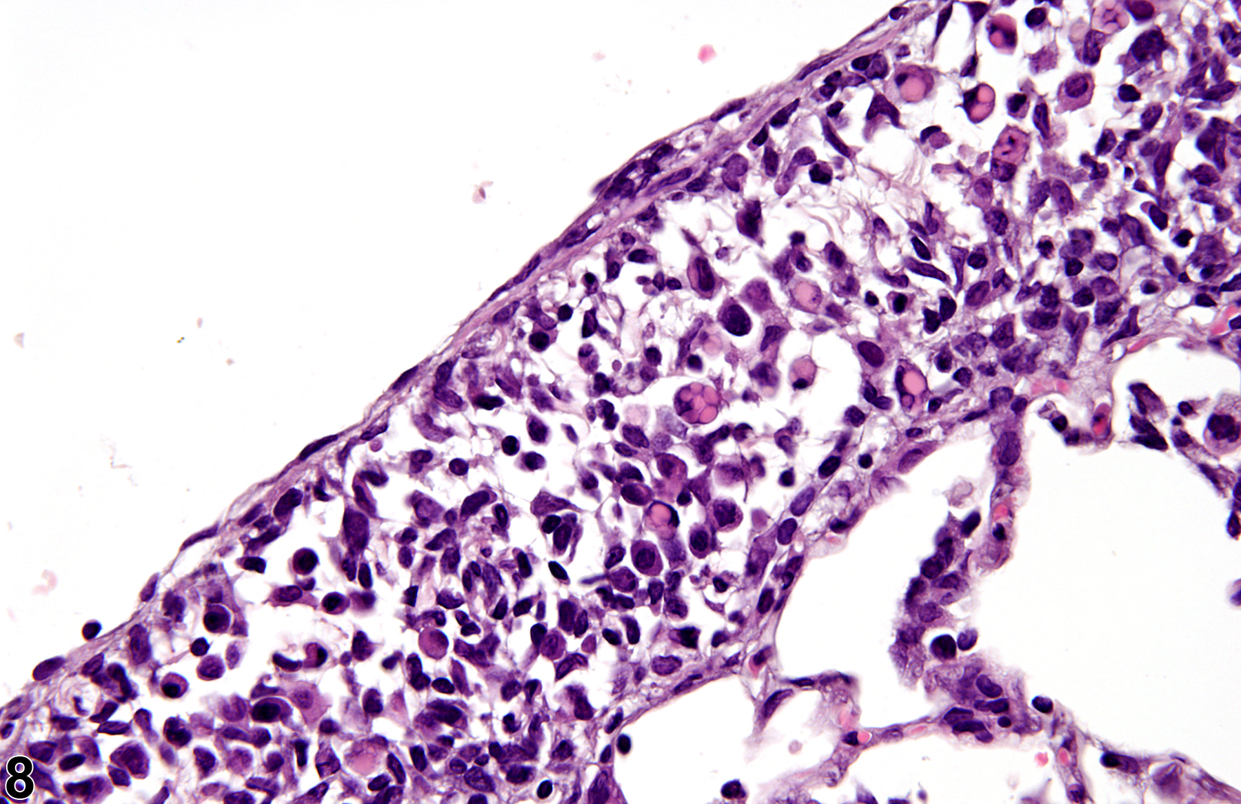
Lung - Inflammation, Chronic in a male F344/N rat from a subchronic study. These focal, subpleural lesions are a common background finding.

Lung - Inflammation, Chronic in a male F344/N rat from a subchronic study. This is a slightly more severe example of the common background lesion shown in Figure 9, with thickening of the alveolar septa.

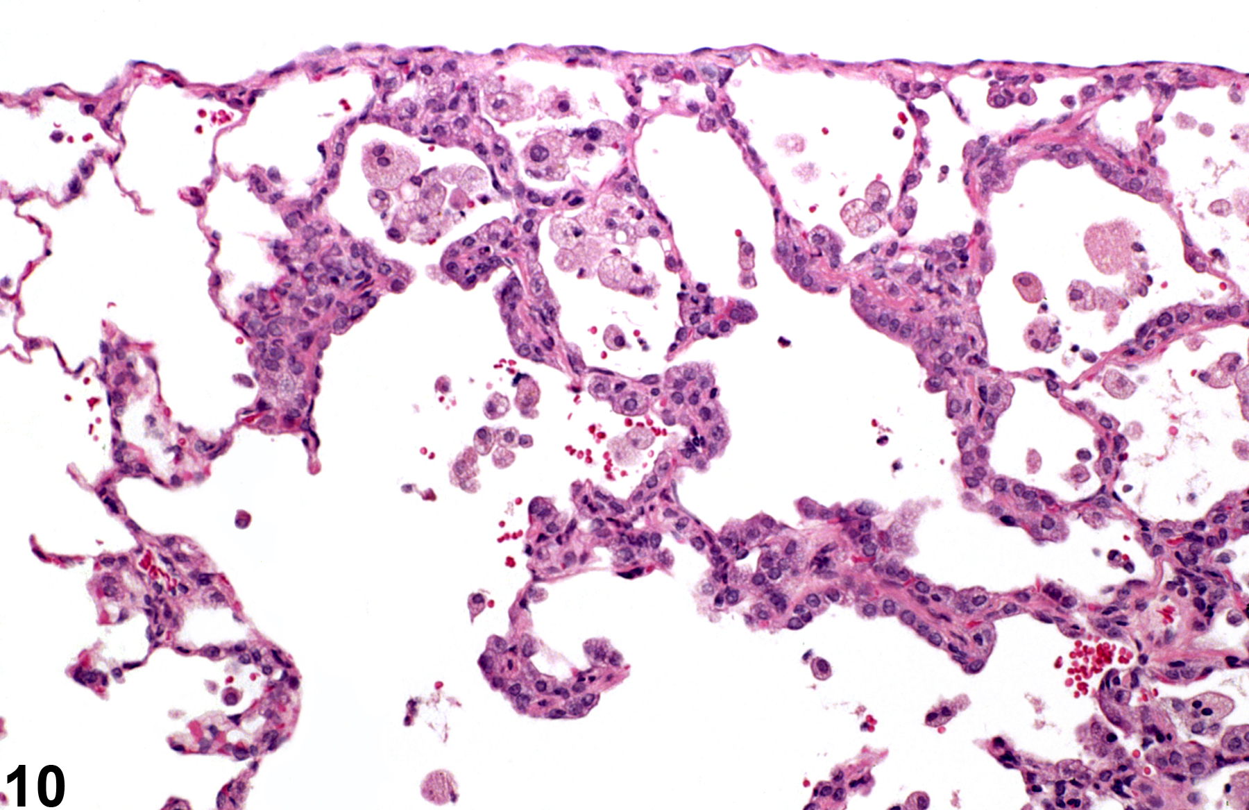
Lung - Inflammation, Chronic in a female B6C3F1/N mouse from a chronic study. Numerous cholesterol clefts and pigmented macrophages are present in this inflammatory lesion.

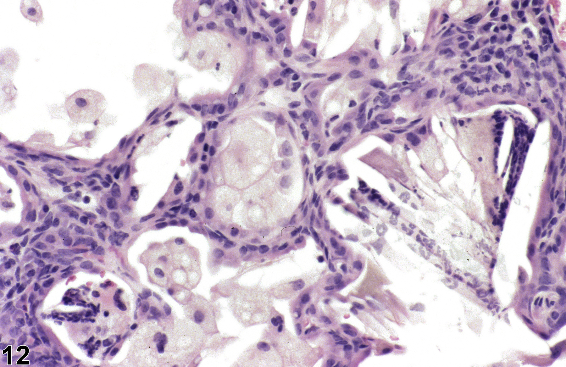
Lung - Inflammation, Chronic in a female B6C3F1/N mouse from a chronic study (higher magnification of Figure 11). The macrophages are large, foamy, and activated.
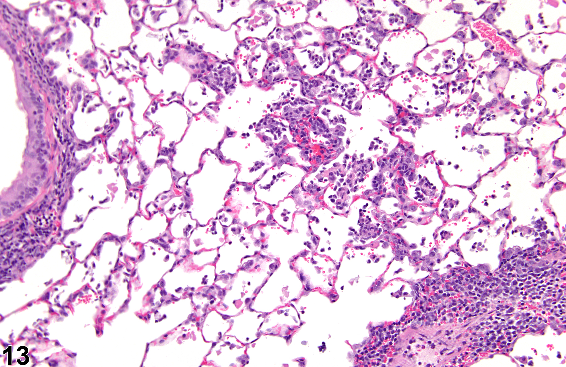
Lung - Inflammation, Chronic active in a male F344/N rat from a subchronic study. There is a mixture of lymphocytes, macrophages, and neutrophils.

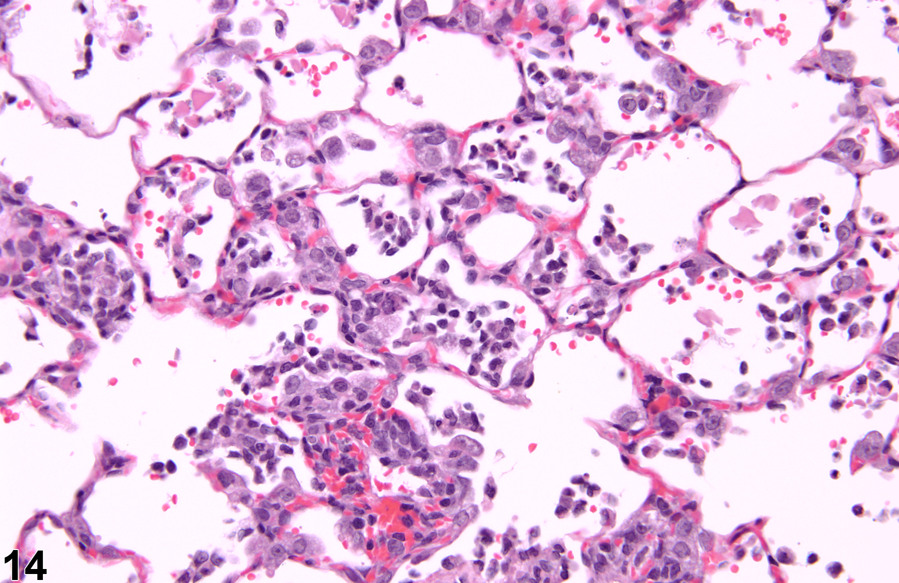
Lung - Inflammation, Chronic active in a male F344/N rat from a subchronic study (higher magnification of Figure 13). There is a mixture of lymphocytes, macrophages, and neutrophils, with a small amount of alveolar hemorrhage.

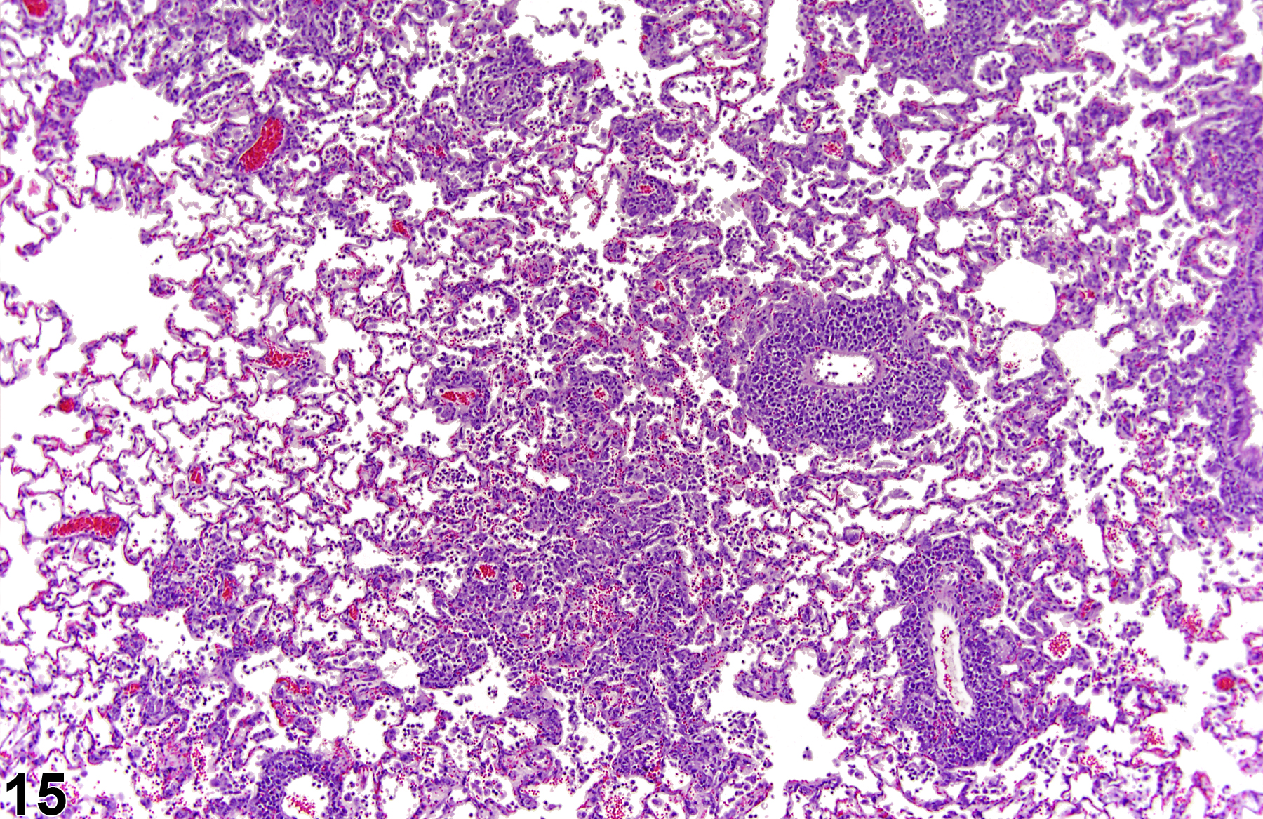
Lung - Inflammation, Chronic active in a male F344/NTac rat from a subchronic study. The perivascular and interstitial inflammation in this control rat is consistent with Pneumocystis carinii infection (formerly rat respiratory virus).

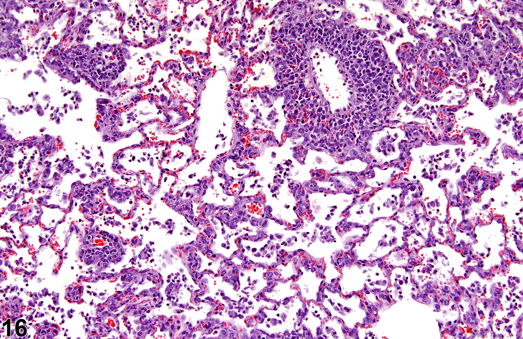
Lung - Inflammation, Chronic active in a male F344/NTAC rat from a subchronic study (higher magnification of Figure 15). The lesion is consistent with Pneumocystis carinii infection (formerly rat respiratory virus).

Lung - Inflammation, Chronic active in a male B6C3F1/N mouse from a subchronic study. There is a mixture of inflammatory cell types, including neutrophils, and alveolar proteinosis.
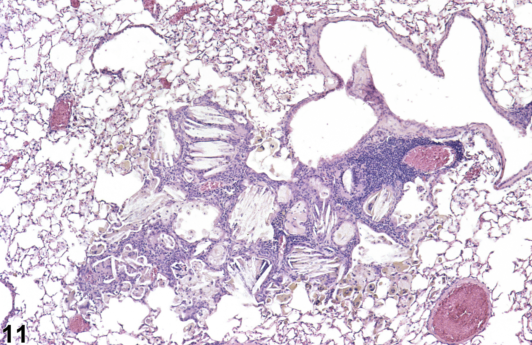
Lung - Inflammation, Granulomatous in a male Wistar Han rat from a chronic study. Clusters of large, foamy macrophages are surrounded by mononuclear cells.

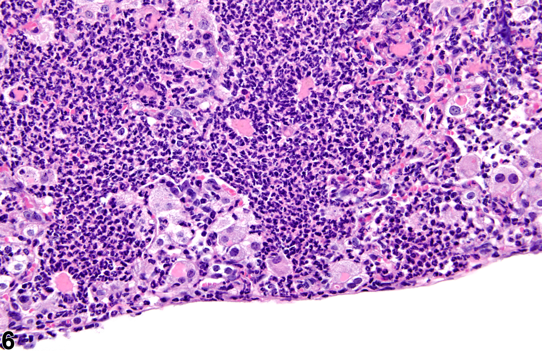
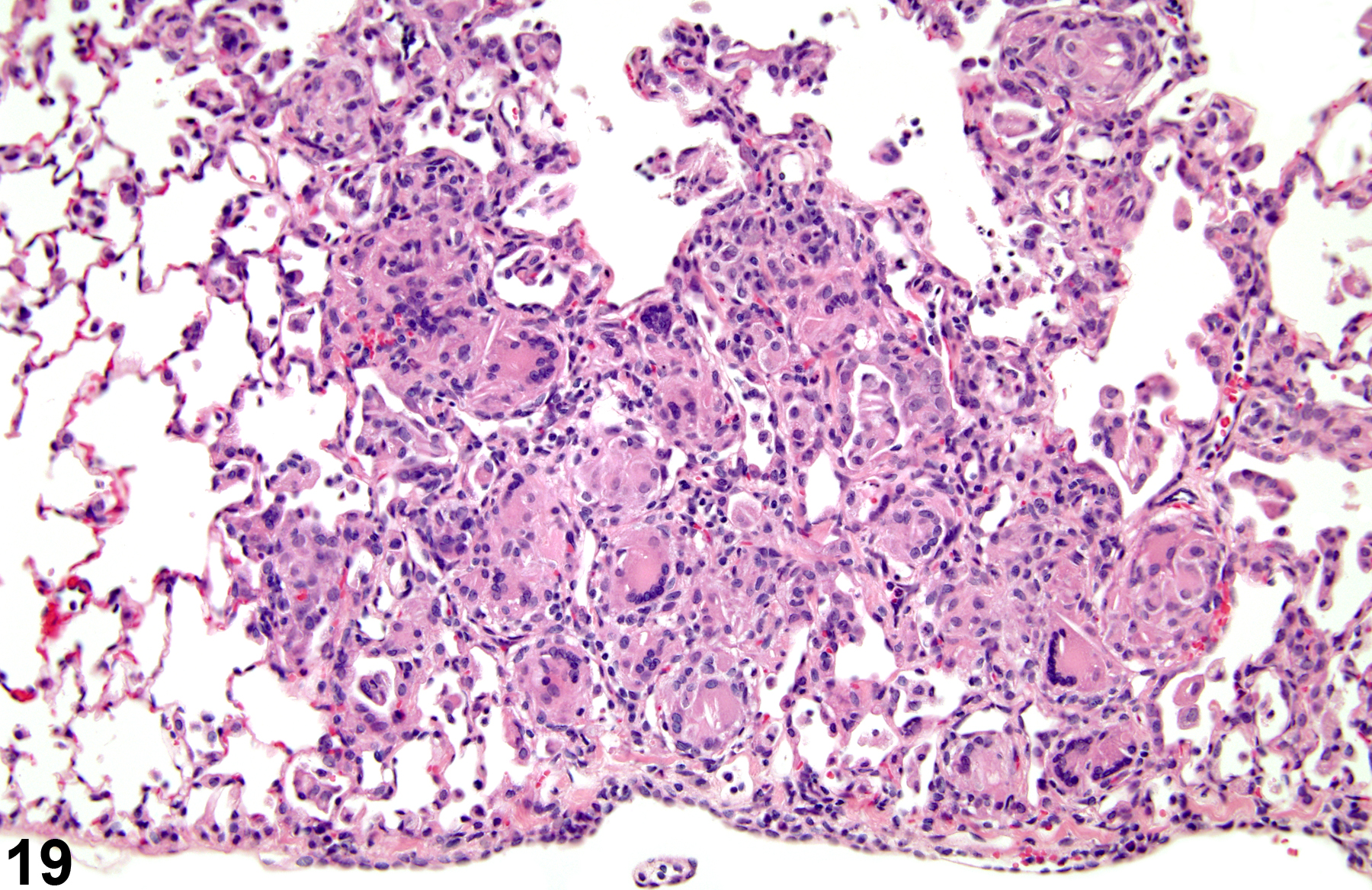
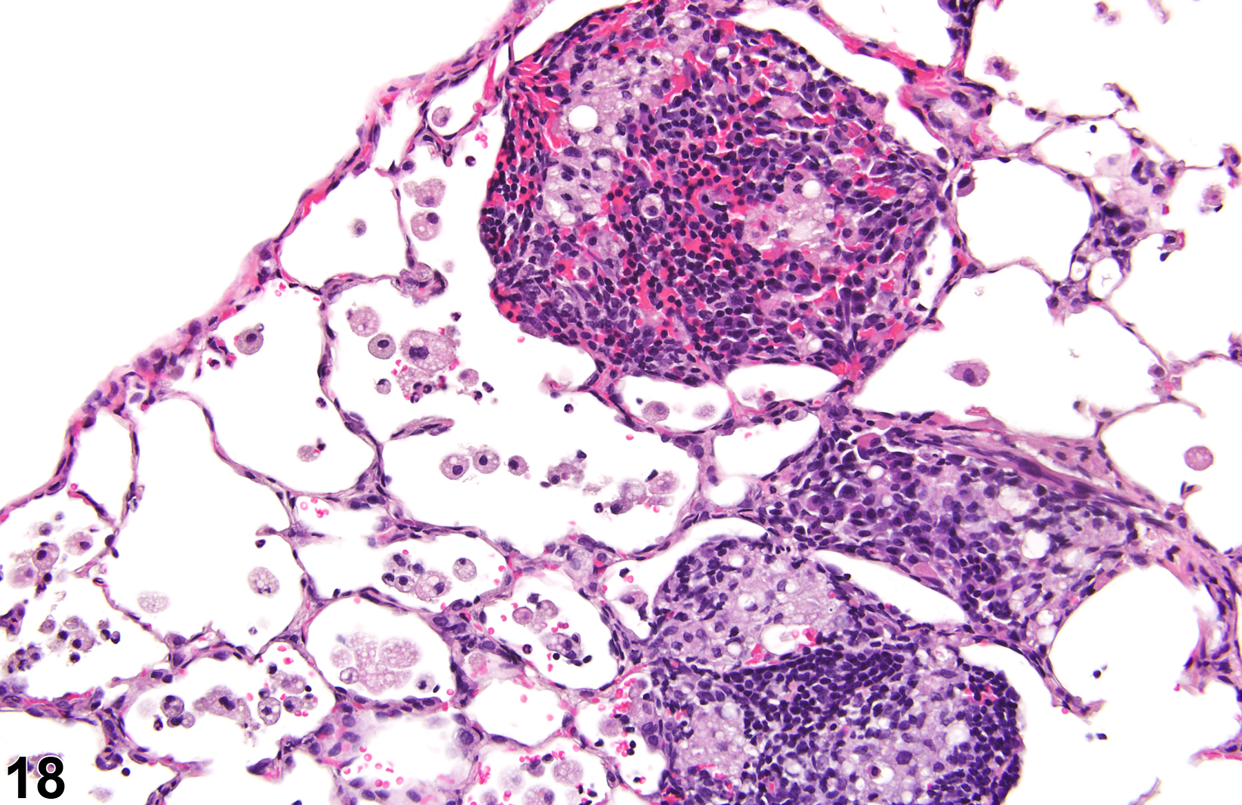
Lung - Inflammation, Granulomatous in a female F344/NTAC rat from a subchronic study. There are numerous multinucleated giant cells in this lesion.

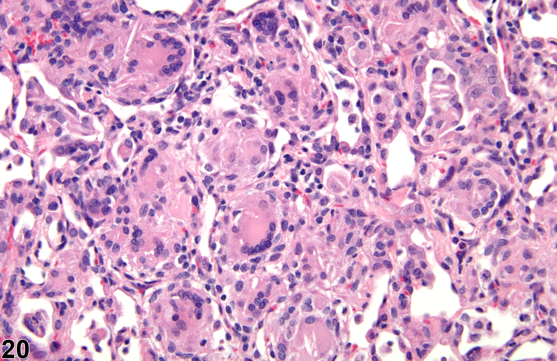
Lung - Inflammation, Granulomatous from a female F344/NTac rat in a subchronic study (higher magnification of Figure 19). The multinucleated giant cells are of the Langhans type.