

Respiratory System
Lung - Metaplasia, Goblet Cell
Narrative
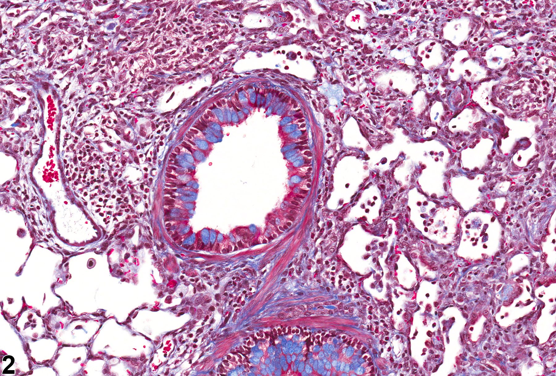
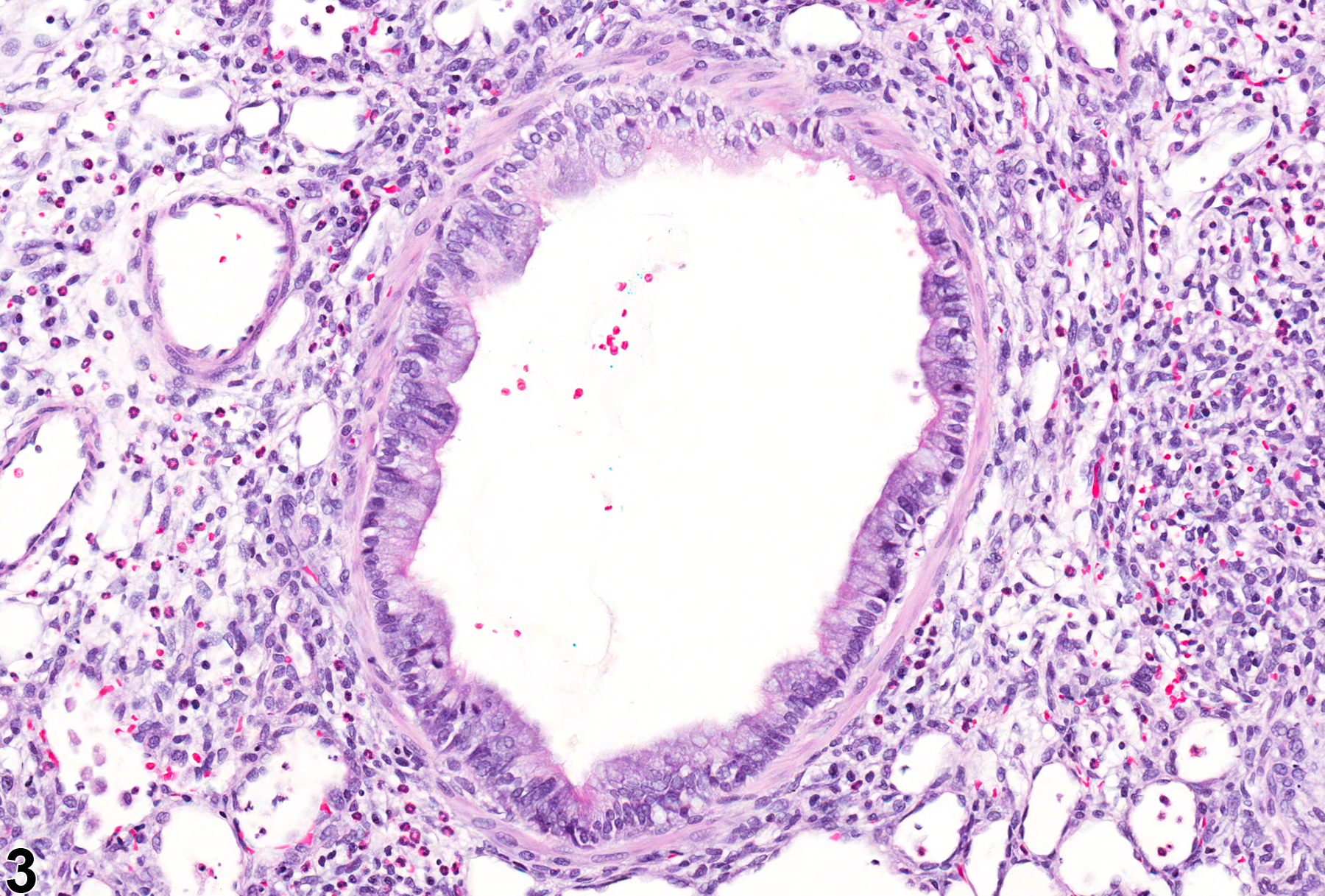
Goblet cell metaplasia is characterized by the presence of goblet cells (above the normal number; see above) in the epithelium lining the bronchi or bronchioles (Figure 1, Figure 2, and Figure 3). Goblet cell metaplasia may occur in association with infectious agents or exposure to inhaled irritants in both rats and mice. It is frequently seen concurrently with inflammation and, less frequently, fibrosis, though it may be seen in the absence of these lesions. Mucus can be stained with the Alcian blue method, which can facilitate the identification of this lesion. Mucus also stains blue with the Mason''s trichrome staining method (Figure 2), but this stain is not typically used to identify goblet cells, and Alcian blue is preferred.
Boorman GA, Eustis SL. 1990. Lung. In: Pathology of the Fischer Rat: Reference and Atlas (Boorman GA, Eustis SL, Elwell MR, Montgomery CA, MacKenzie WF, eds). Academic Press, San Diego, CA, 339-367.
Dixon D, Herbert RA, Sills RC, Boorman GA. 1999. Lungs, pleura, and mediastinum. In: Pathology of the Mouse: Reference and Atlas (Maronpot RR, Boorman GA, Gaul BW, eds). Cache River Press, Vienna, IL, 293-332.
Mariassy AT. 1992. Epithelial cells of the trachea and bronchi. In: Treatise on Pulmonary Toxicology: Comparative Biology of the Normal Lung, Vol 1 (Parent RA, ed). CRC Press, Boca Raton, FL, 63-76.
Plopper CG, Hyde DM. 1992. Epithelial cells of bronchioles. In: Treatise on Pulmonary Toxicology: Comparative Biology of the Normal Lung, Vol 1 (Parent RA, ed). CRC Press, Boca Raton, FL, 85-92.
Renne, R, Brix A, Harkema J, Herbert R, Kittle B, Lewis D, March T, Nagano K, Pino M, Rittinghausen S, Rosenbruch M, Tellier P, Wohrmann T. 2009. Proliferative and nonproliferative lesions of the rat and mouse respiratory tract. Toxicol Pathol 37(suppl):5S-73S.
Abstract: https://www.ncbi.nlm.nih.gov/pubmed/20032296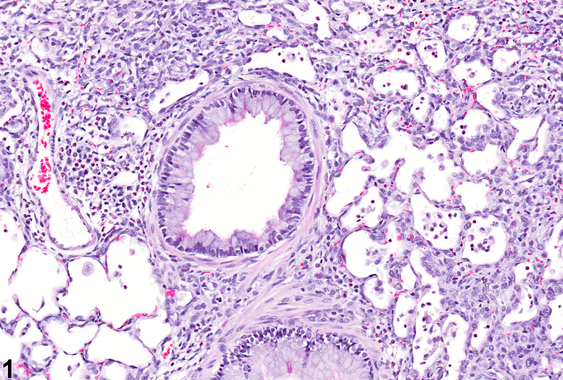
Lung, Bronchiole - Metaplasia, Goblet cell from a male Sprague-Dawley rat in an acute study. The majority of the cells in this airway are goblet cells. Image provided courtesy of Dr. J. Bonner.
All Images

Lung, Bronchiole - Metaplasia, Goblet cell from a male Sprague-Dawley rat in an acute study. The majority of the cells in this airway are goblet cells. Image provided courtesy of Dr. J. Bonner.


Lung, Bronchiole - Metaplasia, Goblet cell from a male Sprague-Dawley rat in an acute study. Scattered cells contain a single, large, cytoplasmic vacuole. Image provided courtesy of Dr. J. Bonner.