U.S. flag

An official website of the United States government

Dot gov

The .gov means it's official.
Federal government websites often end in .gov or .mil. Before sharing sensitive information, make sure you're on a federal government site.

Https

The site is secure.
The https:// ensures that you are connecting to the official website and that any information you provide is encrypted and transmitted securely.

Skip to Main Navigation
Skip to Page Content
Skip to Atlas Navigation

Lung - Metaplasia, Squamous

Image of alveolar squamous metaplasia in the lung from a female Harlan Sprague-Dawley rat in a subchronic study
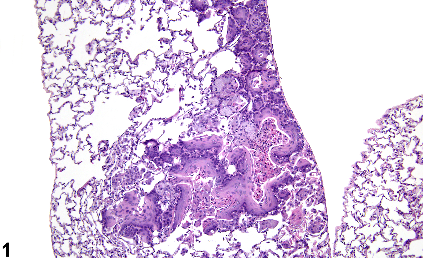
Lung, Alveolus - Metaplasia, Squamous in a female Harlan Sprague-Dawley rat from a subchronic study. The normal alveolar epithelium has been replaced by squamous epithelium.
Figure 1 of 8
Image of alveolar squamous metaplasia in the lung from a female Harlan Sprague-Dawley rat in a subchronic study
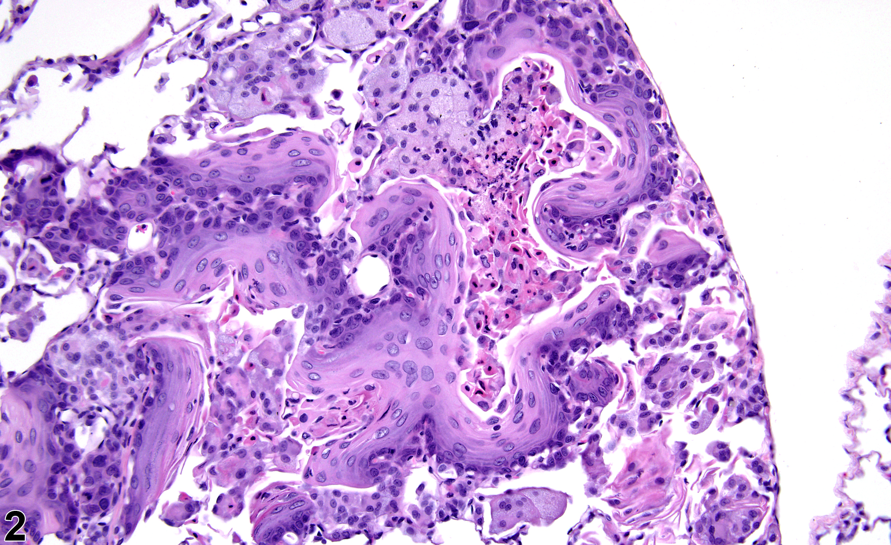
Lung, Alveolus - Metaplasia, Squamous in a female Harlan Sprague-Dawley rat from a subchronic study (higher magnification of Figure 1). The metaplastic epithelium matures normally from basal cells to squamous epithelial cells.
Figure 2 of 8
Image of alveolar squamous metaplasia in the lung from a female Harlan Sprague-Dawley rat in a chronic study
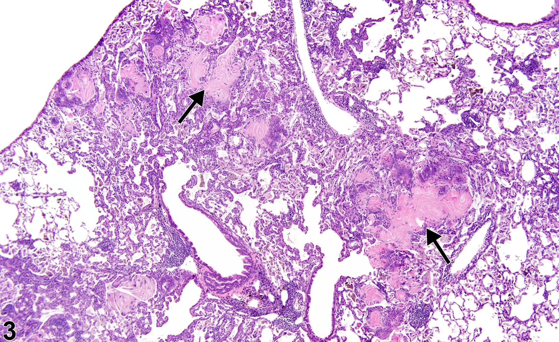
Lung, Alveolus - Metaplasia, Squamous in a female Harlan Sprague-Dawley rat from a chronic study. The normal alveolar epithelium has been replaced by squamous epithelium, which can be identified by the lakes of eosinophilic keratin (arrows).
Figure 3 of 8
Image of alveolar squamous metaplasia in the lung from a female Harlan Sprague-Dawley rat in a chronic study
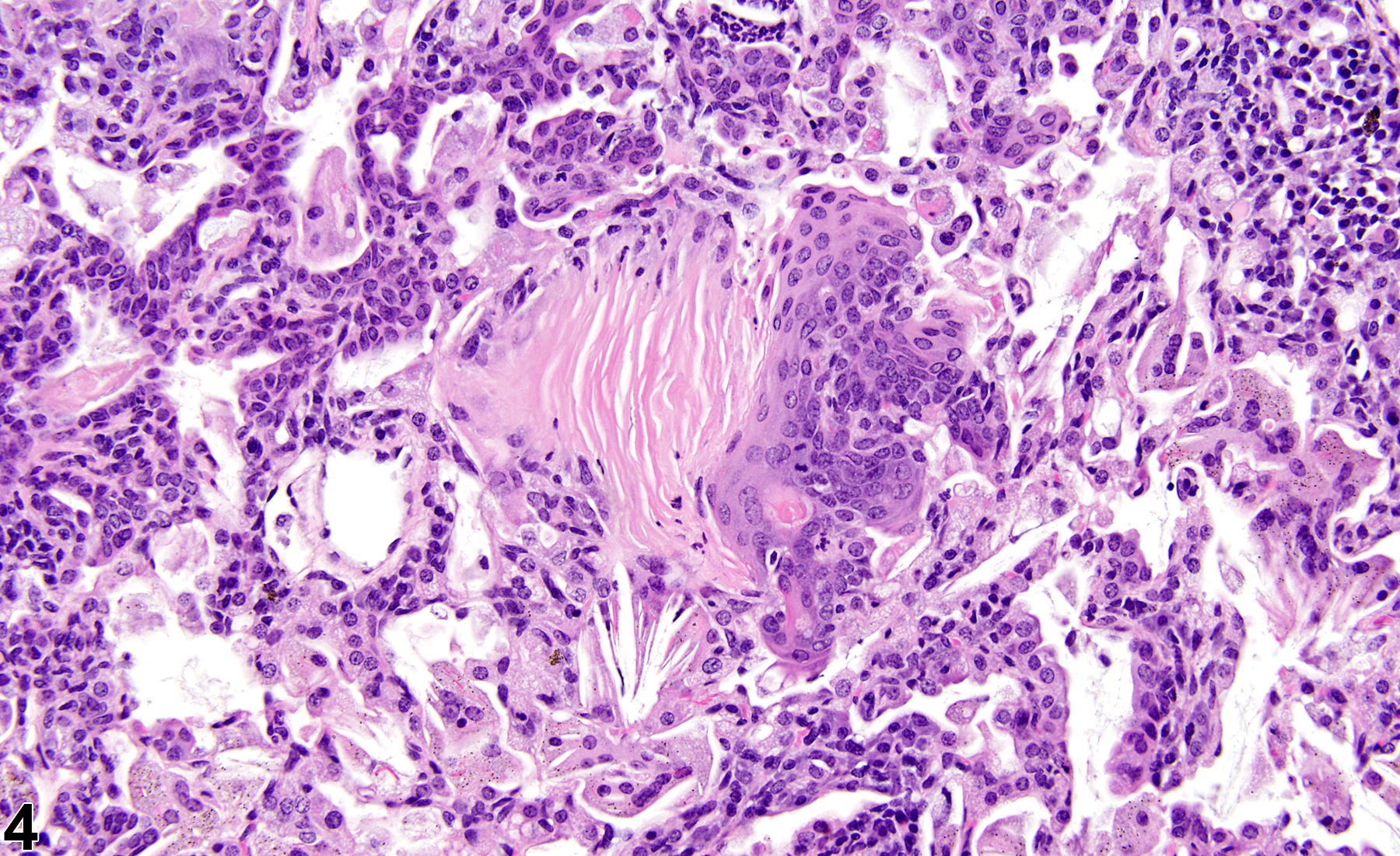
Lung, Alveolus - Metaplasia, Squamous in a female Harlan Sprague-Dawley rat from a chronic study (higher magnification of Figure 3). The metaplastic epithelium is keratinized; there is also hyperplasia of the alveolar epithelium.
Figure 4 of 8
Image of bronchiolar squamous metaplasia in the lung from a male F344/N rat in a chronic study
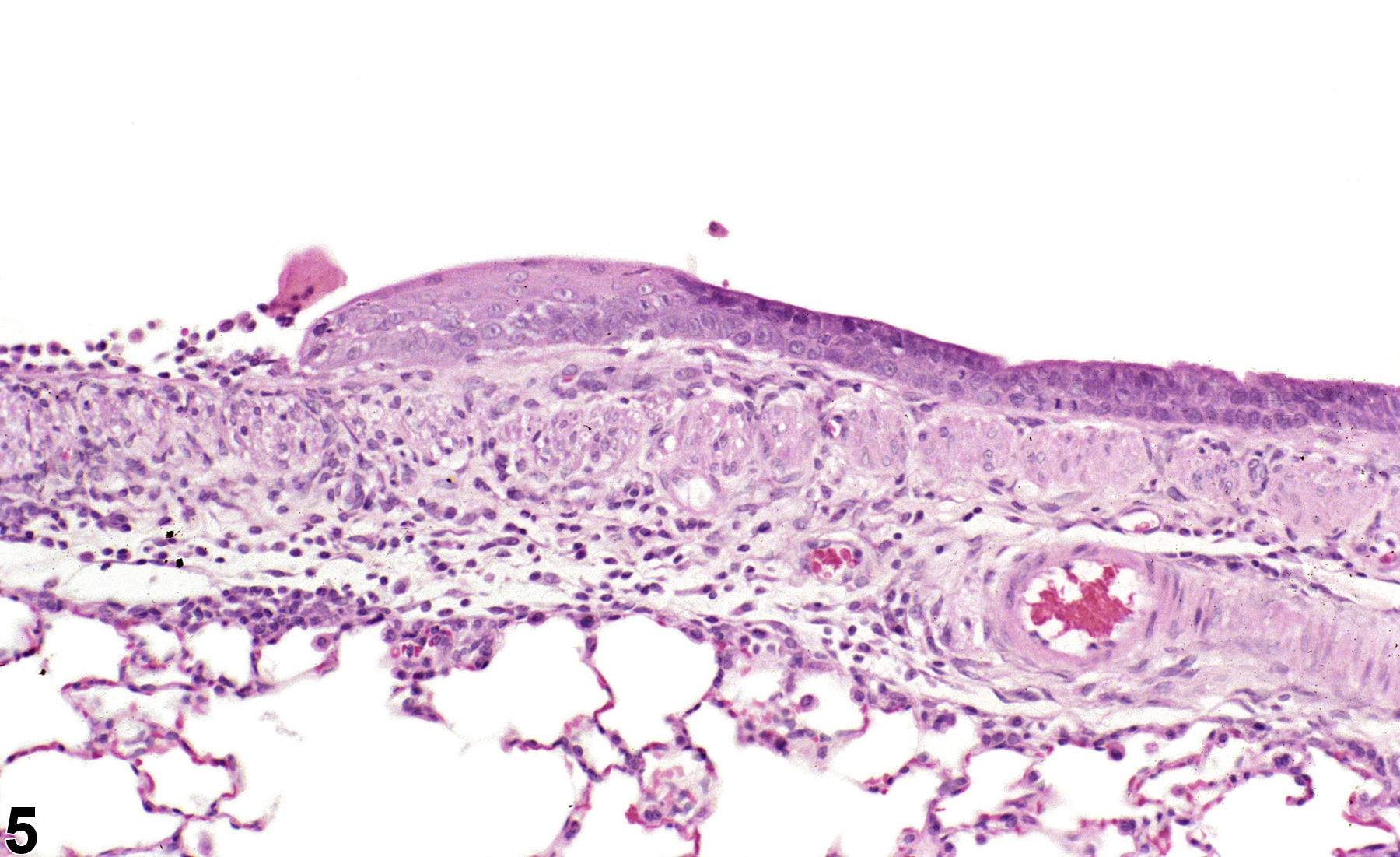
Lung, Bronchiole - Metaplasia, Squamous in a male F344/N rat from an acute study. The normal bronchiolar epithelium has been replaced by squamous epithelium adjacent to an ulcer.
Figure 5 of 8
Image of bronchial squamous metaplasia in the lung from a male F344/N rat in a subchronic study
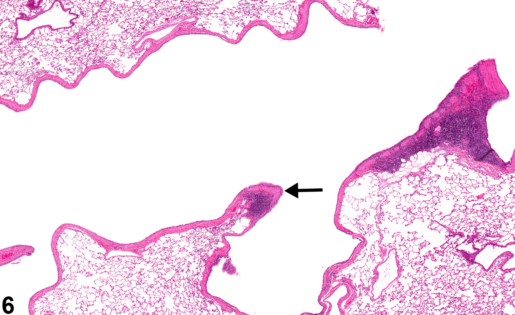
Lung, Bronchus - Metaplasia, Squamous in a male F344/N rat from a subchronic study. The normal bronchiolar epithelium at the bifurcation of an airway has been replaced by squamous epithelium (arrow).
Figure 6 of 8
Image of bronchial squamous metaplasia in the lung from a male F344/N rat in a subchronic study
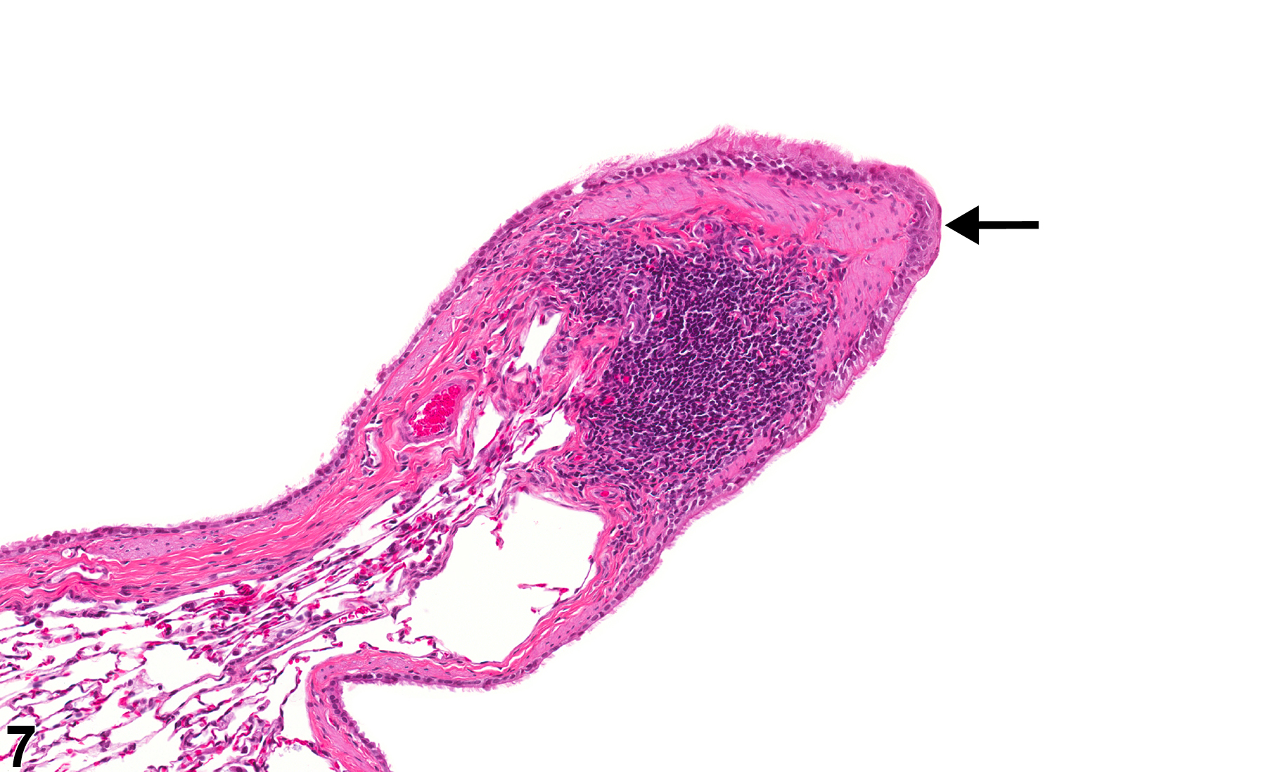
Lung, Bronchus - Metaplasia, Squamous in a male F344/N rat from a subchronic study (higher magnification of Figure 6). The normal bronchiolar epithelium has been replaced by squamous epithelium (arrow).
Figure 7 of 8
Image of bronchiolar squamous metaplasia in the lung from a male B6C3F1/N mouse in a subchronic study
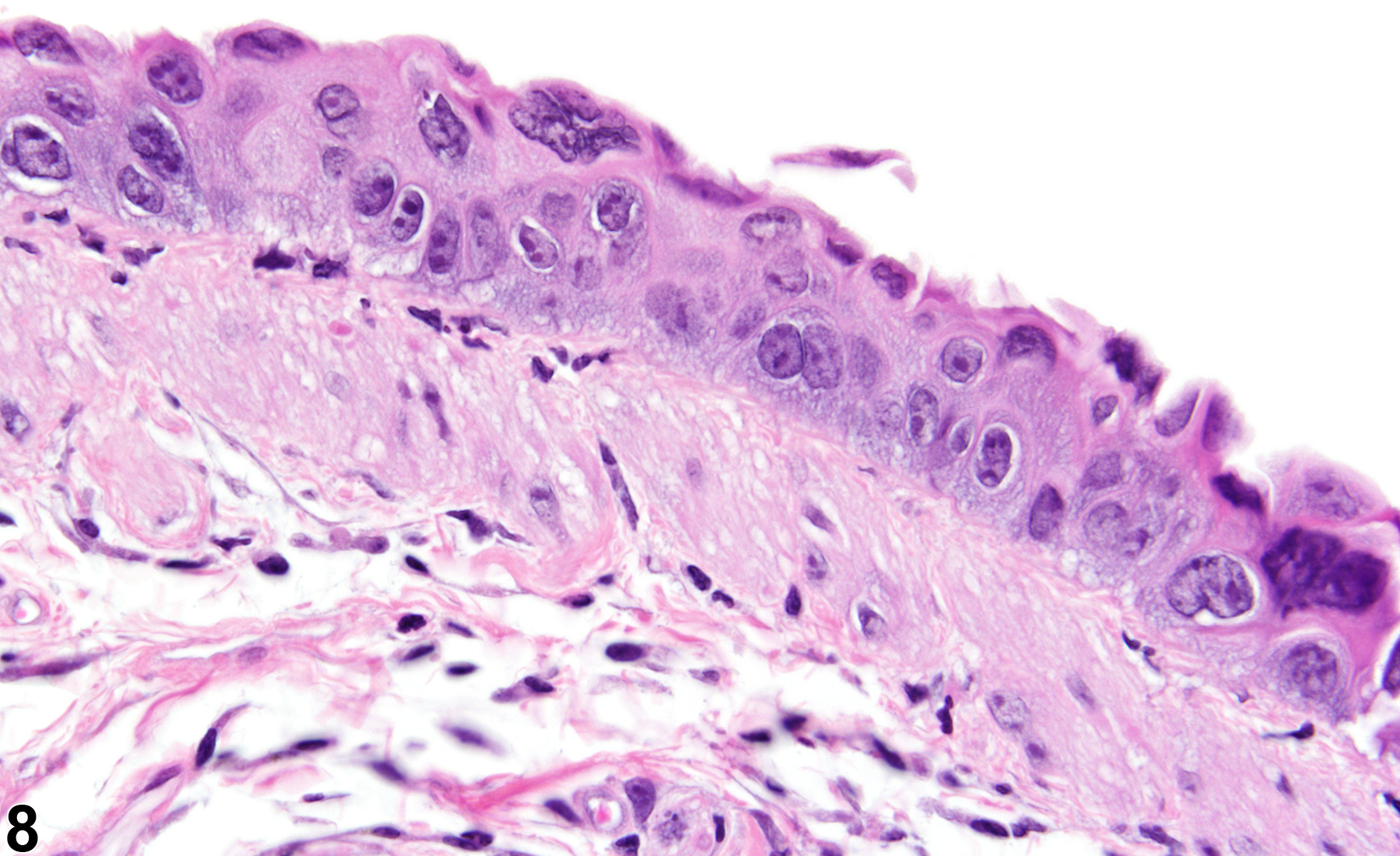
Lung, Bronchiole - Metaplasia, Squamous, Atypical from a male B6C3F1 mouse in a subchronic study. The cells on the epithelial surface are squamous, but the cells in the deeper layers are disorganized, and there is anisocytosis and anisokaryosis.
Figure 8 of 8
next arrow

comment:

Squamous metaplasia of the alveolar epithelium ( Figure 1image opens in a pop-up window , Figure 2image opens in a pop-up window , Figure 3image opens in a pop-up window , Figure 4image opens in a pop-up window , Figure 5image opens in a pop-up window , Figure 6image opens in a pop-up window , Figure 7image opens in a pop-up window , and Figure 8image opens in a pop-up window ) is characterized by replacement of type I and type II pneumocytes with squamous epithelium. Squamous metaplasia of the airway epithelium is characterized by replacement of bronchiolar or bronchial epithelium with squamous epithelium. Squamous epithelium is a stratified epithelium with orderly progression of cell maturation from cuboidal basal cells at the basement membrane to flattened squamous cells at the surface. The epithelium may be keratinized, and the cells may contain keratohyalin granules. Early squamous metaplasia, where only the surface cells are flattened, can be difficult to identify. Squamous metaplasia should not be confused with regeneration, since regenerating epithelial cells can assume a flattened morphology (stretching out to cover a denuded basement membrane). Generally, more than one layer of cells is present in squamous metaplasia. Squamous metaplasia can be seen as a response to chronic irritation or injury and is thought to be an adaptive change, as the squamous epithelium is more resistant to injury than is respiratory or alveolar epithelium. Consequently, squamous metaplasia is often accompanied by inflammation, fibrosis, necrosis, ulceration, and areas of epithelial hyperplasia. Though not typically considered preneoplastic, under some conditions squamous metaplasia may give rise to neoplasia (cystic keratinizing epithelioma or squamous cell carcinoma). Metaplastic epithelium in which there are atypical cells ( Figure 8image opens in a pop-up window ) may be preneoplastic.

recommendation:

Lung - Metaplasia, Squamous should be diagnosed whenever present and assigned a severity grade. A site modifier (i.e., alveolus, bronchiole, or bronchus) should be included in the diagnosis to indicate the location of the lesion. Associated lesions, such as inflammation, fibrosis, necrosis, or hyperplasia, should be diagnosed separately. If atypia is present, then the modifier "atypical" should be added to the diagnosis.

references:

Boorman GA, Eustis SL. 1990. Lung. In: Pathology of the Fischer Rat: Reference and Atlas (Boorman GA, Eustis SL, Elwell MR, Montgomery CA, MacKenzie WF, eds). Academic Press, San Diego, CA, 339-367.

Renne, R, Brix A, Harkema J, Herbert R, Kittle B, Lewis D, March T, Nagano K, Pino M, Rittinghausen S, Rosenbruch M, Tellier P, Wohrmann T. 2009. Proliferative and nonproliferative lesions of the rat and mouse respiratory tract. Toxicol Pathol 37(suppl):5S-73S.
Abstract: https://www.ncbi.nlm.nih.gov/pubmed/20032296

Sells DM, Brix AE, Nyska A, Jokinen MP, Orzech DP, Walker NJ. 2007. Respiratory tract lesions in noninhalation studies. Toxicol Pathol 35:170-177.
Full Text: https://www.ncbi.nlm.nih.gov/pmc/articles/PMC3433271/

Walker NJ, Yoshizawa K, Miller RA, Brix AE, Sells DM, Jokinen MP, Wyde ME, Easterling M, Nyska A. 2007. Pulmonary lesions in female Harlan Sprague-Dawley rats following two-year oral treatment with dioxin-like compounds. Toxicol Pathol 35:880-889.