Lung - Pigment

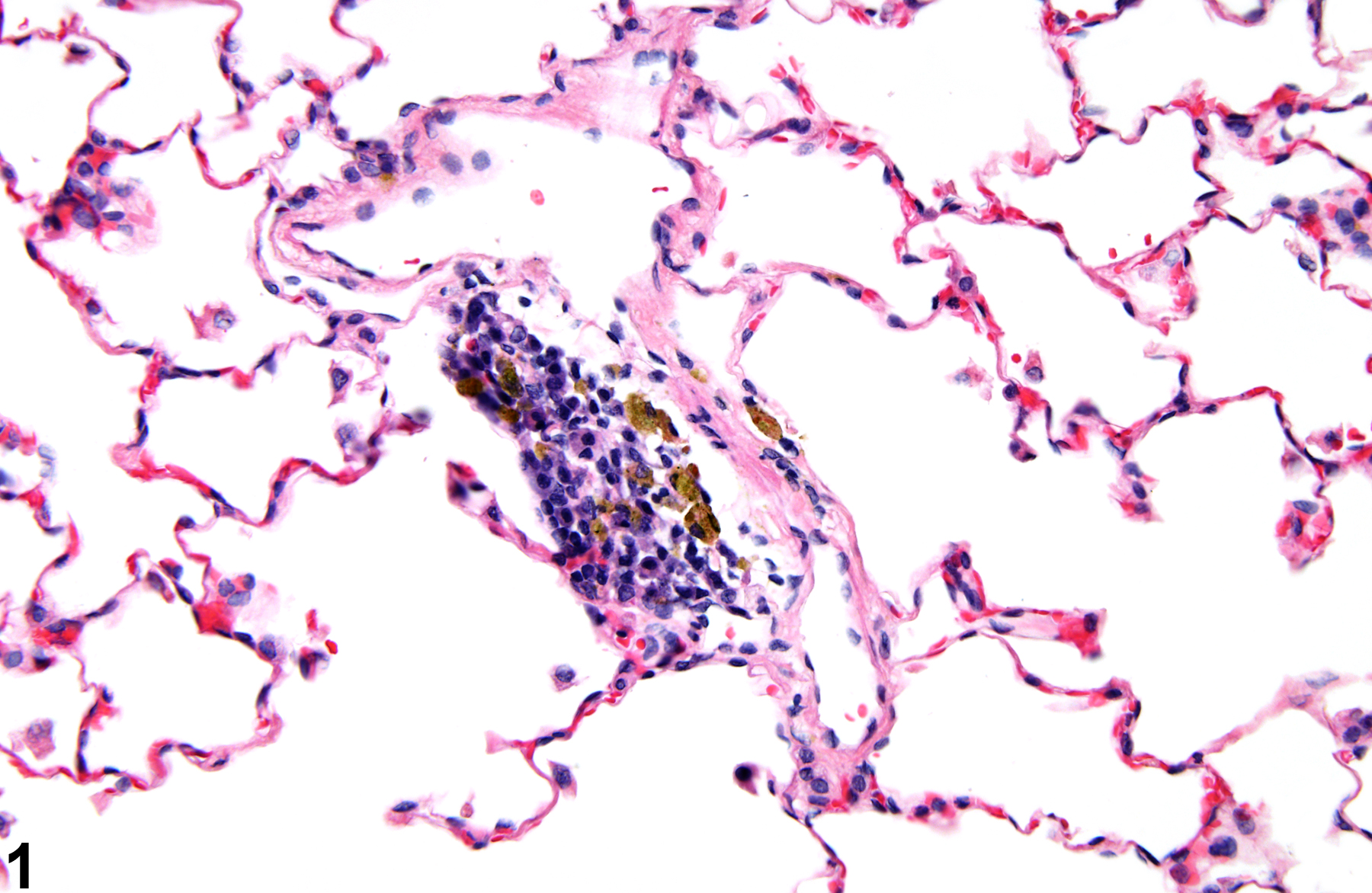
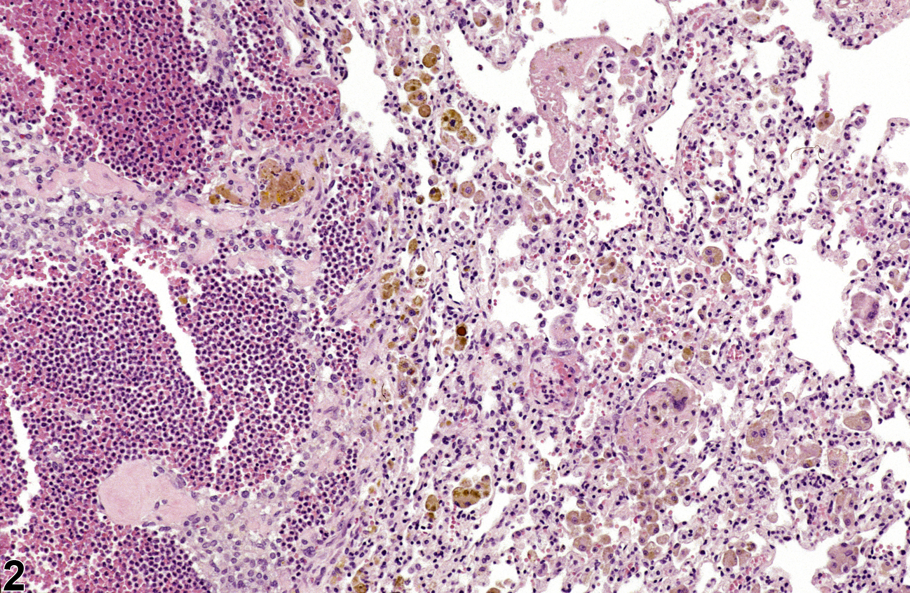
Lung, Alveolus - Pigment in a female Harlan Sprague-Dawley rat from a chronic study. The pigment is most likely hemosiderin and is contained within macrophages.
Figure 1 of 2
comment:
The term "pigment" is reserved for endogenous substances. Various types of pigment can be seen in the lungs, including hemosiderin ( Figure 1

recommendation:
Lung - Pigment should be diagnosed when it is considered to be relevant to the study (or if it is unclear whether it is relevant to the study) or if it is unusually prominent. Not all pigments have to be diagnosed, as some are ubiquitous in aging animals or are secondary to another disease process (e.g., hemorrhage) and not toxicologically meaningful. The pathologist should use his or her judgment in deciding whether or not the pigment should be diagnosed. When pigment is diagnosed, it should be given a severity grade, and if appropriate, a site modifier (e.g., bronchus, bronchiole) should be used to identify the location of the lesion. Definitive pigment identification is often difficult in histologic sections, even with a battery of special stains. Therefore, a diagnosis of pigment (as opposed to diagnosing the type of pigment, e.g., hemosiderin or lipofuscin) is most appropriate. The pathology narrative should include a description of the morphologic features of the pigment, its distribution within the tissue, and the cell type affected. Associated lesions, such as hemorrhage, alveolar macrophage infiltration, or inflammation, should be diagnosed separately.related links:
Lung - Foreign Materialreferences:
Kumar V, Abbas AK, Fausto N. 2005. Cellular adaptations, cell injury, and cell death. In: Pathologic Basis of Disease, 7th ed. Elsevier Saunders, Philadelphia, 39-40.
Pizzolato P. 1976. Formalin pigment (acid hematin) and related pigments. Am J Med Technol 42:436-440. Abstract: https://www.ncbi.nlm.nih.gov/pubmed/790956
Web page last updated on: December 03, 2015