

Respiratory System
Lung - Proteinosis
Narrative
Boorman GA, Eustis SL. 1990. Lung. In: Pathology of the Fischer Rat: Reference and Atlas (Boorman GA, Eustis SL, Elwell MR, Montgomery CA, MacKenzie WF, eds). Academic Press, San Diego, CA, 339-367.
Dixon D, Herbert RA, Sills RC, Boorman GA. 1999. Lungs, pleura, and mediastinum. In: Pathology of the Mouse: Reference and Atlas (Maronpot RR, Boorman GA, Gaul BW, eds). Cache River Press, Vienna, IL, 293-332.
Hook GER. 1991. Alveolar proteinosis and phospholipodoses of the lungs. Toxicol Pathol 19:482-513.
Abstract: http://tpx.sagepub.com/content/19/4-1/482.refs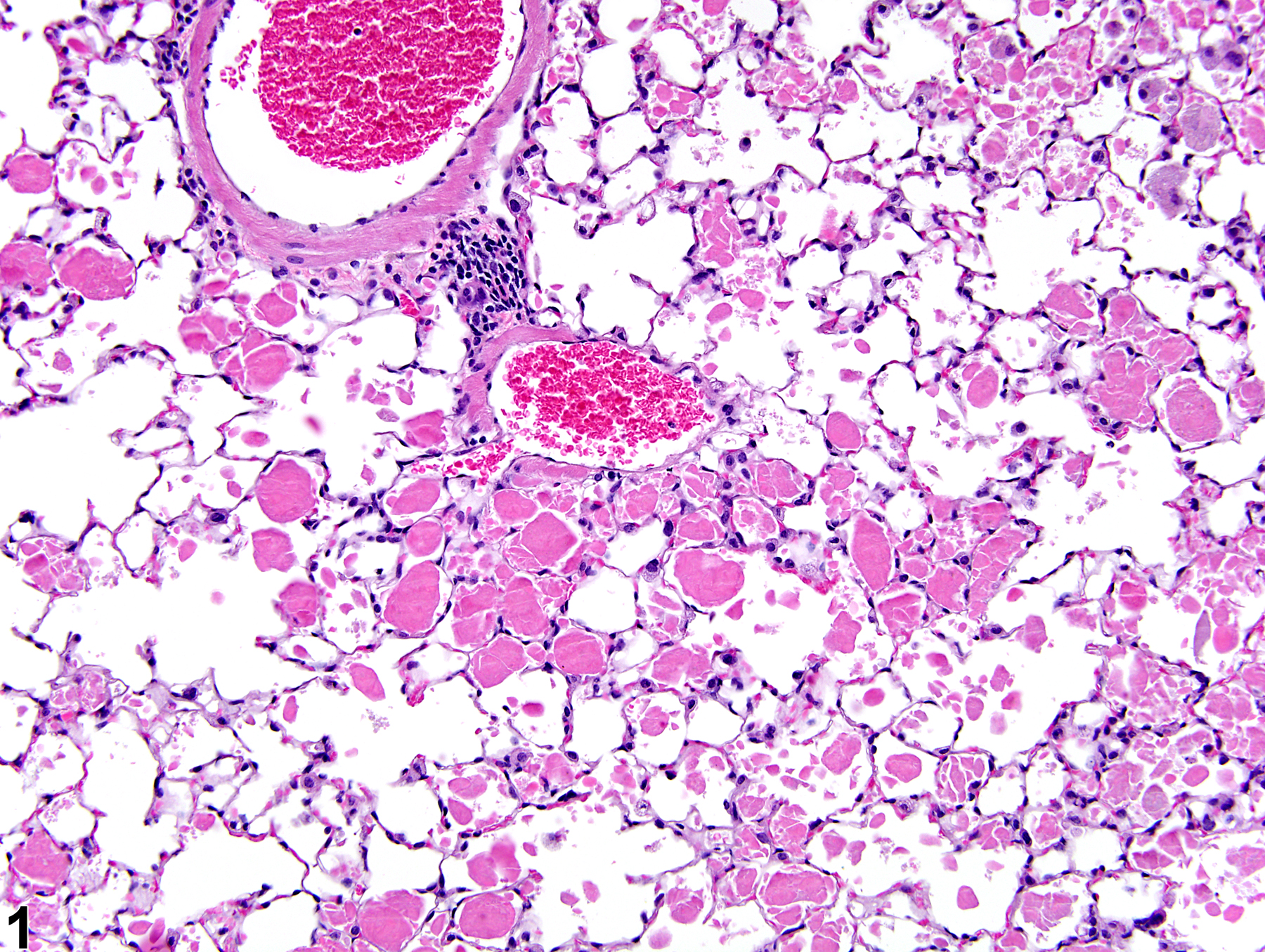
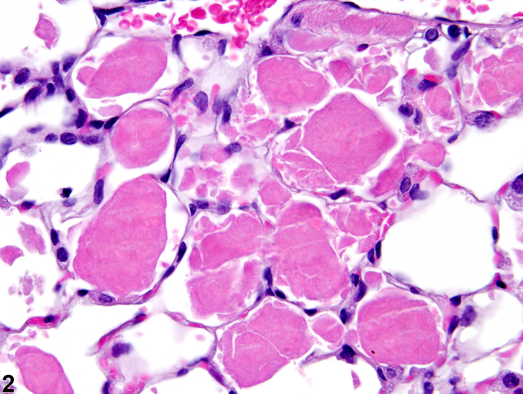
Lung, Alveolus - Proteinosis in a male B6C3F1/N mouse from a chronic study. There is amorphous, brightly eosinophilic material (proteinosis) within the alveoli.
All Images

Lung, Alveolus - Proteinosis in a male B6C3F1/N mouse from a chronic study. There is amorphous, brightly eosinophilic material (proteinosis) within the alveoli.

Lung, Alveolus - Proteinosis in a male B6C3F1/N mouse from a chronic study (higher magnification of Figure 1). Little to no inflammation is associated with the amorphous, brightly eosinophilic material (proteinosis) in the alveoli.

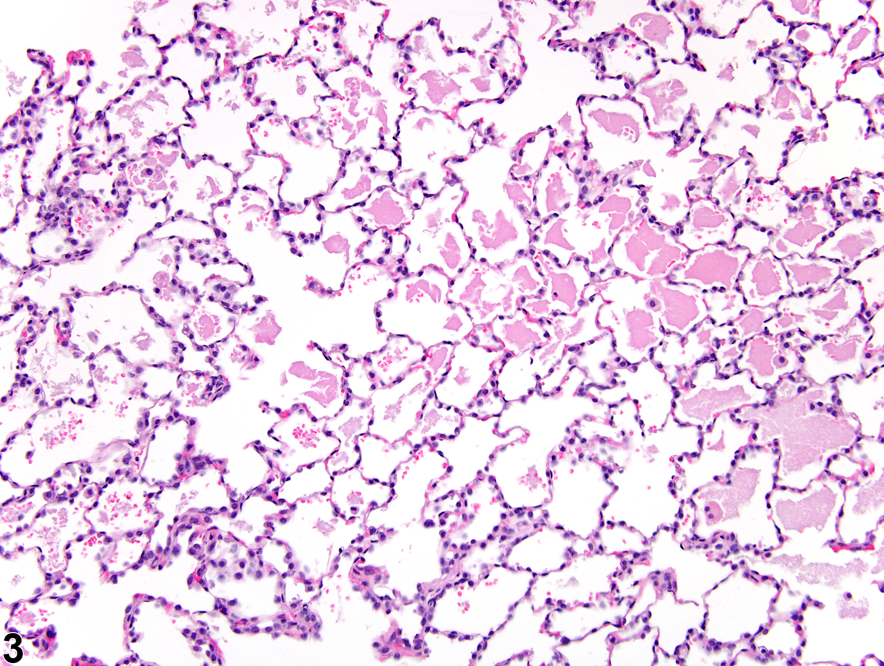
Lung, Alveolus - Proteinosis in a male B6C3F1 mouse from a subchronic study. There is amorphous, eosinophilic material (proteinosis) within the alveoli.

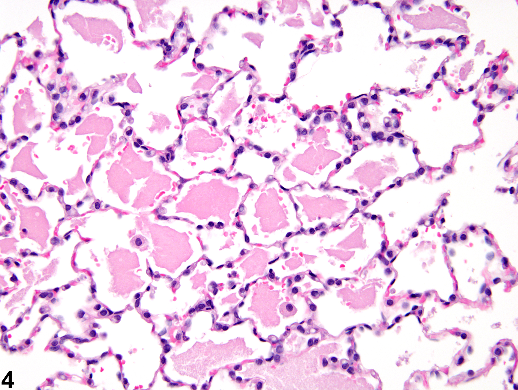
Lung, Alveolus - Proteinosis in a male F344/N rat from a subchronic study (higher magnification of Figure 3). Scattered alveolar macrophages are associated with the amorphous, eosinophilic material (proteinosis) in the alveoli.