

Respiratory System
Lung - Regeneration
Narrative
Boorman GA, Eustis SL. 1990. Lung. In: Pathology of the Fischer Rat: Reference and Atlas (Boorman GA, Eustis SL, Elwell MR, Montgomery CA, MacKenzie WF, eds). Academic Press, San Diego, CA, 339-367.
Dixon D, Herbert RA, Sills RC, Boorman GA. 1999. Lungs, pleura, and mediastinum. In: Pathology of the Mouse: Reference and Atlas (Maronpot RR, Boorman GA, Gaul BW, eds). Cache River Press, Vienna, IL, 293-332.
Renne RA, Dungworth DL, Keenan CM, Morgan KT, Hahn FF, Schwartz LW. 2003. Non-proliferative lesions of the respiratory tract in rats. In: Guides for Toxicologic Pathology. STP/ARP/AFIP, Washington, DC.
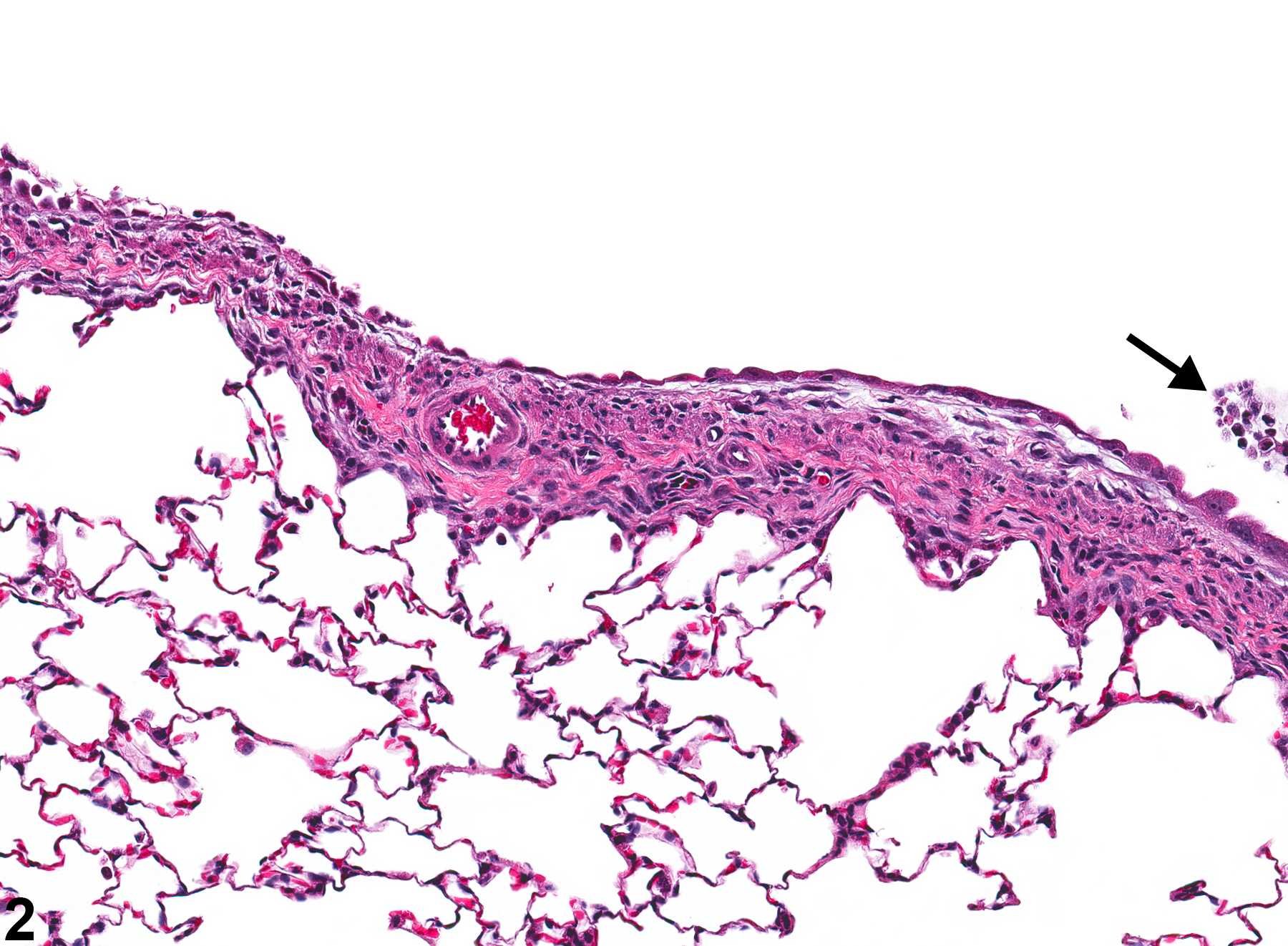
Lung, Bronchiole - Regeneration in a male Wistar Han rat from an acute study. A single, thin layer of flattened epithelial cells covers the bronchiolar surface.
All Images

Lung, Bronchiole - Regeneration in a male Wistar Han rat from an acute study. A single, thin layer of flattened epithelial cells covers the bronchiolar surface.

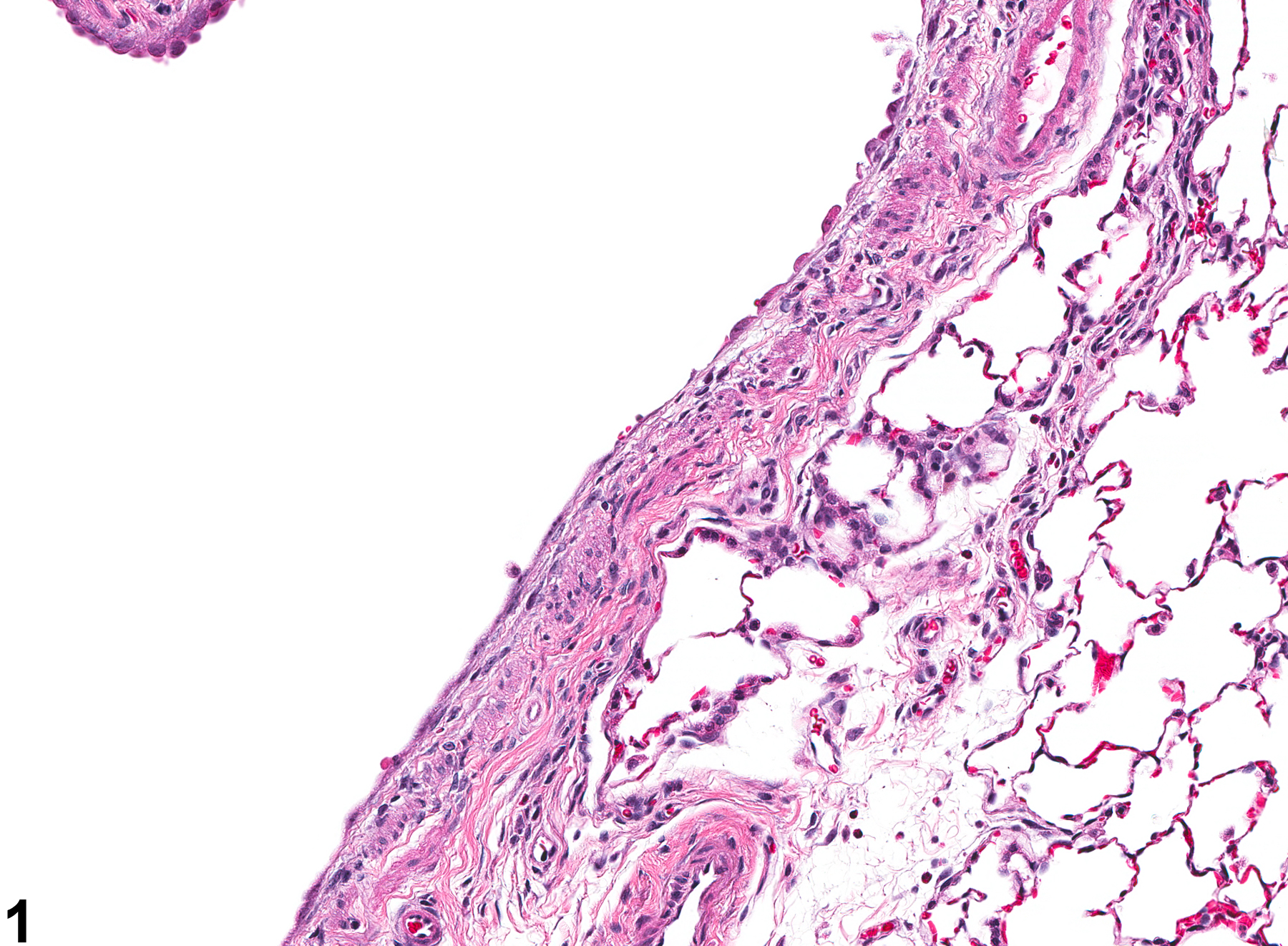
Lung, Bronchiole - Regeneration in a male Wistar Han rat from an acute study. There is a single layer of flattened epithelial cells on the bronchiolar surface; the cell debris (arrow) in the bronchiolar lumen suggests epithelial damage.

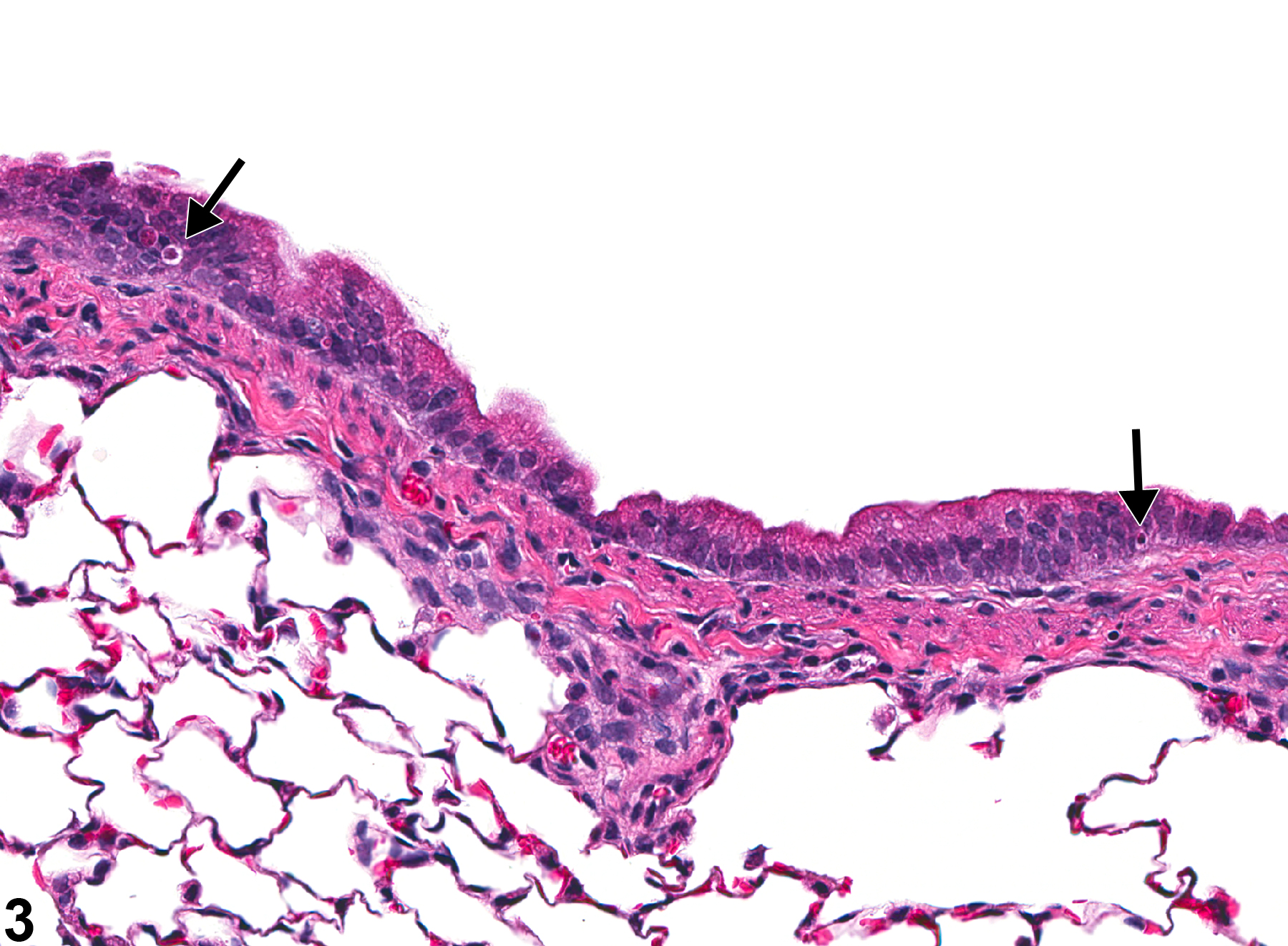
Lung, Bronchiole - Regeneration in a male Wistar Han rat from an acute study. The scattered necrotic cells in the epithelium (arrows) suggest that this hyperplastic response is associated with regeneration secondary to previous damage.