

Respiratory System
Lung - Vacuolization, Cytoplasmic
Narrative
Kumar V, Abbas AK, Fausto N. 2005. Robbins and Cotran Pathologic Basis of Disease, 7th ed. Elsevier Saunders, Philadelphia, 20.
National Toxicology Program. 1991. NTP TOX 5. Toxicity Studies of Cobalt Sulfate Heptahydrate in F344/N Rats and B6C3F1 Mice (Inhalation Studies). NIH Publication No. 91-3124. NTP, Research Triangle Park, NC.
Abstract: https://ntp.niehs.nih.gov/go/11946National Toxicology Program. 2011. NTP TR 564. Toxicology and Carcinogenesis Studies of 1-Bromopropane (CAS No. 106-94-5) in F344/N Rats and B6C3F1 Mice (Inhalation Studies). NTP, Research Triangle Park, NC.
Abstract: https://ntp.niehs.nih.gov/go/34854Plopper CG, Van Winkle LS, Fanucchi MV, Malburg SR, Nishio SJ, Chang A, Buckpitt AR. 2001. Early events in naphthalene-induced acute Clara cell toxicity. II. Comparison of glutathione depletion and histopathology by airway location. Am J Respir Cell Mol Biol 24:272-281.
Abstract: https://www.ncbi.nlm.nih.gov/pubmed/11245626Wallig MA. 2002. Morphological manifestation of toxic cell injury. In: Handbook of Toxicologic Pathology (Haschek WM, Rousseaux CG, Wallig MA, eds). Academic Press, San Diego, CA, 39-65.
Lung, Bronchiole - Vacuolization, Cytoplasmic in a female B6C3F1/N mouse from a chronic study. The bronchiolar epithelial cells have vacuolated cytoplasm.
All Images

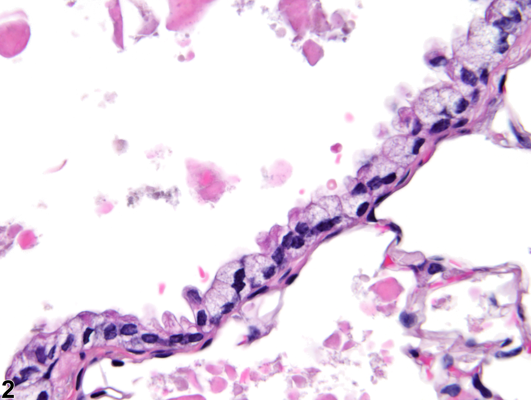
Lung, Bronchiole - Vacuolization, Cytoplasmic in a female B6C3F1/N mouse from a chronic study. The bronchiolar epithelial cells have vacuolated cytoplasm.

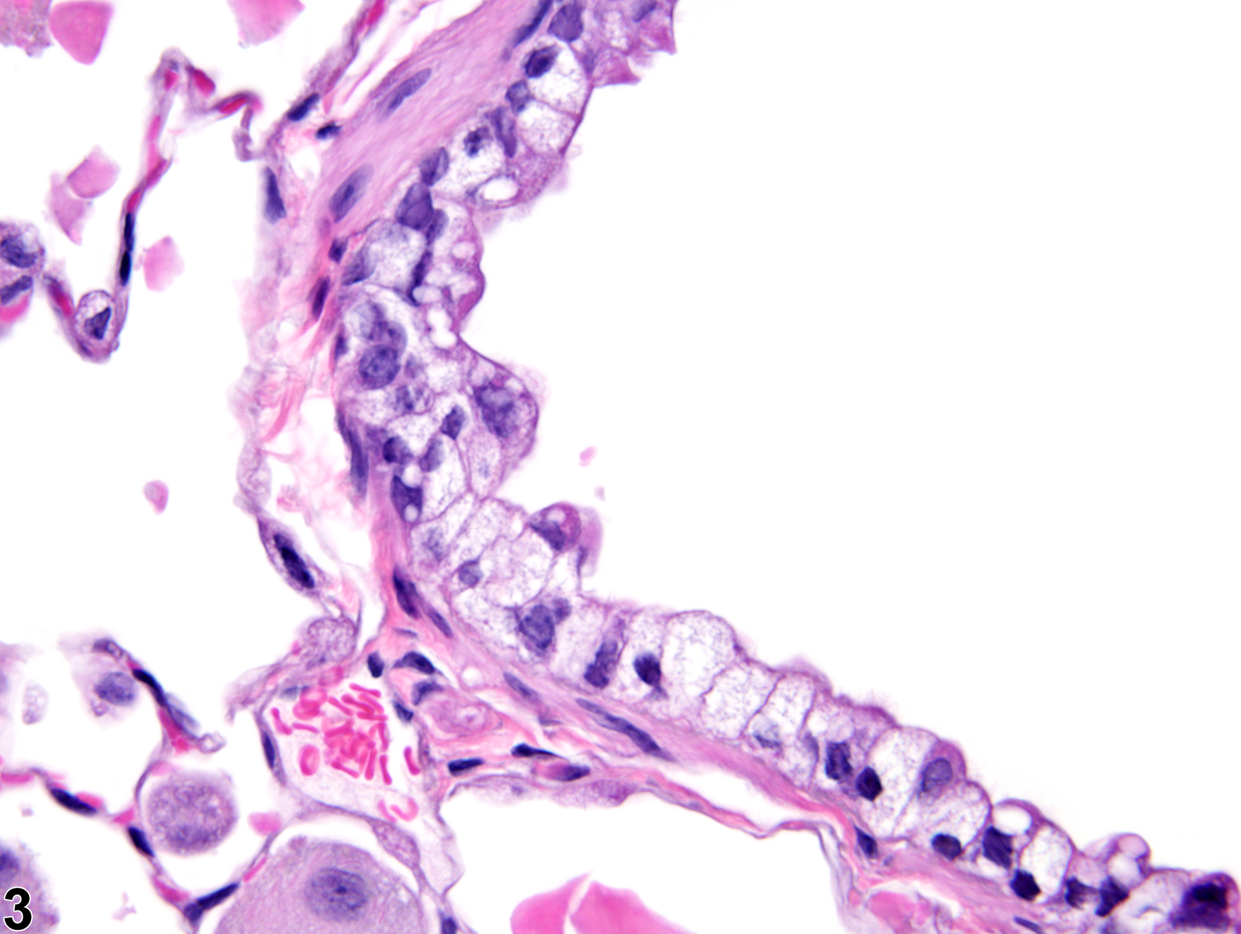
Lung, Bronchiole - Vacuolization, Cytoplasmic in a female B6C3F1/N mouse from a chronic study (higher magnification of Figure 1). Most of the cells have also lost their cilia.

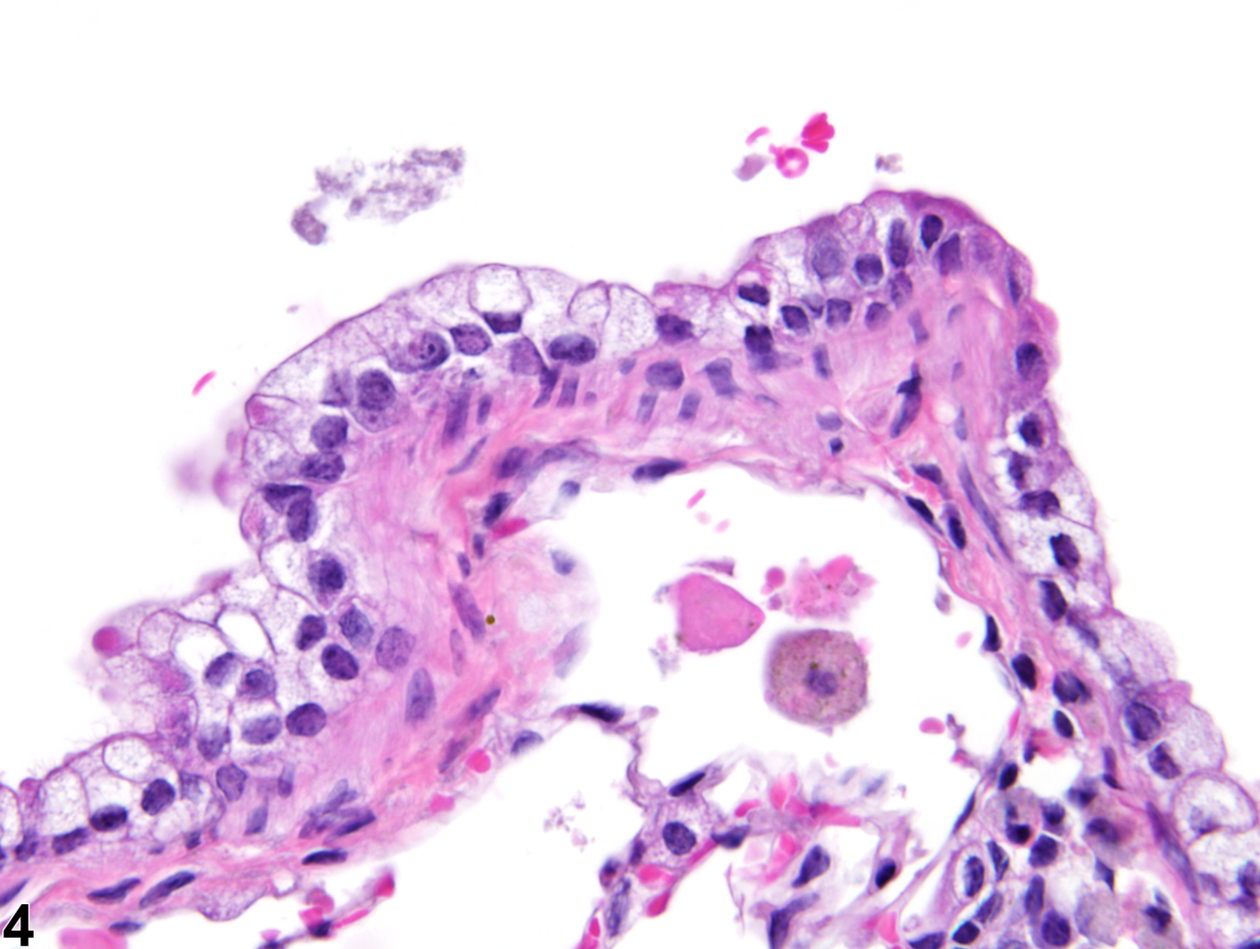
Lung, Bronchiole - Vacuolization, Cytoplasmic in a female B6C3F1/N mouse from a chronic study. The bronchiolar epithelial cells contain clear, intracytoplasmic vacuoles.

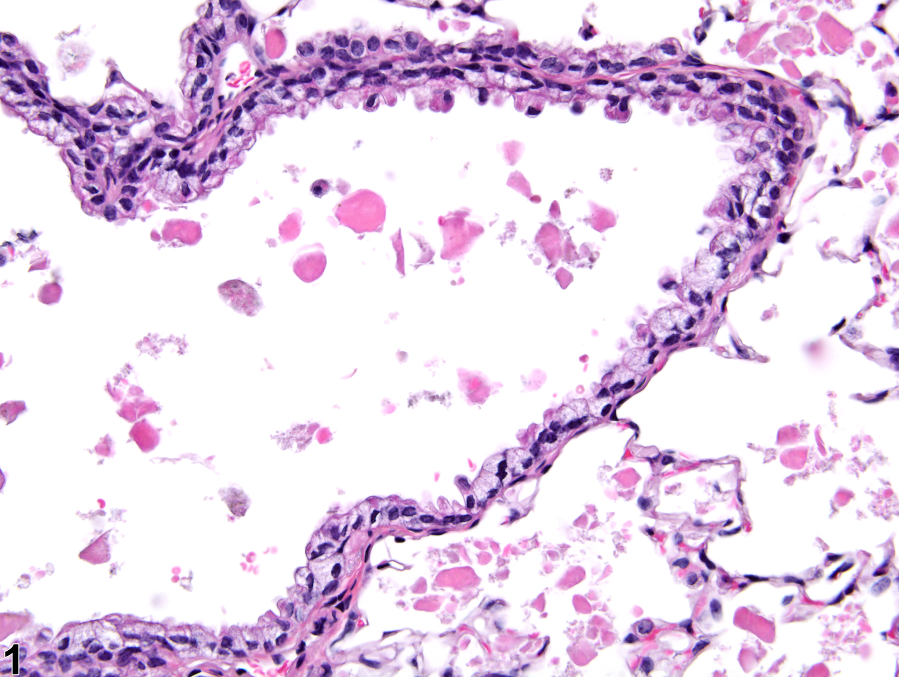
Lung, Bronchiole - Vacuolization, Cytoplasmic in a female B6C3F1/N mouse from a chronic study. The bronchiolar epithelial cells contain clear, intracytoplasmic vacuoles and have lost their cilia.