U.S. flag

An official website of the United States government

Dot gov

The .gov means it's official.
Federal government websites often end in .gov or .mil. Before sharing sensitive information, make sure you're on a federal government site.

Https

The site is secure.
The https:// ensures that you are connecting to the official website and that any information you provide is encrypted and transmitted securely.

Skip to Main Navigation
Skip to Page Content
Skip to Atlas Navigation

Nose, Epithelium - Degeneration

Image of degeneration in the nose, olfactory epithelium from a male F344/N rat in a chronic study
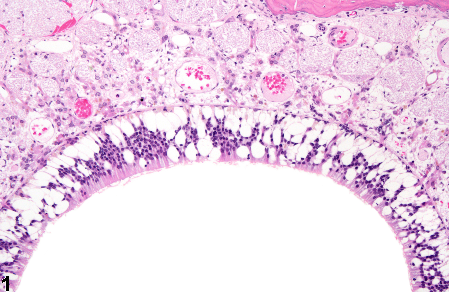
Nose, Olfactory epithelium - Degeneration in a male F344/N rat from a chronic study. Numerous vacuoles are present in the olfactory mucosa.
Figure 1 of 4
Image of degeneration in the nose, olfactory epithelium from a female B6C3F1/N mouse in a chronic study
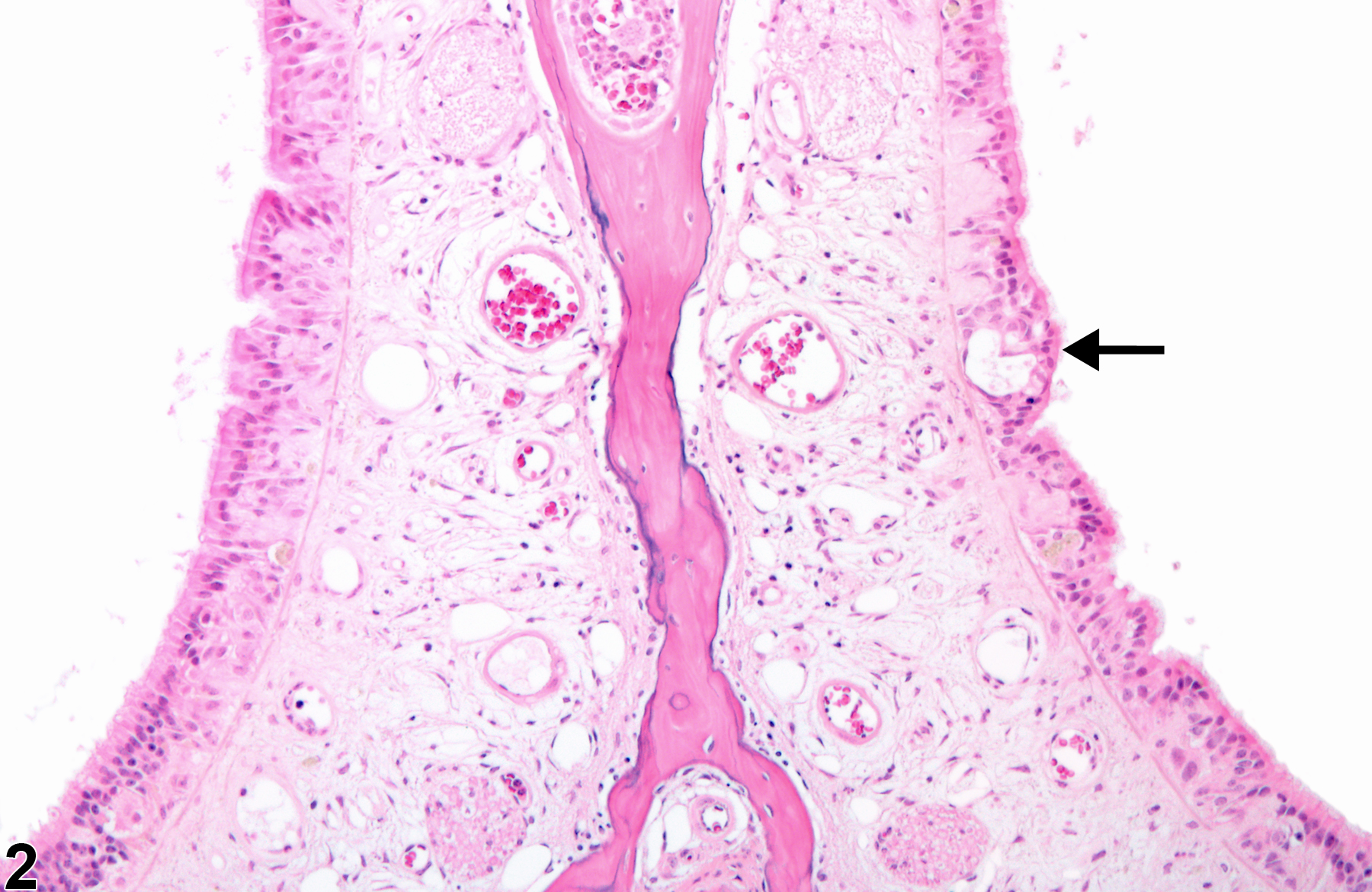
Nose, Olfactory epithelium - Degeneration in a female B6C3F1/N mouse from a chronic study. A clear vacuole (arrow), disorganization, and homogeneous cytoplasmic change are present in the mucosa.
Figure 2 of 4
Image of degeneration in the nose, respiratory epithelium from a male B6C3F1/N mouse in a chronic study
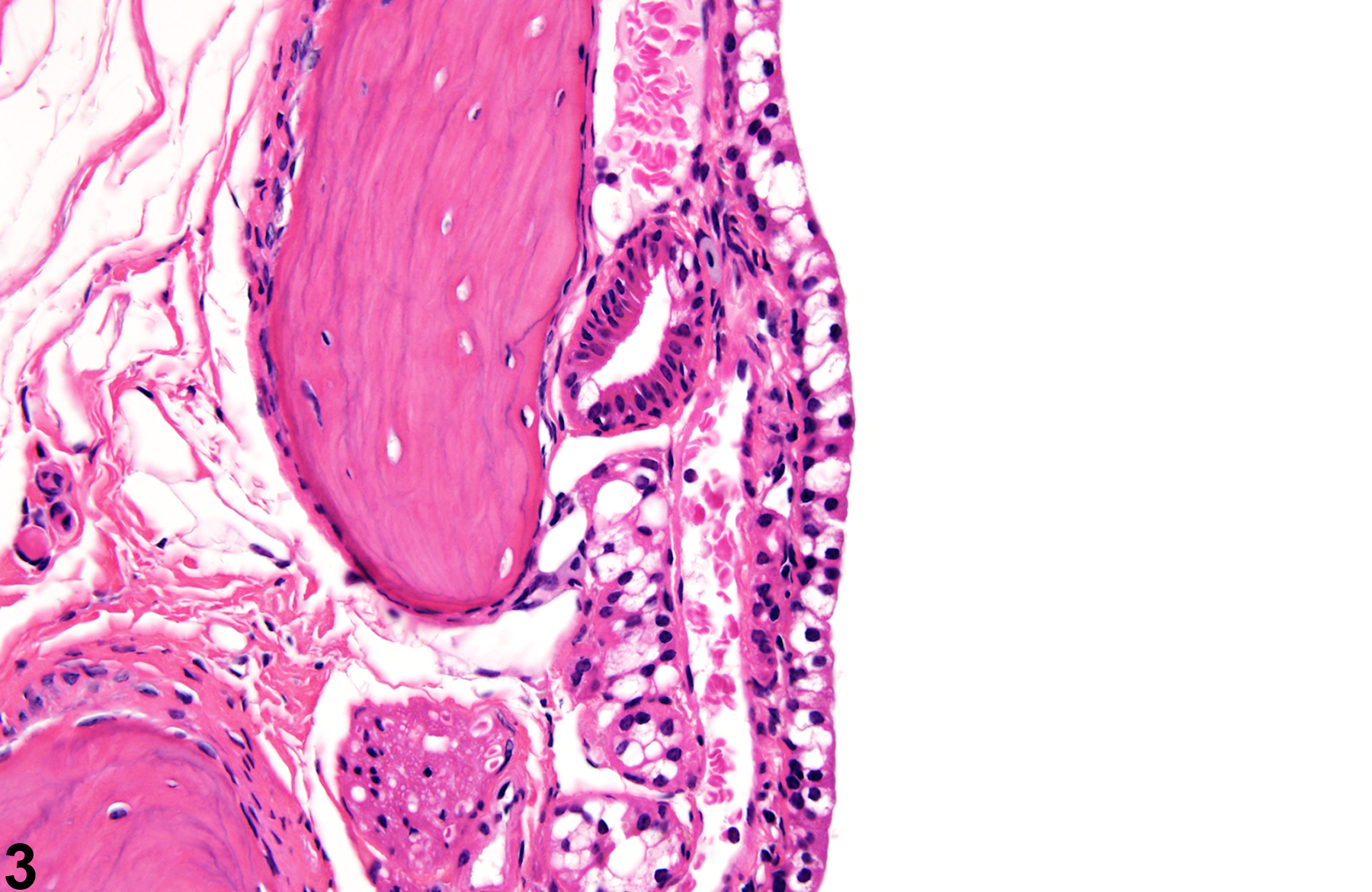
Nose, Respiratory epithelium - Degeneration in a male B6C3F1/N mouse from a chronic study. Cytoplasmic vacuoles are present in the respiratory epithelium lining a turbinate.
Figure 3 of 4
Image of degeneration in the nose, respiratory epithelium from a male B6C3F1/N mouse in a chronic study
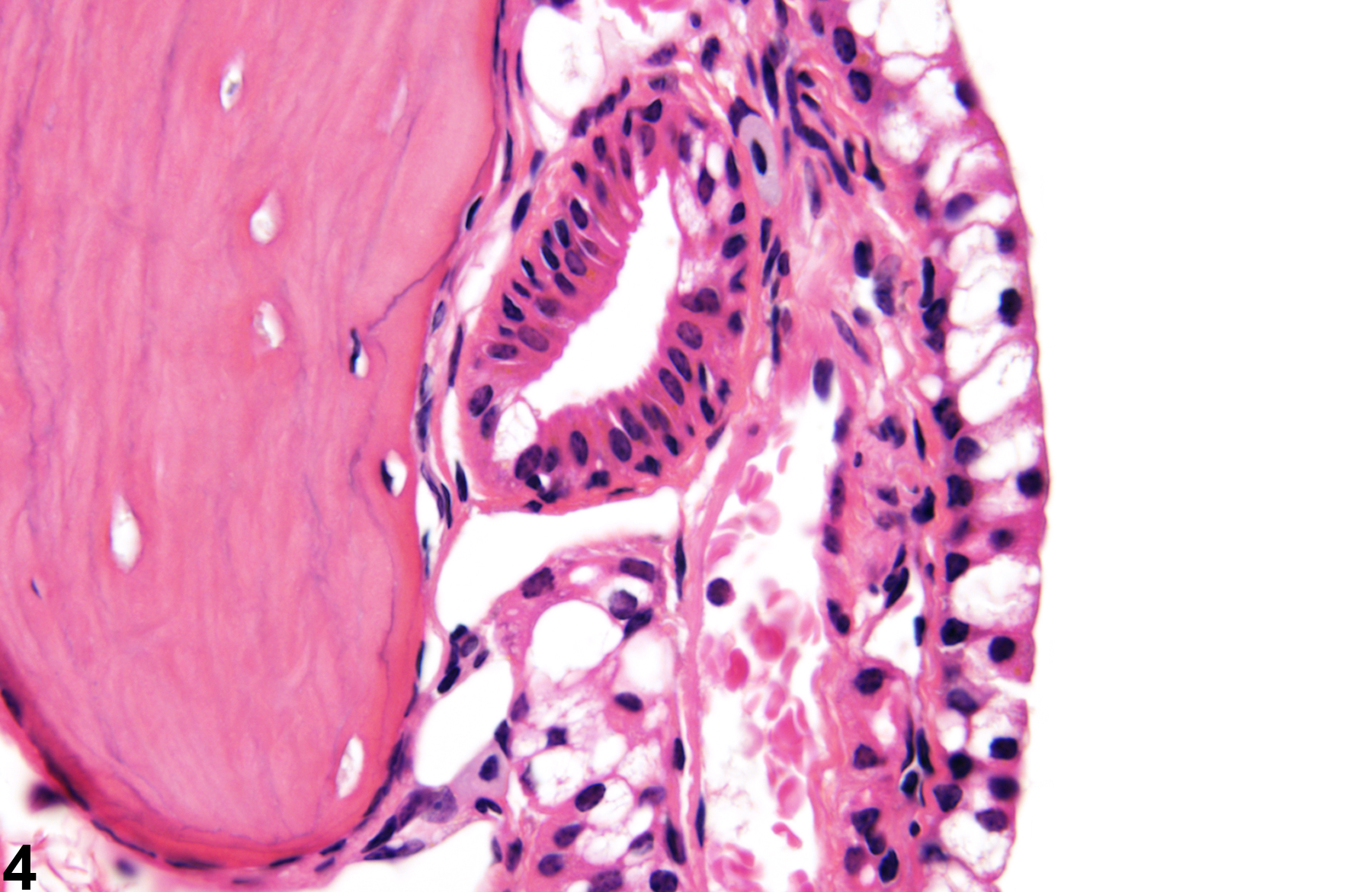
Nose, Respiratory epithelium - Degeneration in a male B6C3F1/N mouse from a chronic study (higher magnification of Figure 3). Cytoplasmic vacuoles are present in the respiratory epithelium.
Figure 4 of 4
next arrow

comment:

Degeneration of the nasal epithelium ( Figure 1image opens in a pop-up window , Figure 2image opens in a pop-up window , Figure 3image opens in a pop-up window , and Figure 4image opens in a pop-up window ) is characterized by one or more of the following: loss of cilia, epithelial cell bleb formation, cellular disorientation, cellular eosinophilia, swelling, loss of cell-to-cell contact, or vacuolar changes within the epithelium. Degeneration may imply a current, ongoing process, so when the process seems to be active, the term "degeneration" may be appropriate. Degenerative changes may also occur in the epithelia of accessory nasal structures, possibly in the absence of degenerative changes in the lining epithelia. "Cystic degeneration" is reserved for epithelial-lined structures within the mucosa or lamina propria. Such cysts usually contain homogeneous or granular material. Epithelial degeneration is a common treatment-related lesion in toxicity studies but may also be seen in control animals.

recommendation:

Epithelial degeneration should be diagnosed and assigned a severity grade whenever present. The type of epithelium should be included in the diagnosis as a site modifier (e.g., Nose, Olfactory epithelium - Degeneration). A thorough description of the lesion should be included in the report narrative, including identification of the target cell (e.g., sensory or sustentacular cells in the olfactory epithelium) if possible. Degenerative changes in the epithelium of an accessory nasal organ, in the absence of degeneration in other epithelial sites, should be diagnosed separately; if they occur as part of a more widespread degenerative epithelial process, then they should not be diagnosed separately but considered a component of that process and simply described in the pathology narrative. A diagnosis of cystic degeneration is appropriate when there are epithelial-lined structures in the nasal mucosa; this change should be given a severity grade and site identification. Associated lesions, such as inflammation, should be diagnosed separately if warranted by severity.

references:

Boorman GA, Morgan KT, Uraih LC. 1990. Nose, larynx, and trachea. In: Pathology of the Fischer Rat: Reference and Atlas (Boorman GA, Eustis SL, Elwell MR, eds). Academic Press, San Diego, 315-337.

Herbert RA, Leninger JR. 1999. Nose, larynx, and trachea. In: Pathology of the Mouse: Reference and Atlas (Maronpot RR, ed). Cache River Press, Vienna, IL, 259-292.

Monticello TM, Morgan KT, Uraih LC. 1990. Nonneoplastic nasal lesions in rats and mice. Environ Health Perspect 85:249-274.
Full Text: https://www.ncbi.nlm.nih.gov/pmc/articles/PMC1568333/

Renne R, Brix A, Harkema J, Kittel B, Lewis D, March T, Nagano K, Pino M, Rittinghausen S, Rosenbruch M, Tellier P, Wohrmann T. 2009. Proliferative and nonproliferative lesions of the rat and mouse respiratory tract. Toxicol Pathol 37(7 suppl):5S-73S.
Abstract: https://www.ncbi.nlm.nih.gov/pubmed/20032296