

Respiratory System
Nose, Epithelium - Fibrosis
Narrative
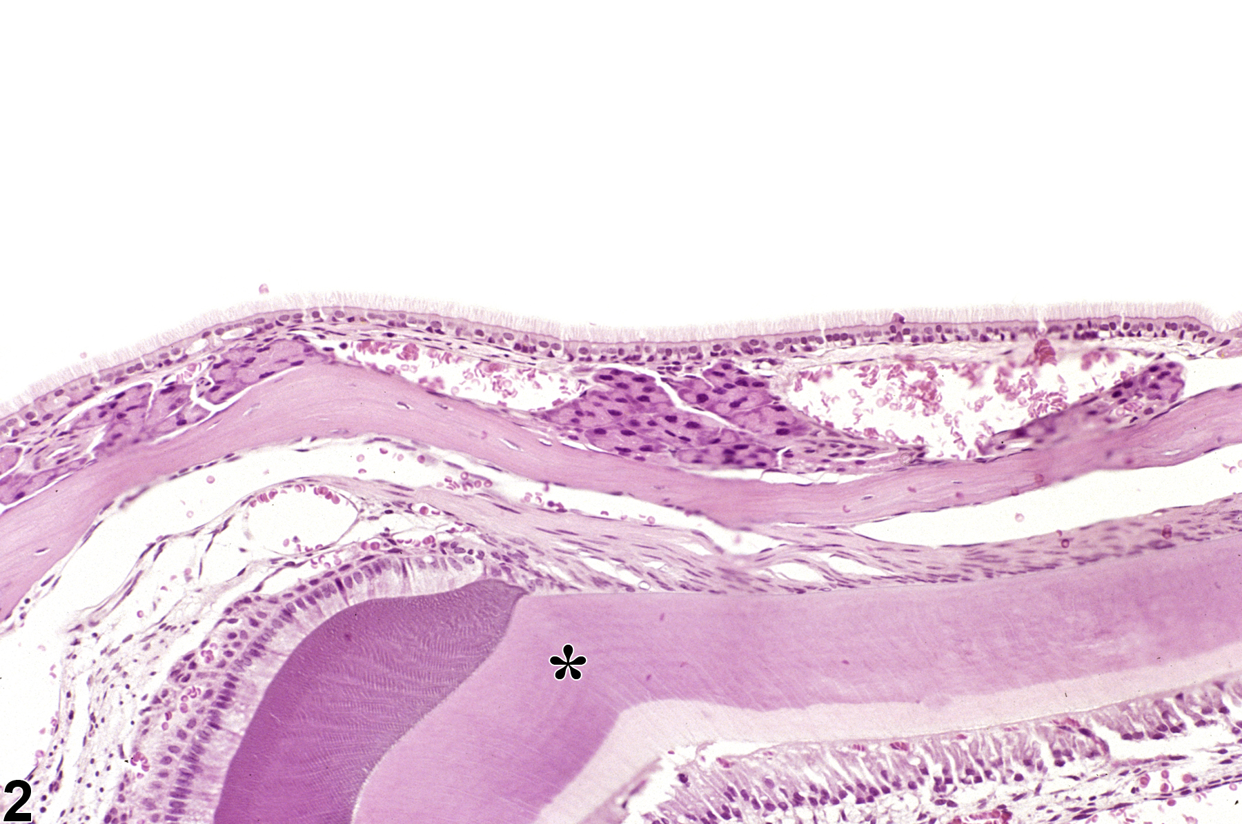
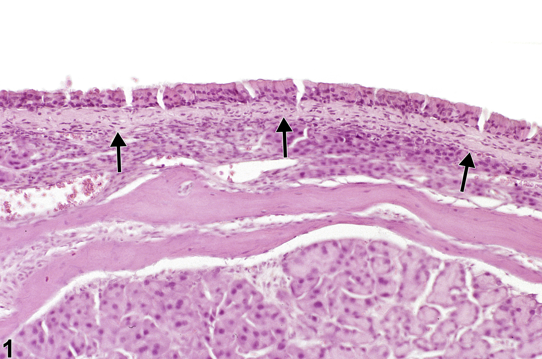
Fibrosis appears microscopically as an increase in the amount of fibrous connective tissue that either replaces or expands normal structures. Because it is often a response to severe tissue damage, fibrosis is often associated with inflammation and/or necrosis. Fibrosis in the lamina propria of the nose (Figure 1; compare with control shown in Figure 2) is uncommon.
References not listed.
Nose, Transitional epithelium - Fibrosis in a female B6C3F1/N mouse from a chronic study. Eosinophilic fibrillar material expands the lamina propria, separating the epithelium from the glands in the lamina propria.
All Images

Nose, Transitional epithelium - Fibrosis in a female B6C3F1/N mouse from a chronic study. Eosinophilic fibrillar material expands the lamina propria, separating the epithelium from the glands in the lamina propria.

Nose, Transitional epithelium - Normal in a male B6C3F1/N mouse from a chronic study. This normal lateral wall in the nose is presented for comparison with Figure 2. A tooth (asterisk) is visible in the section.