U.S. flag

An official website of the United States government

Dot gov

The .gov means it's official.
Federal government websites often end in .gov or .mil. Before sharing sensitive information, make sure you're on a federal government site.

Https

The site is secure.
The https:// ensures that you are connecting to the official website and that any information you provide is encrypted and transmitted securely.

Skip to Main Navigation
Skip to Page Content
Skip to Atlas Navigation

Nose, Epithelium - Hyperplasia, Atypical

Image of hyperplasia, atypical in the nose, respiratory epithelium from a male B6C3F1/N mouse in a chronic study
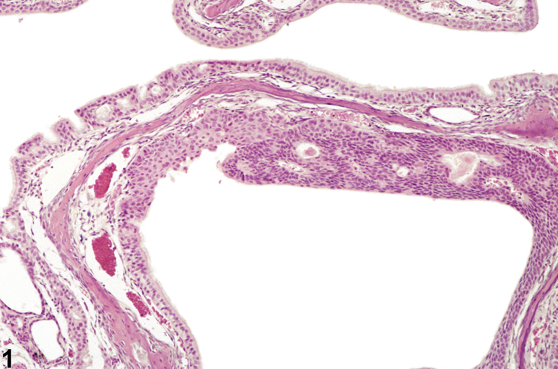
Nose, Respiratory epithelium - Hyperplasia, Atypical in a male B6C3F1/N mouse from a chronic study. An abnormal proliferation of basal cells has replaced respiratory epithelium on the turbinate.
Figure 1 of 5
Image of hyperplasia, atypical in the nose, respiratory epithelium from a female B6C3F1/N mouse in a chronic study
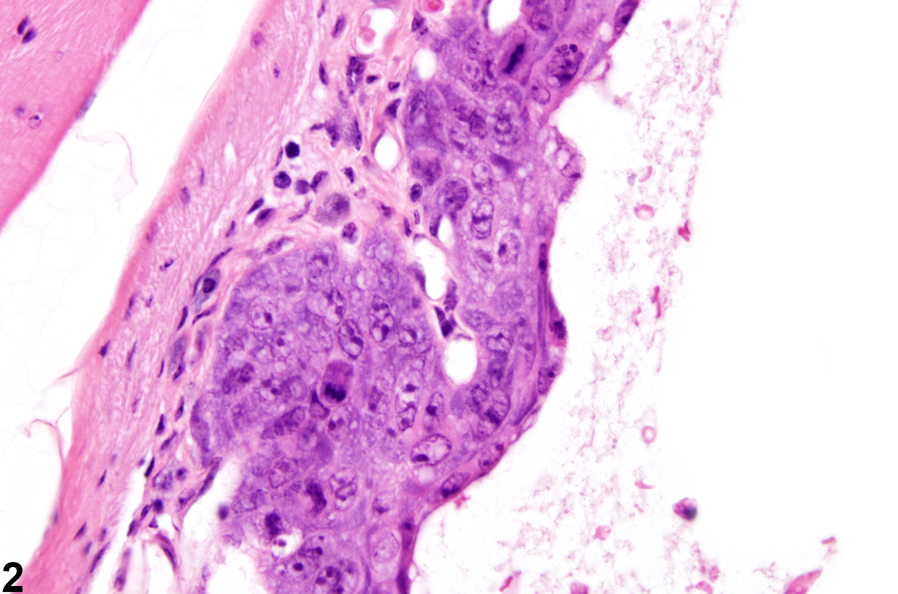
Nose, Respiratory epithelium - Hyperplasia, Atypical in a female B6C3F1/N mouse from a chronic study. Proliferation of atypical and poorly organized epithelium is present on the turbinate.
Figure 2 of 5
Image of hyperplasia, atypical in the nose, squamous epithelium from a female Osborne Mendel rat in a chronic study
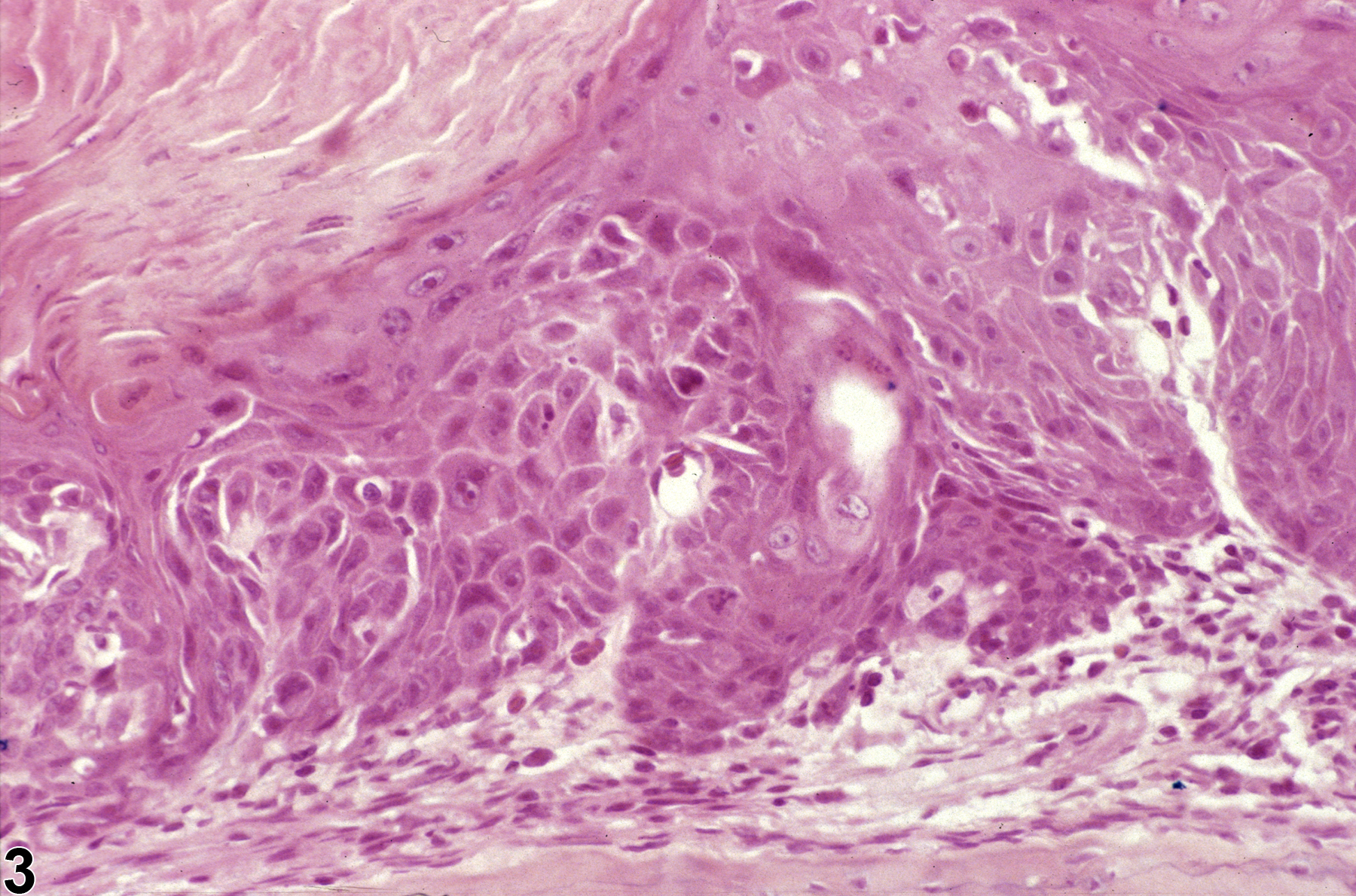
Nose, Squamous epithelium - Hyperplasia, Atypical in a female Osborne Mendel rat from a chronic study. There is some disorganization of the squamous cells and rete peg-like structures.
Figure 3 of 5
Image of hyperplasia, atypical in the nose, olfactory epithelium from a male F344/N rat in a chronic study
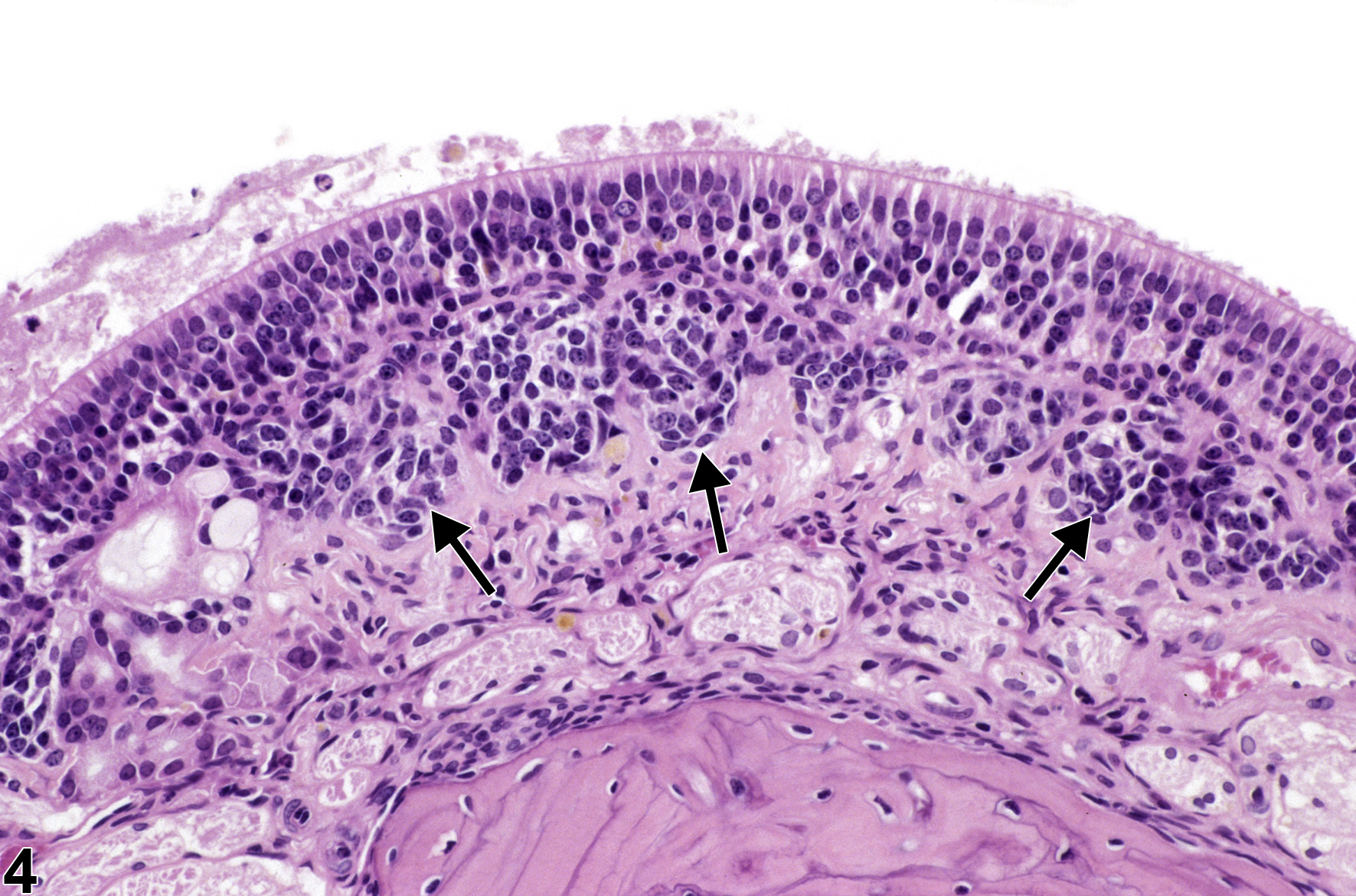
Nose, Olfactory epithelium - Hyperplasia, Atypical in a male F344/N rat from a chronic study. Nests of proliferative cells (arrows) have extended beneath the basement membrane.
Figure 4 of 5
Image of hyperplasia, atypical in the nose, olfactory epithelium from a male F344/N rat in a chronic study
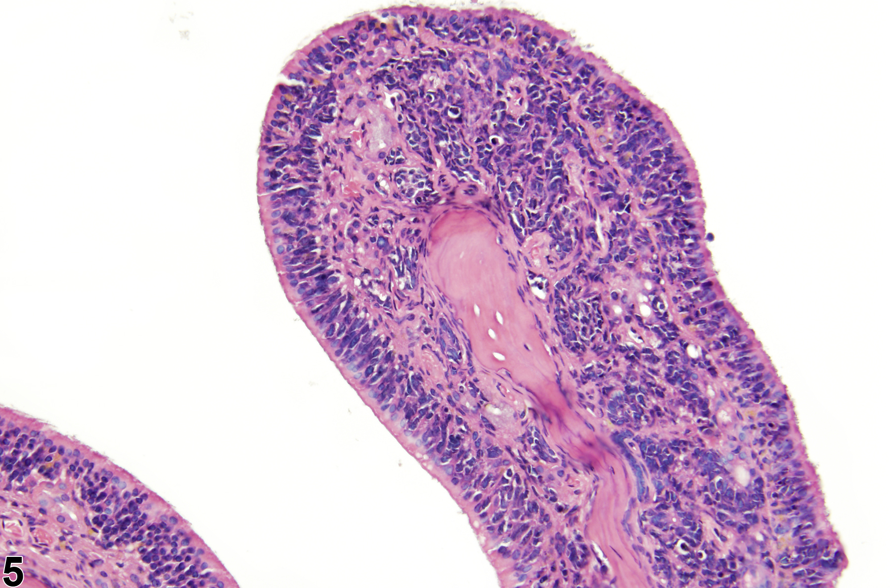
Nose, Olfactory epithelium - Hyperplasia, Atypical in a male F344/N rat from a chronic study. A proliferative band of neuronal-like cells is present beneath the basement membrane, and the overlying olfactory epithelium is disorganized.
Figure 5 of 5
next arrow

comment:

Atypical hyperplasia ( Figure 1image opens in a pop-up window , Figure 2image opens in a pop-up window , Figure 3image opens in a pop-up window , Figure 4image opens in a pop-up window , and Figure 5image opens in a pop-up window ) is a focal or multifocal hyperplastic change (i.e., increased numbers of cells) with features of cellular atypia, which suggests a more aggressive behavior. "Cellular atypia" refers to the presence of morphologic features that are consistent with altered differentiation (cellular pleomorphism, anaplasia) or growth (disorganization of cells, dyskeratosis, abnormal mitotic figures). Atypical hyperplasia can occur in any of the epithelial cell types found in the nasal cavity or in the glands (septal, Bowman's, or Steno's glands) but is most commonly seen in the transitional or respiratory epithelium. Atypical hyperplasia of the olfactory epithelium often manifests as proliferation of basal epithelial cells that extends below the basement membrane. Atypical hyperplasia is thought to represent a preneoplastic change.

recommendation:

Atypical hyperplasia is an important finding that should be diagnosed whenever present and assigned a severity grade. When atypia occurs within a metaplastic lesion, the metaplasia should be diagnosed, but the modifier "atypical" should be added to the diagnosis (e.g., Nose, Olfactory epithelium - Metaplasia, Respiratory, Atypical). The type of epithelium affected should be included in the diagnosis as a site modifier (i.e., squamous, transitional, respiratory, or olfactory epithelium).

references:

Boorman GA, Morgan KT, Uraih LC. 1990. Nose, larynx, and trachea. In: Pathology of the Fischer Rat: Reference and Atlas (Boorman GA, Eustis SL, Elwell MR, eds). Academic Press, San Diego, 315-337.

Brown HR, Monticello TM, Maronpot RR, Randall HW, Hotchkiss JR, Morgan KT. 1991. Proliferative and neoplastic lesions in the rodent nasal cavity. Toxicol Pathol 19(4, pt 1):358-372.
Abstract: https://www.ncbi.nlm.nih.gov/pubmed/1813982

Herbert RA, Leninger JR. 1999. Nose, larynx, and trachea. In: Pathology of the Mouse: Reference and Atlas (Maronpot RR, ed). Cache River Press, Vienna, IL, 259-292.

Monticello TM, Morgan KT, Uraih LC. 1990. Nonneoplastic nasal lesions in rats and mice. Environ Health Perspect 85:249-274.
Full Text: https://www.ncbi.nlm.nih.gov/pmc/articles/PMC1568333/

Renne R, Brix A, Harkema J, Kittel B, Lewis D, March T, Nagano K, Pino M, Rittinghausen S, Rosenbruch M, Tellier P, Wohrmann T. 2009. Proliferative and nonproliferative lesions of the rat and mouse respiratory tract. Toxicol Pathol 37(7 suppl):5S-73S.
Abstract: https://www.ncbi.nlm.nih.gov/pubmed/20032296