

Respiratory System
Nose - Foreign Material
Narrative
It is uncommon to see the test agent in the nose. There may be little response to small amounts of inert particles, but more reactive materials may elicit an inflammatory response and changes in the nasal epithelium (e.g., degeneration or necrosis, metaplasia, atrophy).
References not listed.
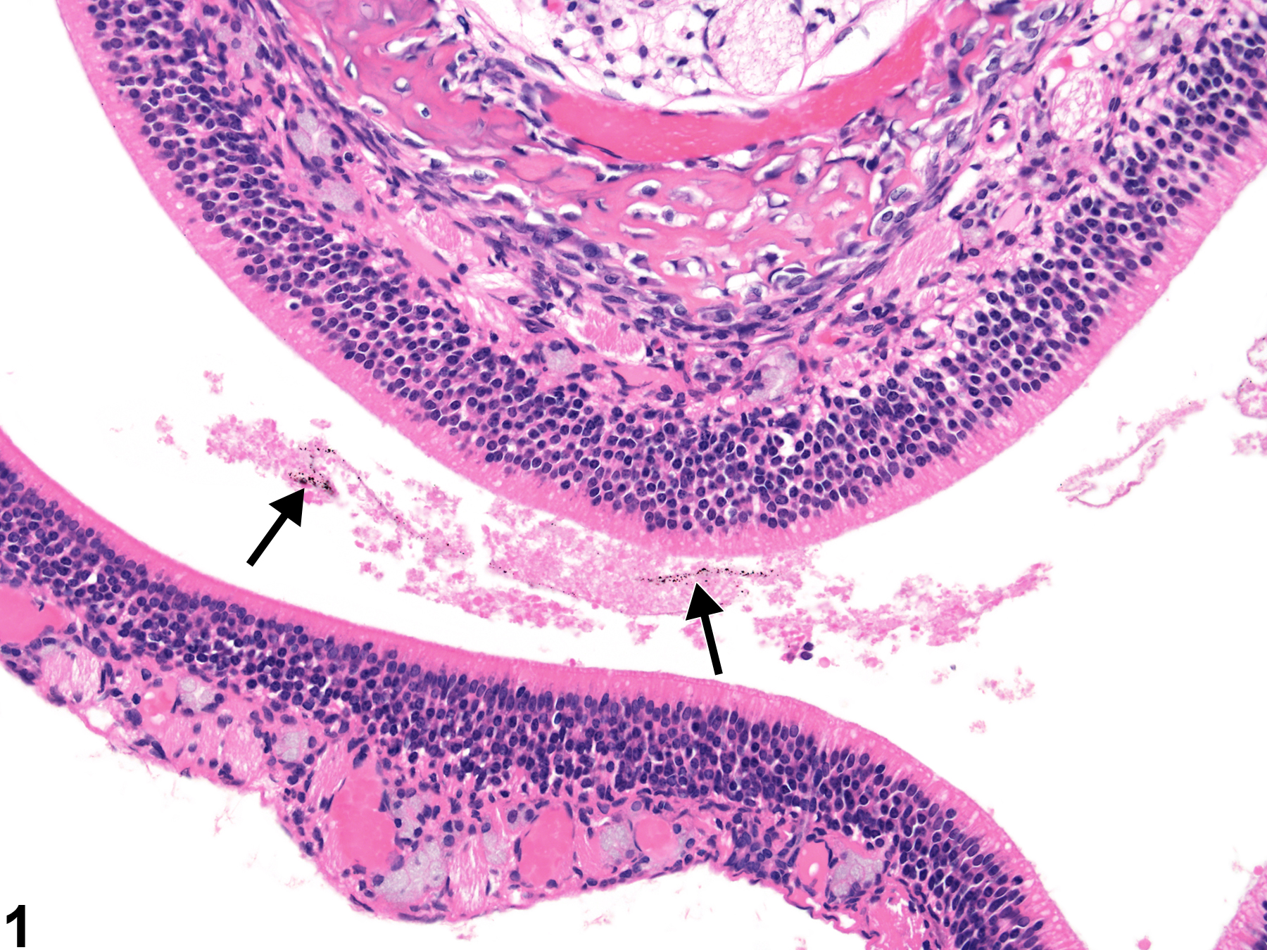
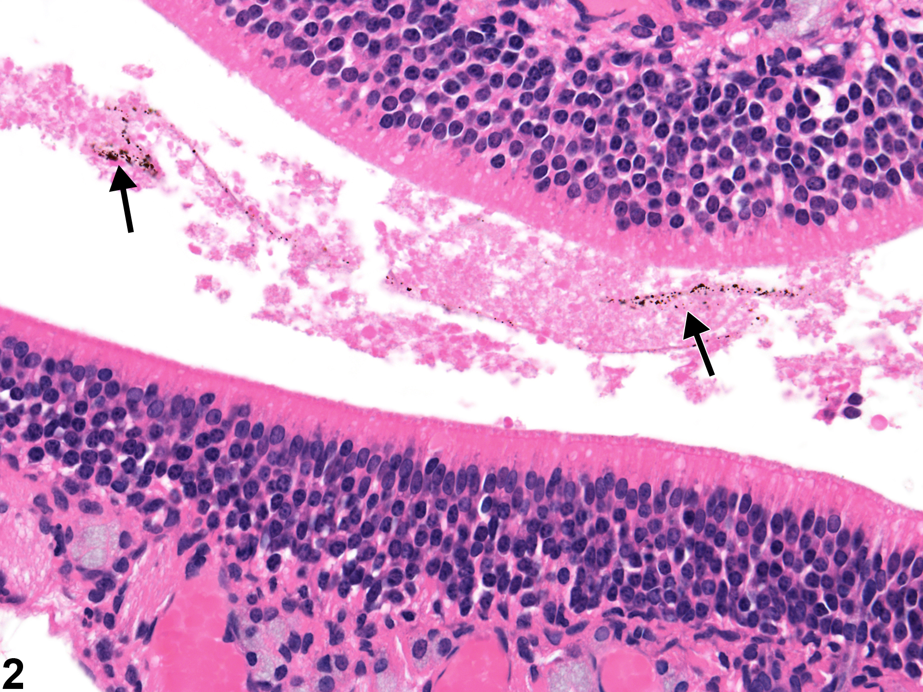
Nose - Foreign material in a female F344/NTac rat from an acute study. Free dark brown particulate material is present in the nasal cavity (arrows).
All Images

Nose - Foreign material in a female F344/NTac rat from an acute study. Free dark brown particulate material is present in the nasal cavity (arrows).

Nose - Foreign material in a female F344/NTac rat from an acute study (higher magnification of Figure 1). The dark brown particulate material (arrows) is associated with proteinaceous material.

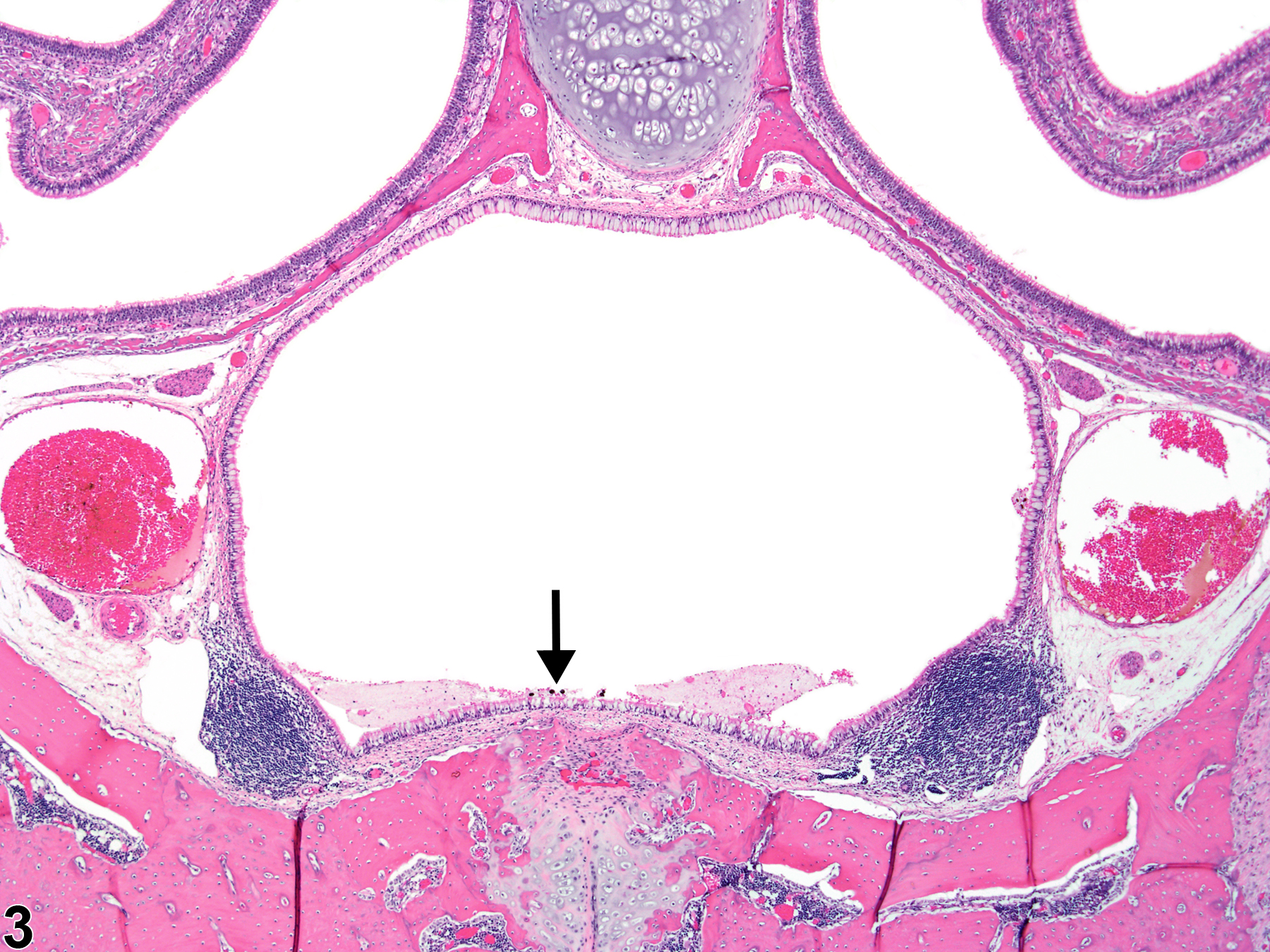
Nose, Nasopharyngeal duct - Foreign material in a female F344/NTac rat from an acute study. Dark brown particulate material is present at the base of the nasopharyngeal duct (arrow).

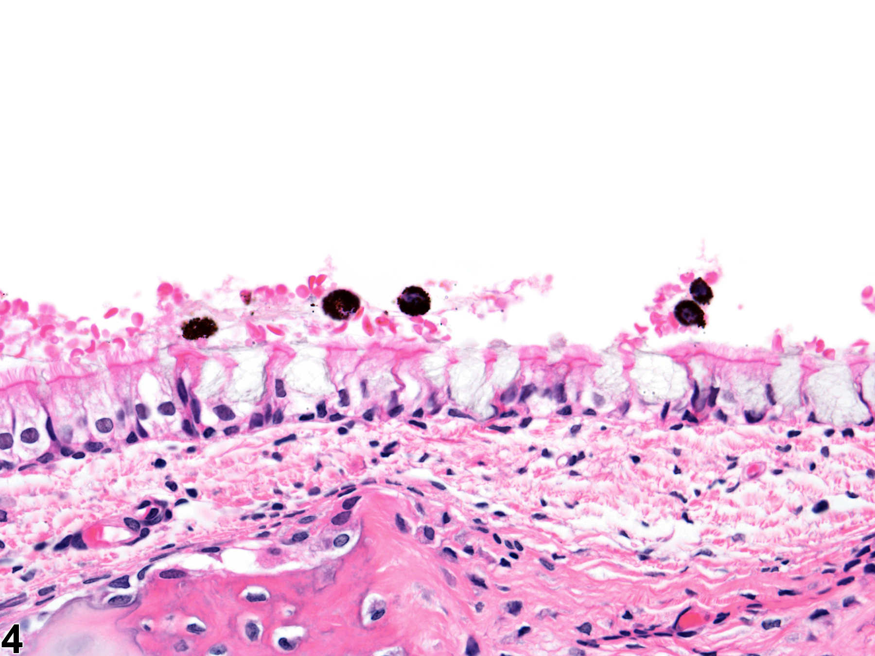
Nose, Nasopharyngeal duct - Foreign material in a female F344/NTac rat from an acute study (higher magnification of Figure 3). The dark brown particulate material is within macrophages.

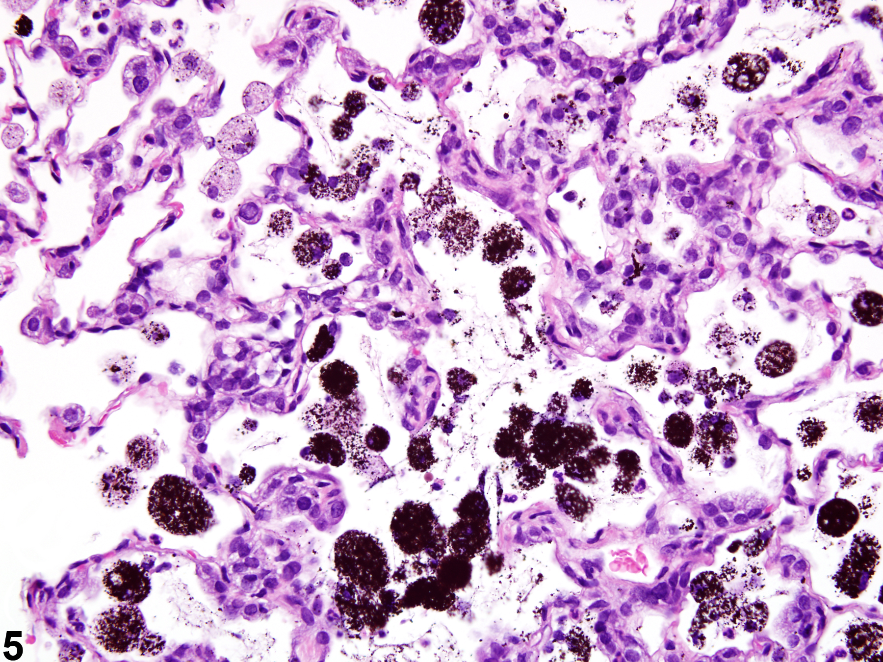
Lung - Foreign material in a female F344/NTac rat from a subchronic study. The appearance of the dark brown particulate material in the lungs from a longer-duration study of the same material is similar to that shown in Figures 1-4.