

Respiratory System
Nose - Fungus
Narrative
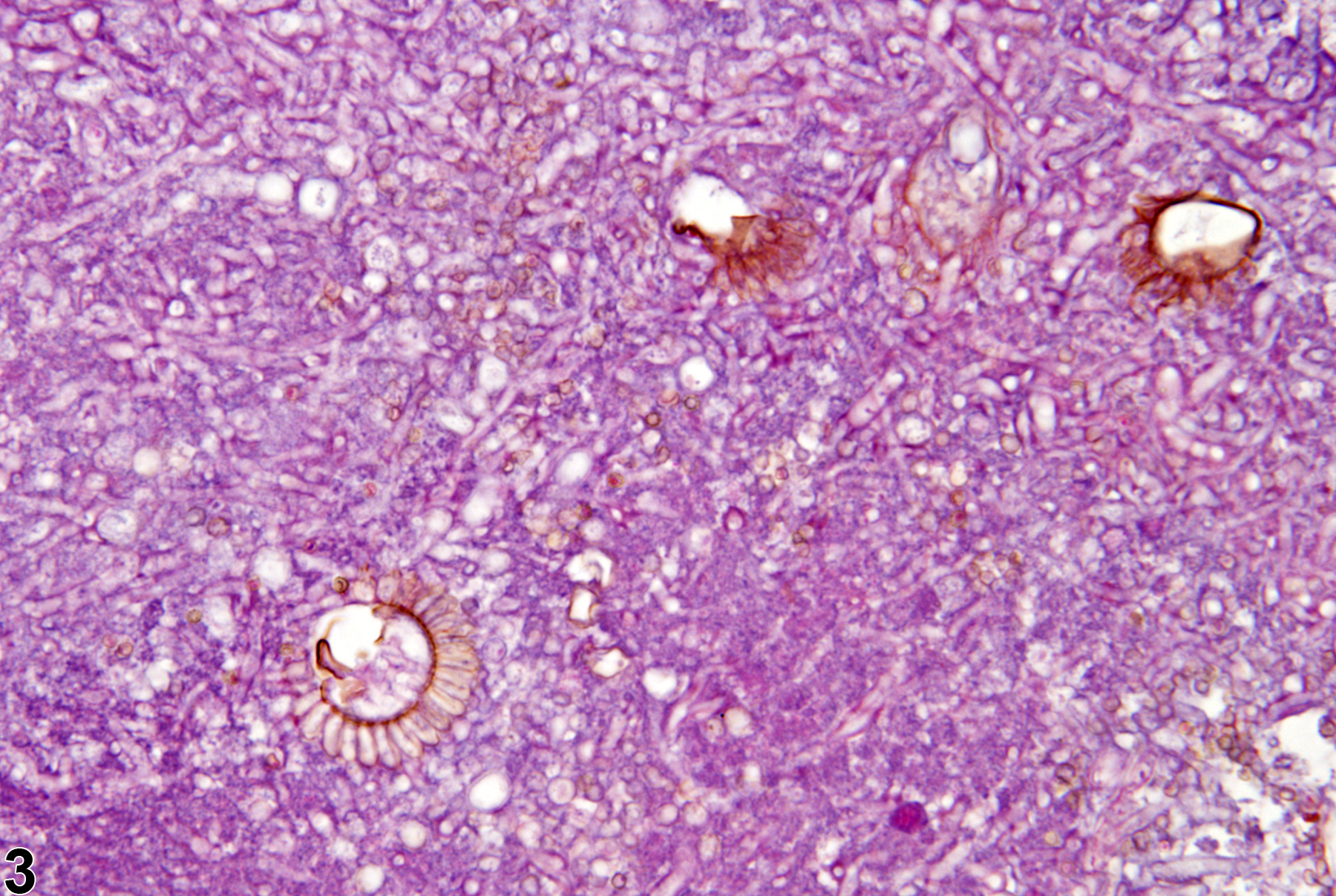
Based on fungal morphology (characteristic fruiting heads and hyphal branching patterns) and reports in the literature, the fungal organisms are usually an Aspergillus sp., with Aspergillus fumigatus being the most common. Aspergillus spp. have characteristic fruiting heads and hyphal branching patterns (Figure 3). The fungi are typically accompanied by suppurative inflammation. Changes such as hyperplasia and metaplasia are usually present in the adjacent epithelium, and there is often acute to chronic-active inflammation in the associated underlying lamina propria.
Boorman GA, Morgan KT, Uraih LC. 1990. Nose, larynx, and trachea. In: Pathology of the Fischer Rat: Reference and Atlas (Boorman GA, Eustis SL, Elwell MR, eds). Academic Press, San Diego, 315-337.
Everitt JI, Richter CB. 1990. Infectious diseases of the upper respiratory tract: Implications for toxicology studies. Environ Health Perspect 85:239-247.
Full Text: https://www.ncbi.nlm.nih.gov/pmc/articles/PMC1568352/Herbert RA, Leninger JR. 1999. Nose, larynx, and trachea. In: Pathology of the Mouse: Reference and Atlas (Maronpot RR, ed). Cache River Press, Vienna, IL, 259-292.
National Toxicology Program. 1986. NTP TR-300. Toxicology and Carcinogenesis Studies of 3-Chloro-2-methylpropene (Technical Grade Containing 5% Dimethylvinyl Chloride) (CAS No. 563-47-3) in F344/N Rats and B6C3F1 Mice (Gavage Studies). NTP, Research Triangle Park, NC.
Abstract: https://ntp.niehs.nih.gov/go/6603Nyska A, Kuttin ES. 1988. Upper respiratory tract infection in mouse and rats. Mycopathologia 101:95-98.
Abstract: https://www.ncbi.nlm.nih.gov/pubmed/2963960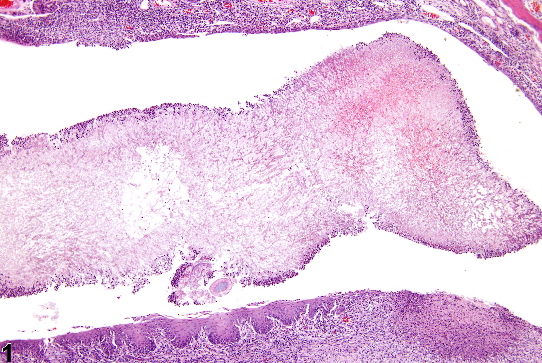
Nose - Fungus in a male F344/N rat from a chronic study. A mat of fungal hyphae is present in the lumen of the nasal cavity and is accompanied by epithelial changes and inflammation of the nasal tissue.
All Images

Nose - Fungus in a male F344/N rat from a chronic study. A mat of fungal hyphae is present in the lumen of the nasal cavity and is accompanied by epithelial changes and inflammation of the nasal tissue.

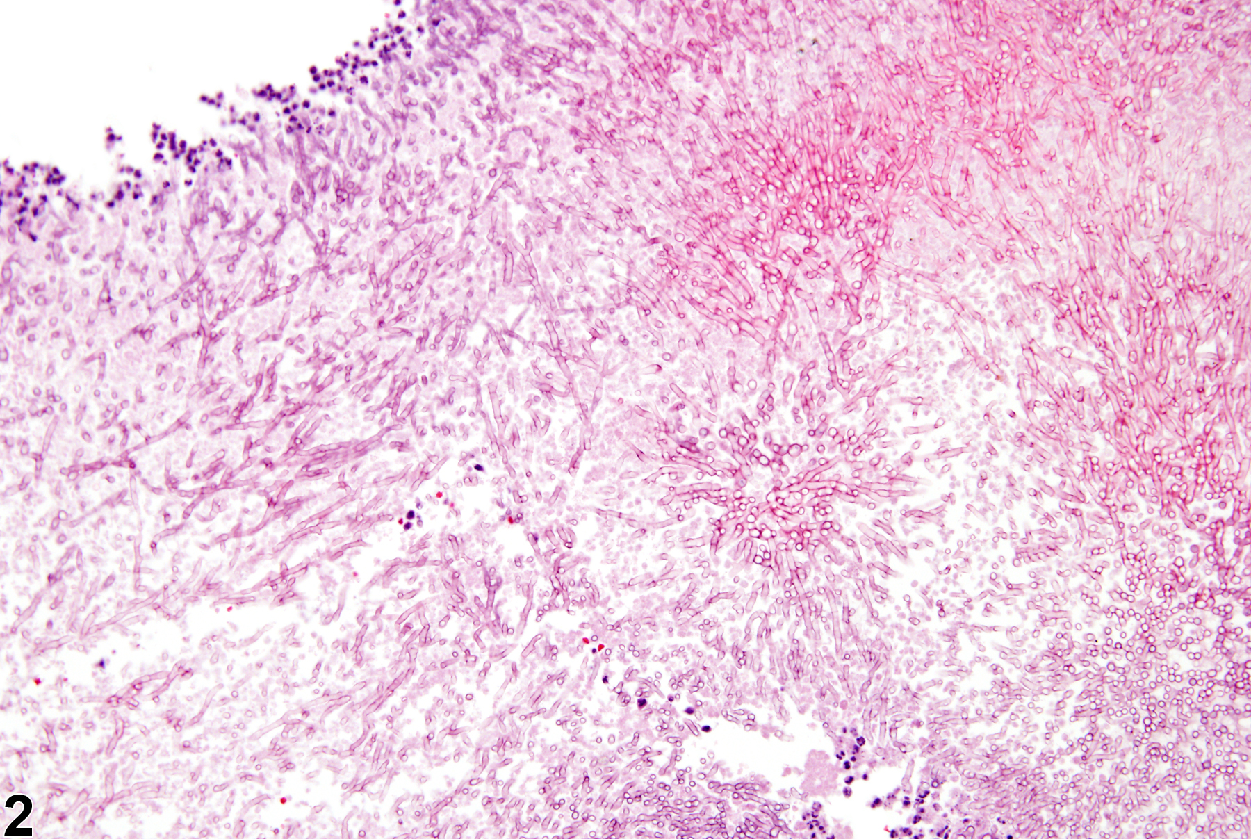
Nose - Fungus in a male F344/N rat from a chronic study (higher magnification of Figure 1). The hyphae are visible in the fungal mat.

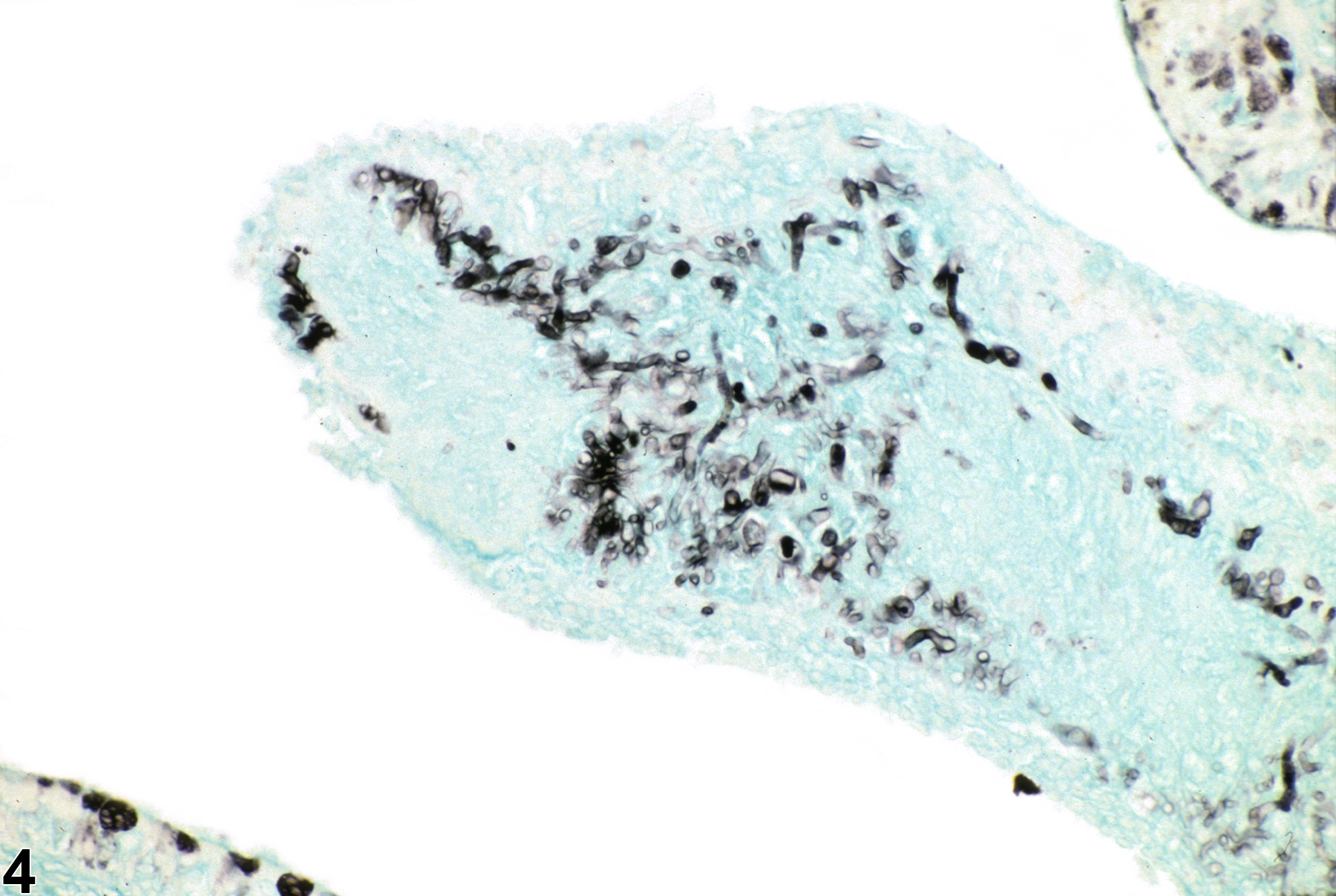
Nose - Fungus in a male F344/N rat from a chronic study. Characteristic pigmented fruiting heads and hyphae are shown in a fungal mat from the nasal cavity.
