

Respiratory System
Nose
Narrative
Documenting nonneoplastic lesions of the nose presents significant challenges, arising from the complex anatomy of the nasal cavity, with numerous cells types and unique anatomic features, and the evaluation of multiple sections, often with multiple concurrent lesions. The diagnoses should include any significant morphologic changes and should document predominant processes present at the time of evaluation.
Nasal olfactory lesions are most commonly seen in inhalation exposures, but they are also seen in feeding studies, gavage studies, and intraperitoneal injection studies. Many factors, such as the route of administration, the physical characteristics of the test agent, and the species of animal, have a significant impact on the anatomic sites affected within the nasal cavity and on the types of lesions observed. For example, the tips of the nasal and maxillary turbinates, which are lined by transitional epithelium, are frequently the initial sites of injury in inhalation studies, but they may be unaffected when nasal lesions are caused by systemic exposure to an agent administered by another route or if enzymatic activation of the agent is required for toxicity. Nasal lesions are generally site specific due to regional deposition of the chemical and regional tissue susceptibility to the chemical, so it is advantageous to subdivide the diagnoses according to site, cell type, or other anatomic designators. The diagnoses should therefore identify the specific cell type(s) or anatomic site(s) involved, and most diagnoses of nasal lesions should include an epithelial type or another relevant anatomic landmark as a site modifier. The epithelial types affected are squamous, transitional, respiratory, and olfactory epithelium.
In order to properly assess the nasal cavity for toxicity, and to allow for comparisons between studies and species, the nasal cavity must be consistently sampled at specific anatomically defined sites. For studies conducted by the National Toxicology Program, the nasal cavity is routinely sectioned at three levels, defined as follows, relative to anatomic landmarks on the maxilla (Figure 1): level I, taken immediately posterior to the upper incisor teeth; level II, through the incisive papilla rostral to the first palatal ridge; and level III, at the middle of the second molar teeth.
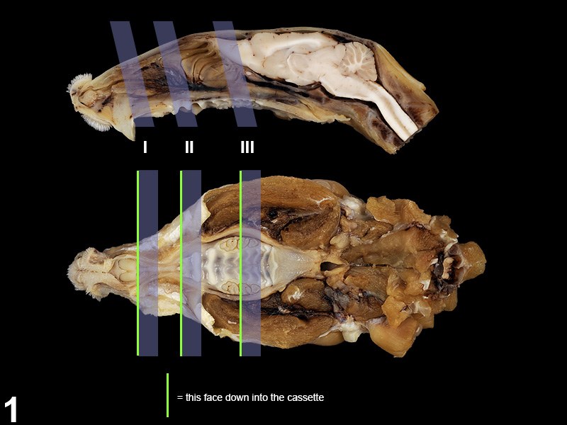
Figure 1. The location of the three levels of the nose routinely examined by the National Toxicology Program.
Histologically, there are four distinct epithelial types in the nasal cavity that vary with the plane of section. At level I, the septum and dorsal meatuses should be lined mainly by respiratory epithelium (pseudostratified, ciliated, columnar) containing some goblet cells (Figure 2 and Figure 3), but they may be lined by olfactory epithelium if the section is taken too far caudally. The lateral wall (Figure 4) should be lined by transitional epithelium (cuboidal or short columnar cells, some of which are ciliated). The transitional epithelium extends dorsally and ventrally from the lateral wall to line the lateral surfaces of the maxilloturbinates and nasoturbinates. The tips of these turbinates are also lined by transitional epithelium. The ventral portion of the ventral meatus at level I (Figure 5) and the nasopalatine ducts are lined by squamous epithelium. The bilateral vomeronasal organ (Figure 5, asterisk) is lined by olfactory-like neuroepithelium medially and by respiratory-like epithelium laterally.
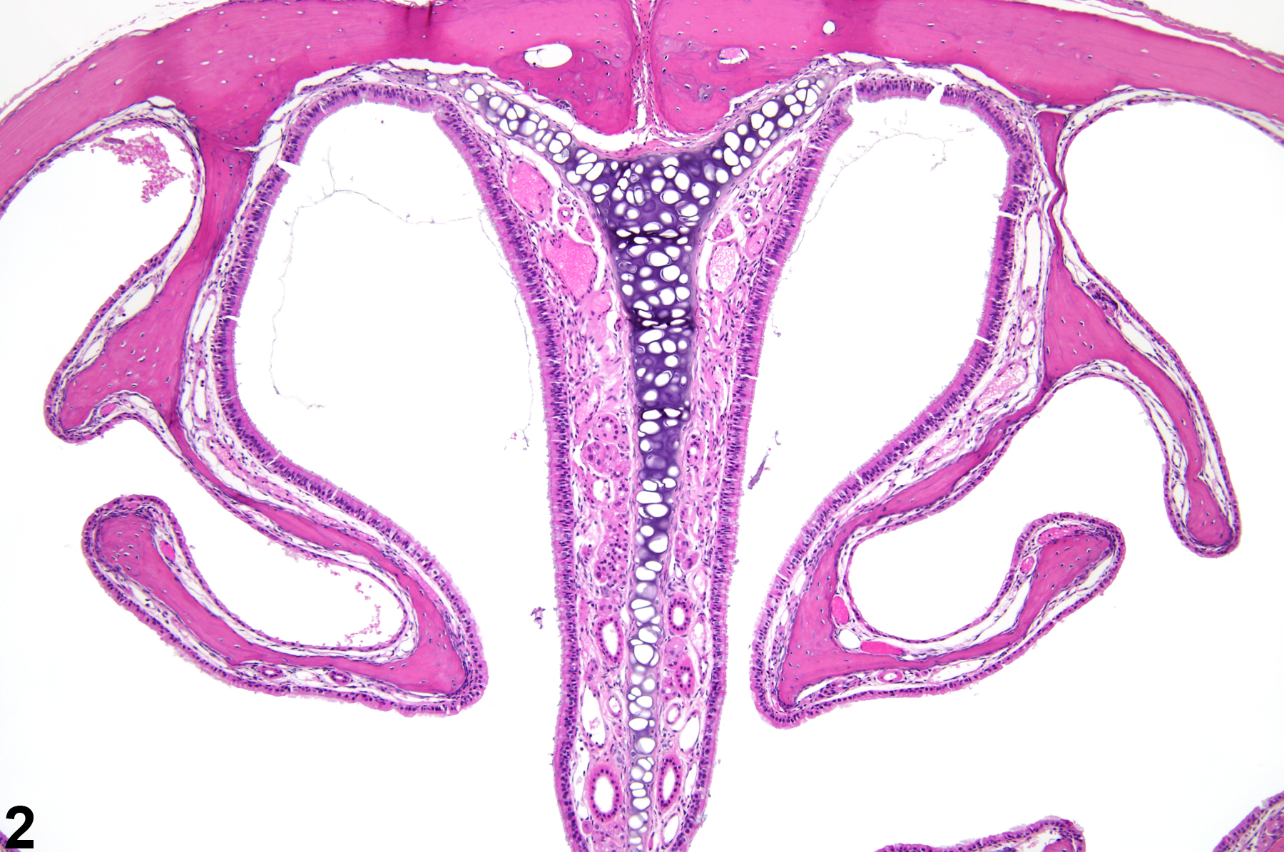
Figure 2. The normal dorsal meatus of level I.

Figure 3. The normal dorsal meatus of level I (higher magnification of Figure 2).
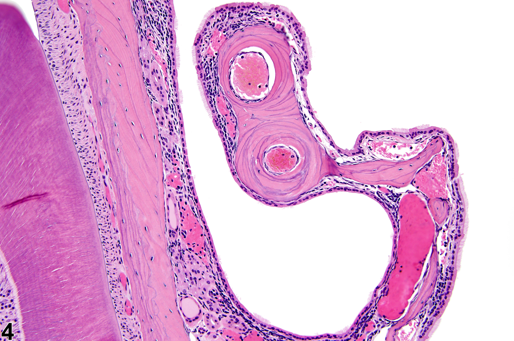
Figure 4. The normal transitional epithelium of the lateral wall at level I.
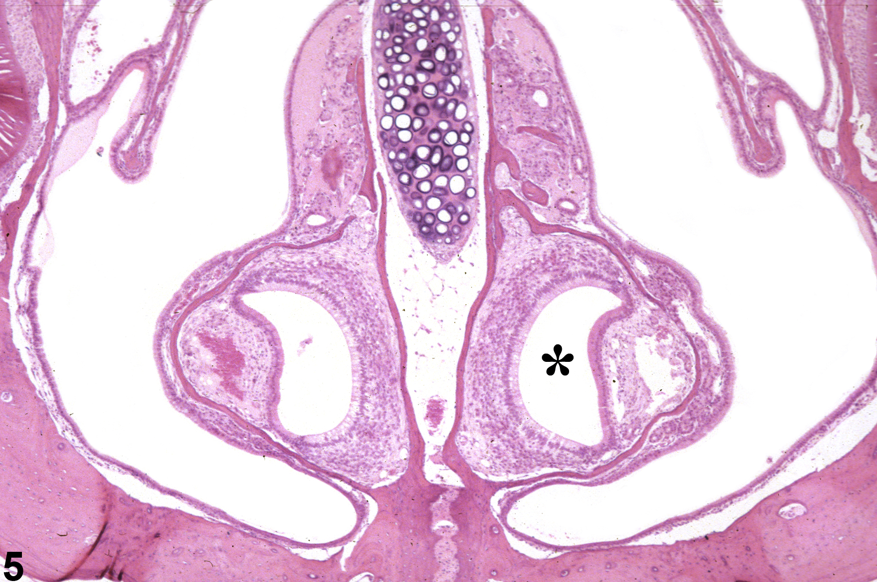
Figure 5. The normal vomeronasal organ (asterisk) at level I.
The dorsal meatus at level II is lined by olfactory epithelium (Figure 6 and Figure 7), which is pseudostratified, columnar epithelium composed primarily of basal cells, specialized bipolar olfactory neurons, and sustentacular (supporting) cells (Figure 7). Olfactory nerves are present in the lamina propria associated with the olfactory epithelium (Figure 7, asterisks). The epithelial types lining the rest of level II are similar to those of level I. Within the laminal propria of the respiratory and olfactory epithelia are various types of glands (Figure 8, arrow) such as the Bowman's glands associated with olfactory epithelium, Steno's glands surrounding the maxillary sinuses, and anterior and posterior septal glands. When diagnosing glandular lesions, the term "glands" should be used as a second site modifier after the epithelial type. For example, "Nose, Olfactory epithelium, Glands" refers to the Bowman’s glands. The type(s) of glands affected should be more thoroughly described in the narrative.
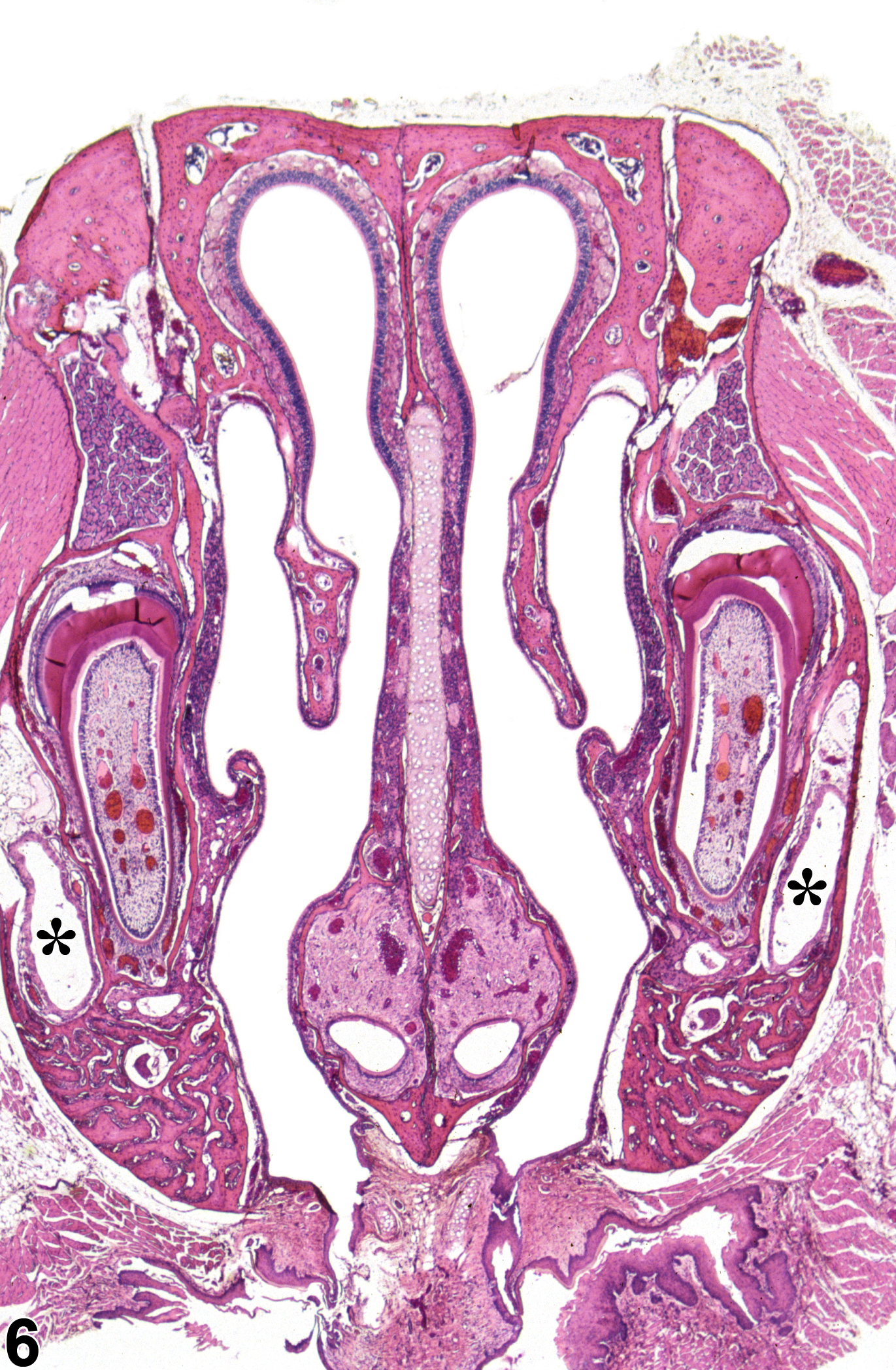
Figure 6. The normal nasal cavity at level II. Asterisks indicate lacrimal ducts.
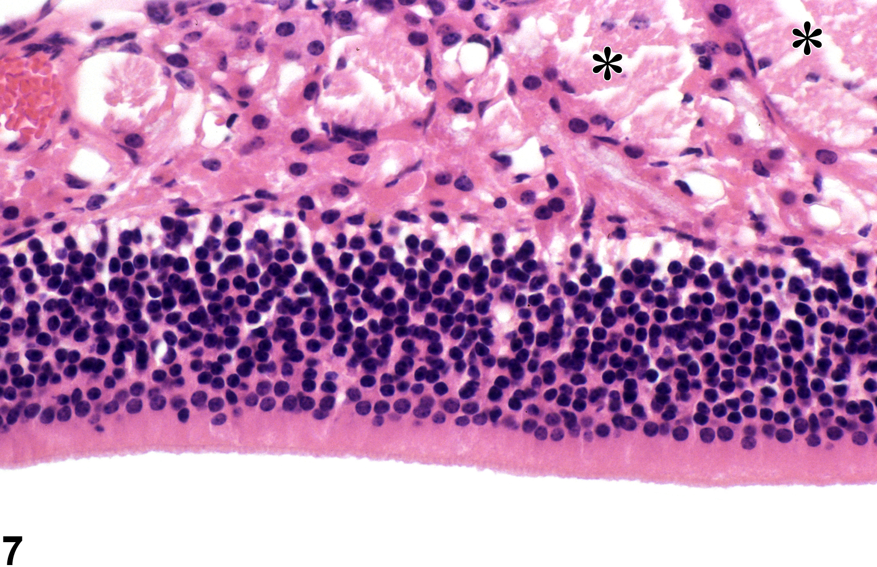
Figure 7. The normal olfactory epithelium and lamina propria at level II. Asterisks indicate olfactory nerves.
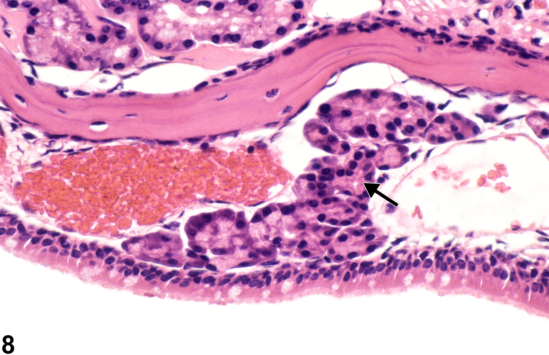
Figure 8. The normal appearance of the respiratory epithelium and the glands in the lamina (arrow) propria at level II.
Level III turbinates are ethmoid turbinates exclusively, which have a more complex structure (Figure 9). The lining epithelium is mainly olfactory epithelium, with some respiratory epithelium ventrally and laterally. Depending on the plane of section, either the septal window or the nasopharyngeal duct (Figure 9, asterisk) can be seen ventral to the nasal septum. Nasopharyngeal duct–associated lymphoid tissue (Figure 9, arrow) is present ventrolateral to the nasopharyngeal duct. The anterior portions of the olfactory lobes of the brain may be present in some sections.
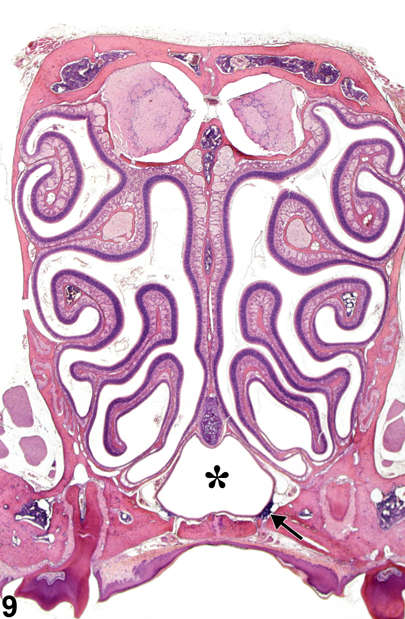
Figure 9. The normal nasal cavity at level III, showing the nasopharyngeal duct (asterisk) and nasal associated lymphoid tissue (NALT, arrow).
Boorman GA, Morgan KT, Uraih LC. 1990. Nose, larynx, and trachea. In: Pathology of the Fischer Rat: Reference and Atlas (Boorman GA, Eustis SL, Elwell MR, eds). Academic Press, San Diego, 315-337.
Herbert RA, Leninger JR. 1999. Nose, larynx, and trachea. In: Pathology of the Mouse: Reference and Atlas (Maronpot RR, ed). Cache River Press, Vienna, IL, 259-292.
Monticello TM, Morgan KT, Uraih LC. 1990. Nonneoplastic nasal lesions in rats and mice. Environ Health Perspect 85:249-274.
Full Text: https://www.ncbi.nlm.nih.gov/pmc/articles/PMC1568333/Renne R, Brix A, Harkema J, Kittel B, Lewis D, March T, Nagano K, Pino M, Rittinghausen S, Rosenbruch M, Tellier P, Wohrmann T. 2009. Proliferative and nonproliferative lesions of the rat and mouse respiratory tract. Toxicol Pathol 37(7 suppl):5S-73S.
Abstract: https://www.ncbi.nlm.nih.gov/pubmed/20032296Uraih LC, Maronpot RR. 1990. Normal histology of the nasal cavity and application of special techniques. Environ Health Perspect 85:187-208.
Full Text: https://www.ncbi.nlm.nih.gov/pmc/articles/PMC1568325/