U.S. flag

An official website of the United States government

Dot gov

The .gov means it's official.
Federal government websites often end in .gov or .mil. Before sharing sensitive information, make sure you're on a federal government site.

Https

The site is secure.
The https:// ensures that you are connecting to the official website and that any information you provide is encrypted and transmitted securely.

Skip to Main Navigation
Skip to Page Content
Skip to Atlas Navigation

Nose, Nerve - Atrophy

Image of atrophy in the nose, nerve from a female F344/N rat in a subchronic study
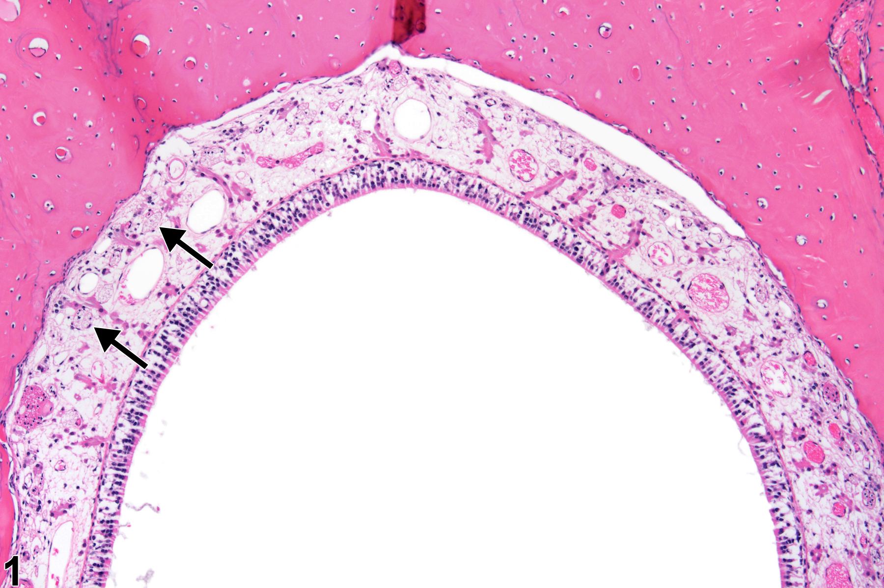
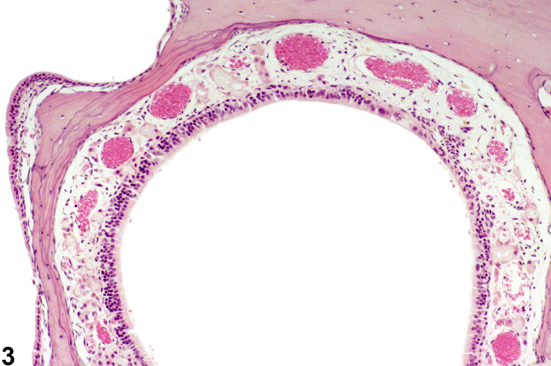
Nose, Nerve - Atrophy in a female F344/N rat from a subchronic study. The olfactory nerves in the lamina propria are small and decreased in number (arrows), and there is loss of cells in the olfactory epithelium.
Figure 1 of 4
Image of atrophy in the nose from a female F344/N rat in a subchronic study
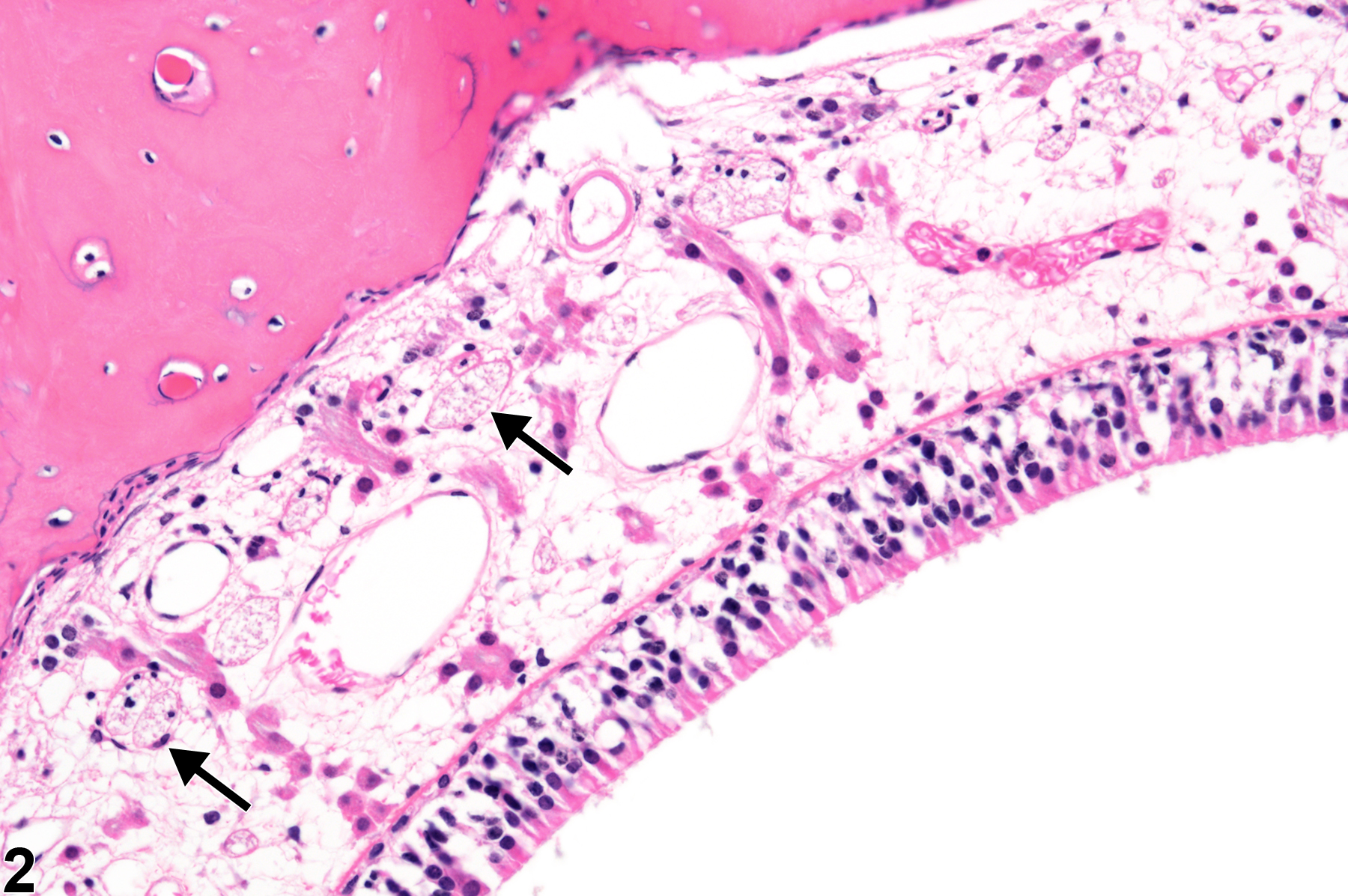
Nose, Nerve - Atrophy in a female F344/N rat from a subchronic study (higher magnification of Figure 1). The olfactory nerves in the lamina propria are decreased in size and number (arrows), and there is loss of cells in the olfactory epithelium.
Figure 2 of 4
Image of atrophy in the nose from a male B6C3F1/N mouse in a chronic study
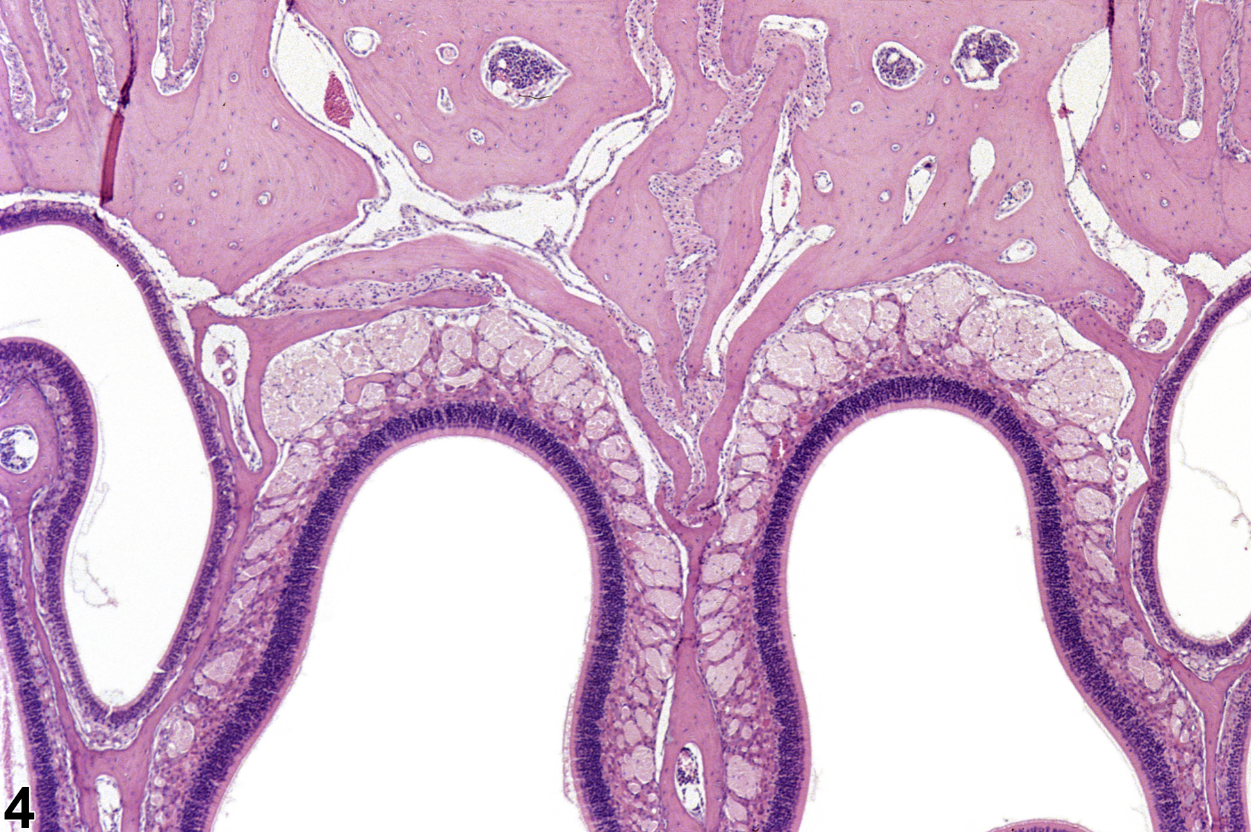
Nose, Nerve - Atrophy in a male B6C3F1/N mouse from a chronic study. No olfactory nerves remain in the lamina propria.
Figure 3 of 4
Image of normal in the nose from a male B6C3F1/N mouse in a chronic study
Nose, Nerve - Normal in a female B6C3F1/N mouse from a chronic study. Normal size and number of olfactory nerves are shown for comparison with Figure 3.
Figure 4 of 4
next arrow

comment:

The olfactory nerves in the lamina propria are bundles of axons from the olfactory neurons whose cell bodies lie in the olfactory epithelium. These nerves are unmyelinated and traverse openings in the ethmoid bone and terminate at the olfactory bulbs. The lesion is characterized by decreased size and number, or complete loss in severe cases ( Figure 3image opens in a pop-up window ), of the olfactory nerves. Atrophy of the olfactory nerves ( Figure 1image opens in a pop-up window , Figure 2image opens in a pop-up window , and Figure 3image opens in a pop-up window ; compare with normal control in Figure 4image opens in a pop-up window ) is secondary to loss of olfactory nerve cells from the olfactory epithelium or to damage to the olfactory bulb of the brain. It is most commonly the result of loss of olfactory neurons in the olfactory epithelium, so the lesion almost always accompanies changes in the olfactory epithelium, such as atrophy or respiratory metaplasia. Since these lesions are usually part of a larger spectrum of lesions in the nasal cavity, inflammation and changes to the respiratory epithelium are often present as well.

recommendation:

Olfactory nerve atrophy should be diagnosed as "Nose, Nerve - Atrophy" whenever present. It should be graded based on the size and number of affected olfactory nerves. Other lesions, including olfactory epithelial atrophy or respiratory metaplasia, should be diagnosed separately. Observation of this lesion should prompt close examination of the olfactory lobe of the brain.

references:

Boorman GA, Morgan KT, Uraih LC. 1990. Nose, larynx, and trachea. In: Pathology of the Fischer Rat: Reference and Atlas (Boorman GA, Eustis SL, Elwell MR, eds). Academic Press, San Diego, 315-337.

Herbert RA, Leninger JR. 1999. Nose, larynx, and trachea. In: Pathology of the Mouse: Reference and Atlas (Maronpot RR, ed). Cache River Press, Vienna, IL, 293-332.

Islam Z, Amuzie CJ, Harkema JR, Pestka JJ. 2007. Neurotoxicity and inflammation in the nasal airways of mice exposed to the macrocyclic trichothecene mycotoxin roridin A: Kinetics and potentiation by bacterial lipopolysaccharide coexposure. Toxicol Sci 98:526-541.
Full Text: http://toxsci.oxfordjournals.org/content/98/2/526.full

Uraih LC, Maronpot RR. 1990. Normal histology of the nasal cavity and application of special techniques. Environ Health Perspect 85:187-208.
Full Text: https://www.ncbi.nlm.nih.gov/pmc/articles/PMC1568325/