

Special Senses System
Ear - Ulcer
Narrative
National Toxicology Program. 1995. NTP TR-444. Initiation/Promotion Study of o-Benzyl-p-Chlorophenol (CAS No. 120-32-1) in Swiss (CD-1®) Mice (Mouse Skin Study). NTP, Research Triangle Park, NC.
Abstract: https://ntp.niehs.nih.gov/go/6032Yoshitomi K, Brown. 1990. HR. Ear and pinna. In: Pathology of the Fischer Rat: Reference and Atlas (Boorman GA, Eustis SL, Elwell MR, Montgomery CA, MacKenzie WF, eds). Academic Press, San Diego, CA, 227-238.
Abstract: https://www.ncbi.nlm.nih.gov/nlmcatalog/9002563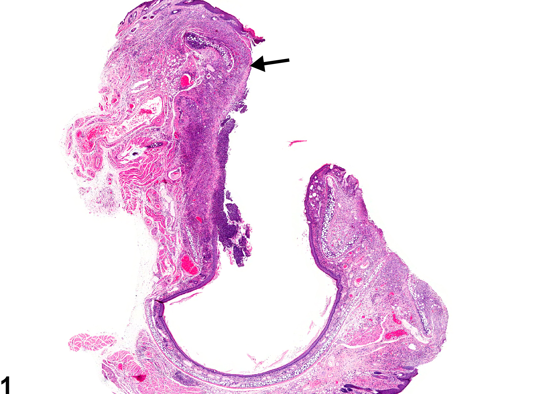
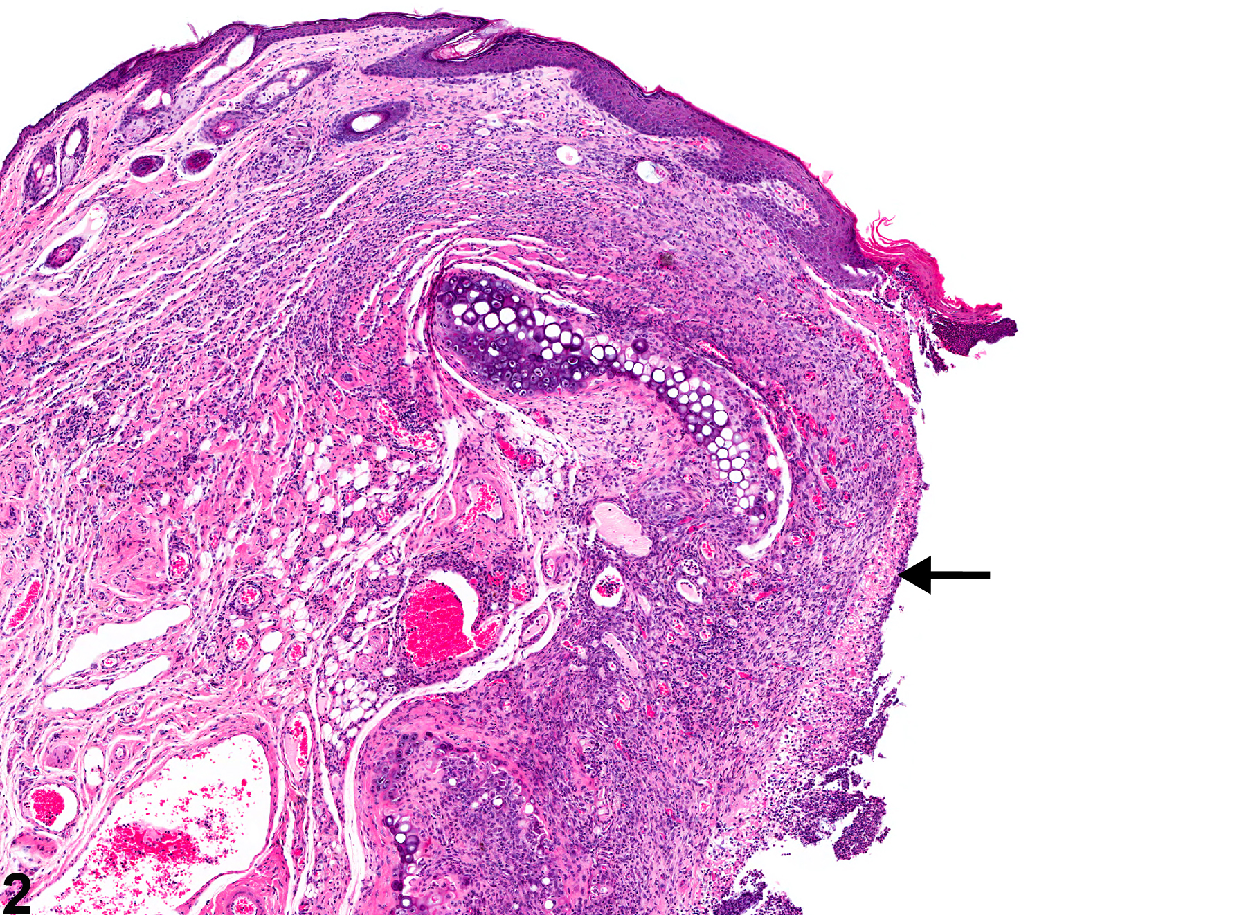
Ear - Ulcer in a male Swiss CD-1 mouse from a chronic study. An ulcer of the external ear canal epithelium; necrotic debris and neutrophils cover much of the ulcer (arrow).
All Images

Ear - Ulcer in a male Swiss CD-1 mouse from a chronic study. An ulcer of the external ear canal epithelium; necrotic debris and neutrophils cover much of the ulcer (arrow).

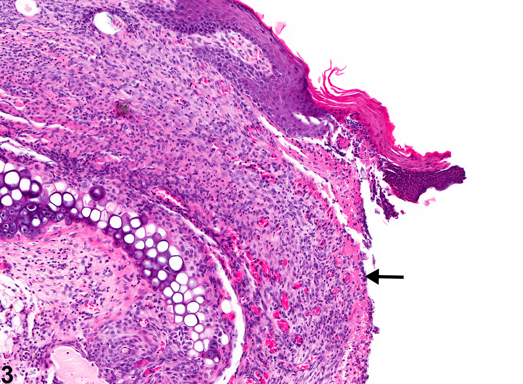
Ear - Ulcer in a male Swiss CD-1 mouse from a chronic study (higher magnification of Figure 1). There is an ulcer (arrow) of the external ear canal epithelium.

Ear - Ulcer in a male Swiss CD-1 mouse from a chronic study (higher magnification of Figure 1). There is an ulcer (arrow) of the external ear canal epithelium; the adjacent epithelium is hyperplastic.