

Special Senses System
Eye, Conjunctiva - Edema
Narrative
Maurer JK, Parker RD. 1996. Light microscopic comparison of surfactant-induced eye irritation in rabbits and rats at three hours and recovery/day 35. Toxicol Pathol 24:403-411.
Abstract: http://tpx.sagepub.com/content/24/4/403.shortMaurer JK, Parker RD, Carr GJ. 1998. Ocular irritation: Microscopic changes occurring over time in the rat with surfactants of known irritancy. Toxicol Pathol 26:217-225.
Abstract: http://tpx.sagepub.com/content/26/2/217.shortNational Toxicology Program. 1994. NTP TR-437. Toxicology and Carcinogenesis Studies of Hexachlorocyclopentadiene (CAS No. 77-47-4) in F344/N Rats and B6C3F1 Mice (Inhalation Studies). NTP, Research Triangle Park, NC.
Abstract: https://ntp.niehs.nih.gov/go/6018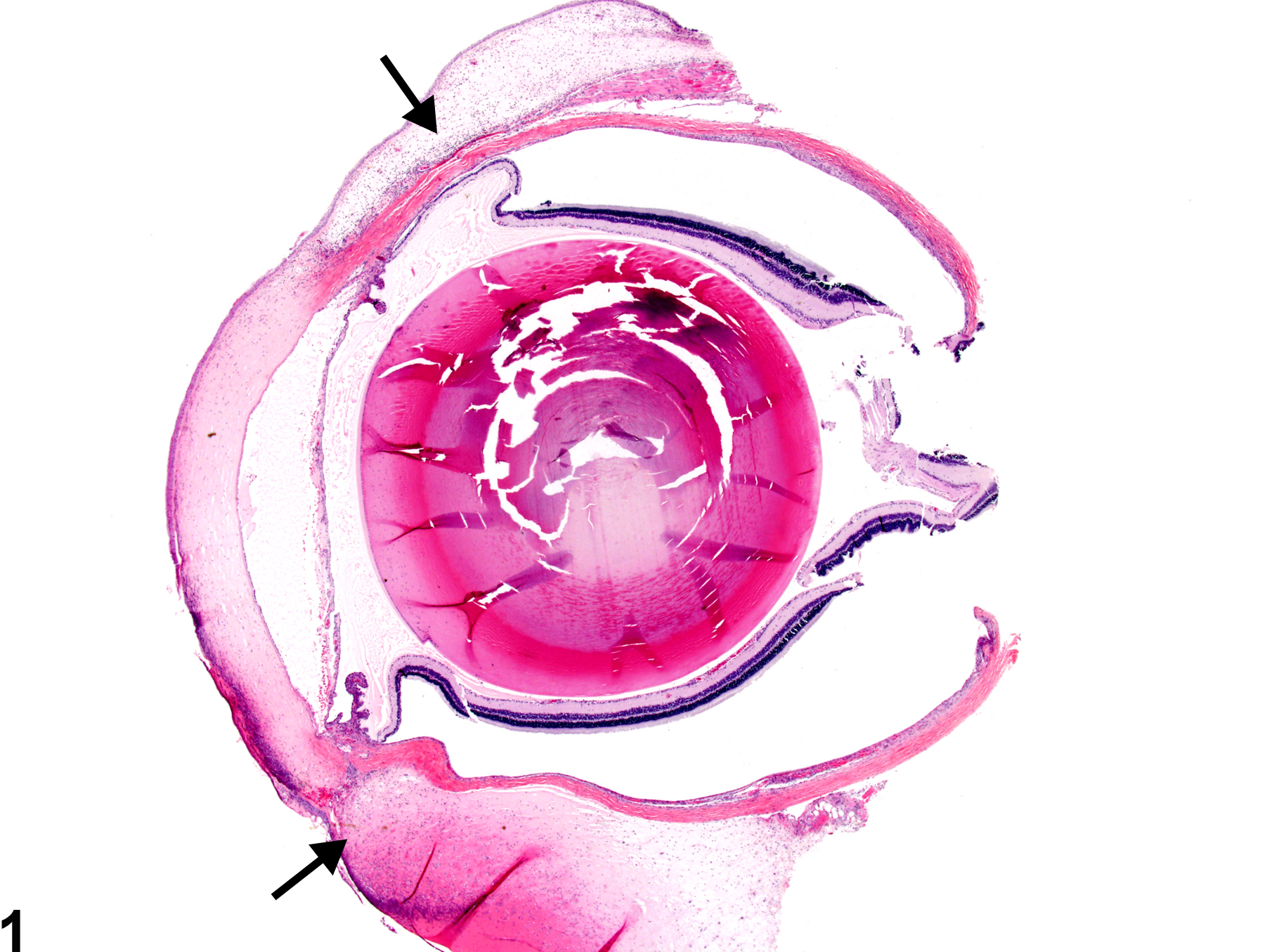
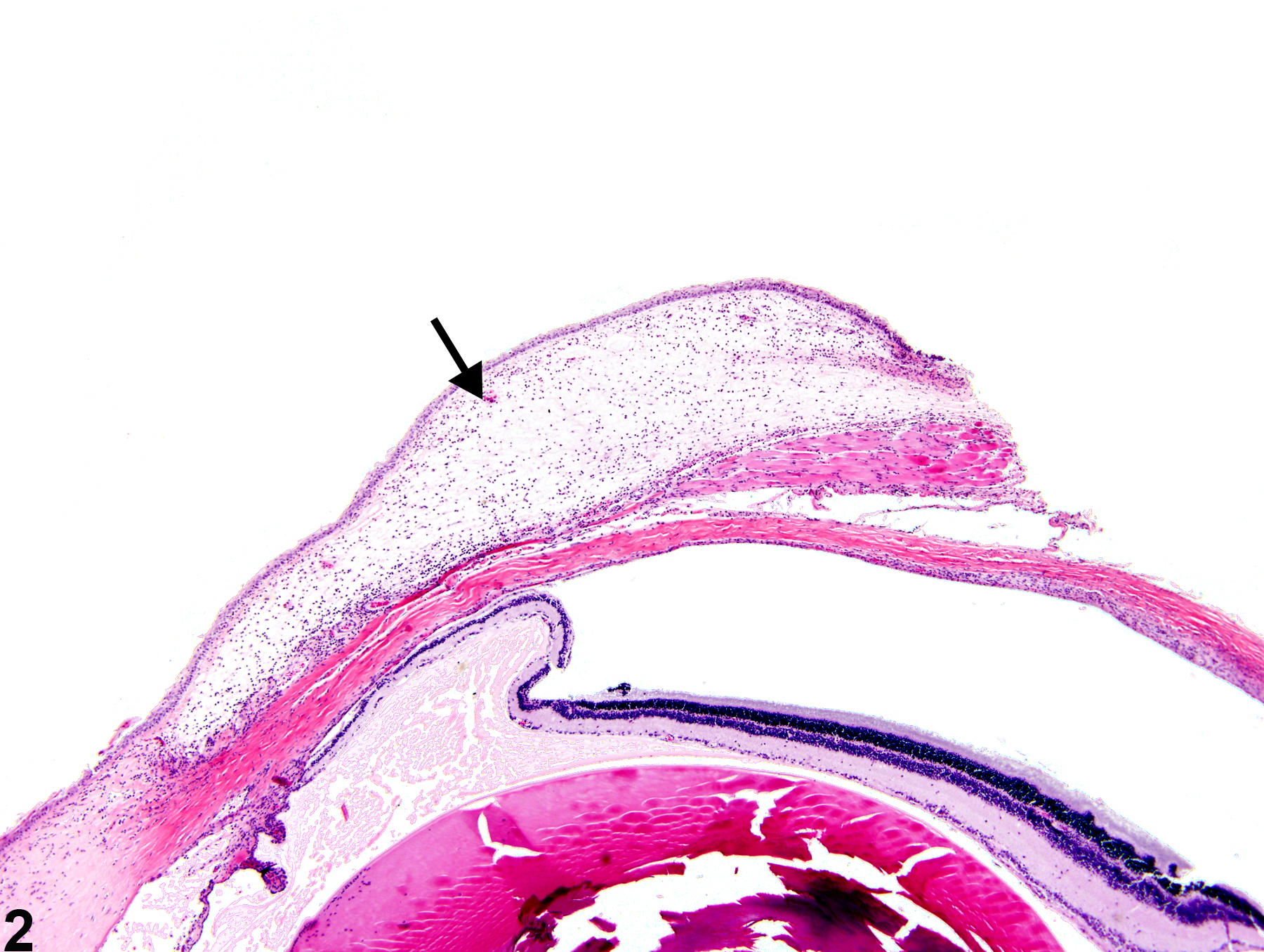
Eye, Conjunctiva - Edema in a female F344/N rat from a chronic study. There is diffuse swelling of the bulbar conjunctiva (arrows) due to accumulation of fluid; inflammatory cells are also present.
All Images

Eye, Conjunctiva - Edema in a female F344/N rat from a chronic study. There is diffuse swelling of the bulbar conjunctiva (arrows) due to accumulation of fluid; inflammatory cells are also present.

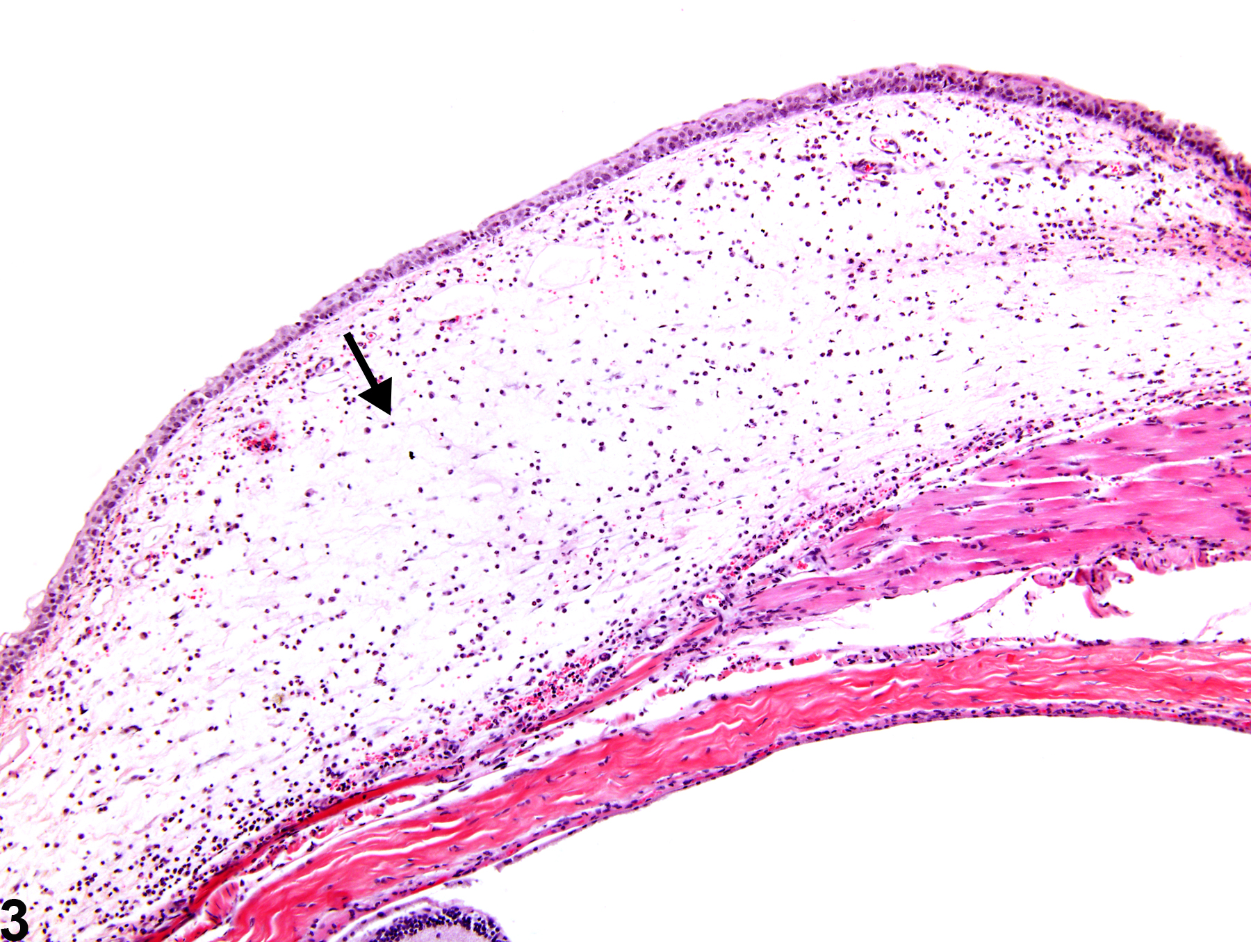
Eye, Conjunctiva - Edema in a female F344/N rat from a chronic study (higher magnification of Figure 1). The bulbar conjunctiva is expanded by clear to pale eosinophilic fluid (arrow); inflammatory cells are also present.

Eye, Conjunctiva - Edema in a female F344/N rat from a chronic study (higher magnification of Figure 1). There is edema of the bulbar conjunctiva characterized by accumulation of clear to pale eosinophilic fluid and inflammatory cells.