

Special Senses System
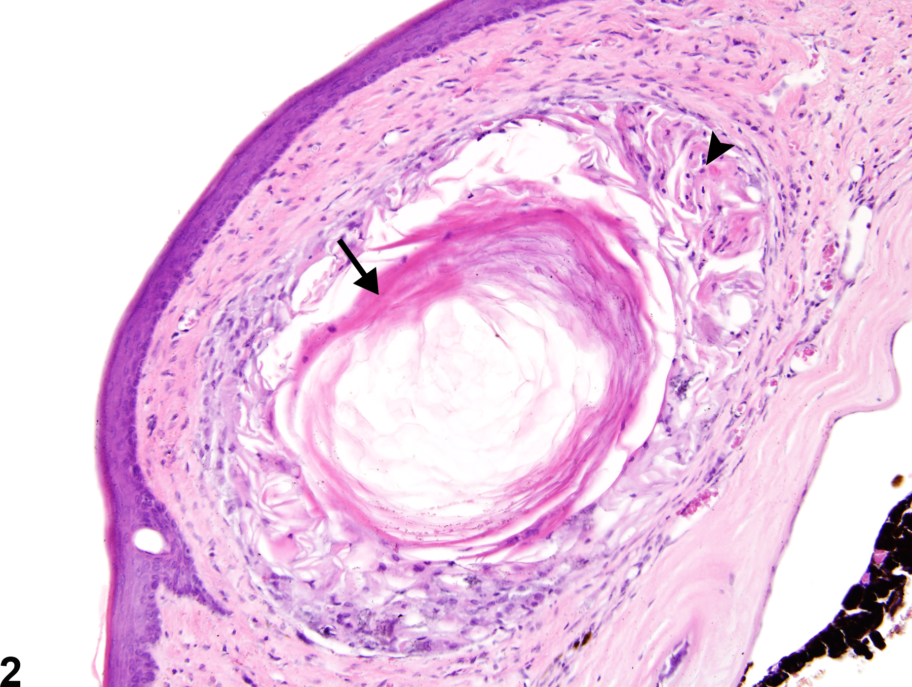
Eye, Cornea - Cyst
Narrative
Geiss V, Yoshitomi K. 1991. Eyes. In: Pathology of the Mouse: Reference and Atlas (Maronpot RR, Boorman GA, Gaul BW, eds). Cache River Press, Vienna, IL, 471-489.
National Toxicology Program. 1990. NTP TR-378. Toxicology and Carcinogenesis Studies of Benzaldehyde (CAS No. 100-52-7) in F344/N Rats and B6C3F1 Mice (Gavage Studies). NTP, Research Triangle Park, NC.
Full Text: https://ntp.niehs.nih.gov/ntp/htdocs/lt_rpts/tr378.pdfYoshitomi K, Boorman GA. 1990. Eye and associated glands. In: Pathology of the Fischer Rat: Reference and Atlas (Boorman GA, Eustis SL, Elwell MR, Montgomery CA, MacKenzie WF, eds). Academic Press, San Diego, CA, 239-260.
Abstract: https://www.ncbi.nlm.nih.gov/nlmcatalog/9002563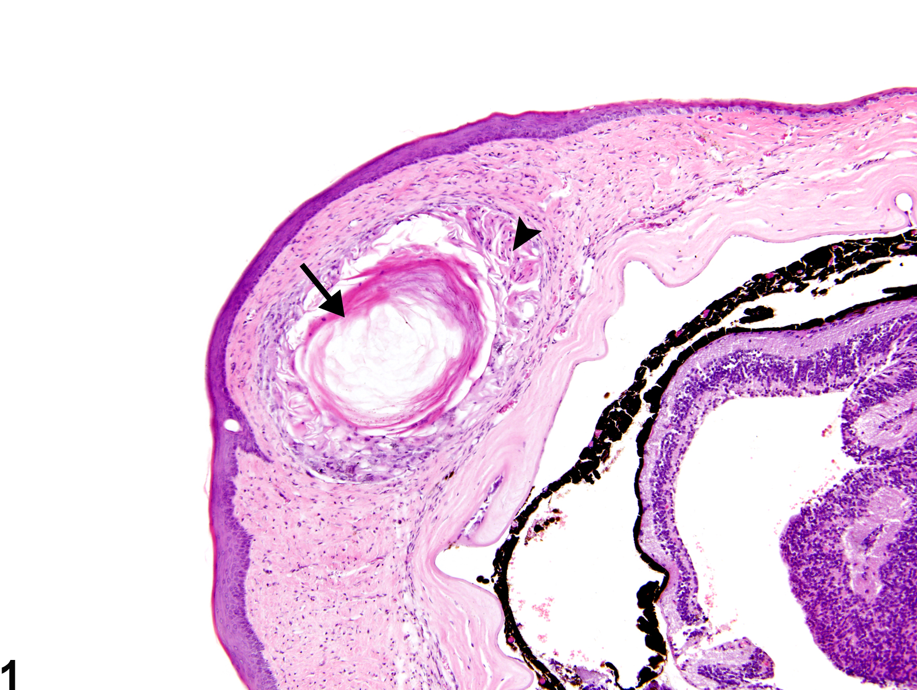
Eye, Cornea - Cyst in a male B6C3F1 mouse from a chronic study. There is a corneal epithelial inclusion cyst (arrow) filled with keratin-like material with secondary granulomatous inflammation (arrowhead).
All Images

Eye, Cornea - Cyst in a male B6C3F1 mouse from a chronic study. There is a corneal epithelial inclusion cyst (arrow) filled with keratin-like material with secondary granulomatous inflammation (arrowhead).

Eye, Cornea - Cyst in a male B6C3F1 mouse from a chronic study (higher magnification of Figure 1). This higher magnification image shows the epithelial inclusion cyst (arrow) and associated granulomatous inflammation (arrowhead) in greater detail.