

Special Senses System
Eye, Cornea - Hyperplasia, Squamous
Narrative
Geiss V, Yoshitomi K. 1991. Eyes. In: Pathology of the Mouse: Reference and Atlas (Maronpot RR, Boorman GA, Gaul BW, eds). Cache River Press, Vienna, IL, 471-489.
Greaves P. 2007. Nervous system and special sense organs. In: Histopathology of Preclinical Toxicity Studies: Interpretation and Relevance in Drug Safety Evaluation, 3rd ed. Academic Press, San Diego, CA, 861-933.
Abstract: http://www.sciencedirect.com/science/book/9780444527714National Toxicology Program. 1988. NTP TR-331. Toxicology and Carcinogenesis Studies of Malonaldehyde, Sodium Salt (3-Hydroxy-2-propenal, Sodium Salt) (CAS No. 24382-04-5) in F344/N Rats and B6C3F1 Mice (Gavage Studies). NTP, Research Triangle Park, NC.
Abstract: https://ntp.niehs.nih.gov/go/8898National Toxicology Program. 1989. NTP TR-342. Toxicology and Carcinogenesis Studies of Dichlorvos (CAS No. 62-73-7) in F344/N Rats and B6C3F1 Mice (Gavage Studies). NTP, Research Triangle Park, NC.
Abstract: https://ntp.niehs.nih.gov/go/10790Roerig DL, Hasegawa AT, Harris GJ, Lynch KL, Wang RIH. 1980. Occurrence of corneal opacities in rats after acute administration of l-α-acetylmethadol. Toxicol Appl Pharmacol 56:156-163.
Abstract: https://www.ncbi.nlm.nih.gov/pubmed/6110254Smith RS, Hawes NL, Kuhlmann SD, Heckenlively JR, Chang B, Roderick TH, Sundberg JP. 1996. Corn1: A mouse model for corneal surface disease and neovascularization. Invest Ophthalmol Vis Sci 37:397-404.
Abstract: https://pubmed.ncbi.nlm.nih.gov/8603845/Wang I-J, Kao CW-C, Liu C-Y, Saika S, Nishina PM, Sundberg JP, Smith RS, Kao WW. 2001. Characterization of corn1 mice: Alteration of epithelial and stromal gene expression. Mol Vis 7:20-26.
Abstract: https://www.ncbi.nlm.nih.gov/pubmed/11182022Yoshitomi K, Boorman GA. 1990. Eye and associated glands. In: Pathology of the Fischer Rat: Reference and Atlas (Boorman GA, Eustis SL, Elwell MR, Montgomery CA, MacKenzie WF, eds). Academic Press, San Diego, CA, 239-260.
Abstract: https://www.ncbi.nlm.nih.gov/nlmcatalog/9002563Yudkin AM, Lambert RM. 1923. Pathogenesis of the ocular lesions produced by a deficiency of vitamin A. J Exp Med 38:17-24.
Full Text: https://www.ncbi.nlm.nih.gov/pmc/articles/PMC2128416/pdf/17.pdf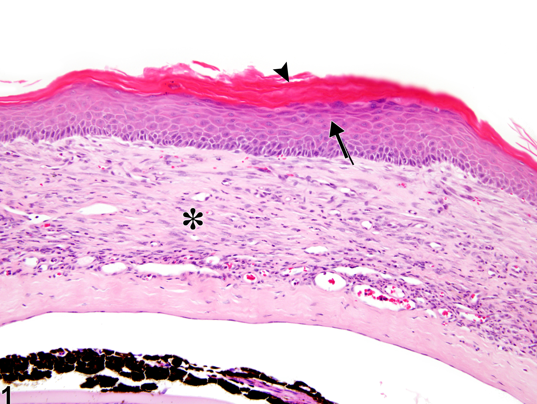
Eye, Cornea - Hyperplasia, Squamous in a male B6C3F1 mouse from a chronic study. There are increased cell layers of the corneal epithelium (arrow) and superficial hyperkeratosis (arrowhead) with inflammation in the underlying stroma (asterisk).
All Images

Eye, Cornea - Hyperplasia, Squamous in a male B6C3F1 mouse from a chronic study. There are increased cell layers of the corneal epithelium (arrow) and superficial hyperkeratosis (arrowhead) with inflammation in the underlying stroma (asterisk).

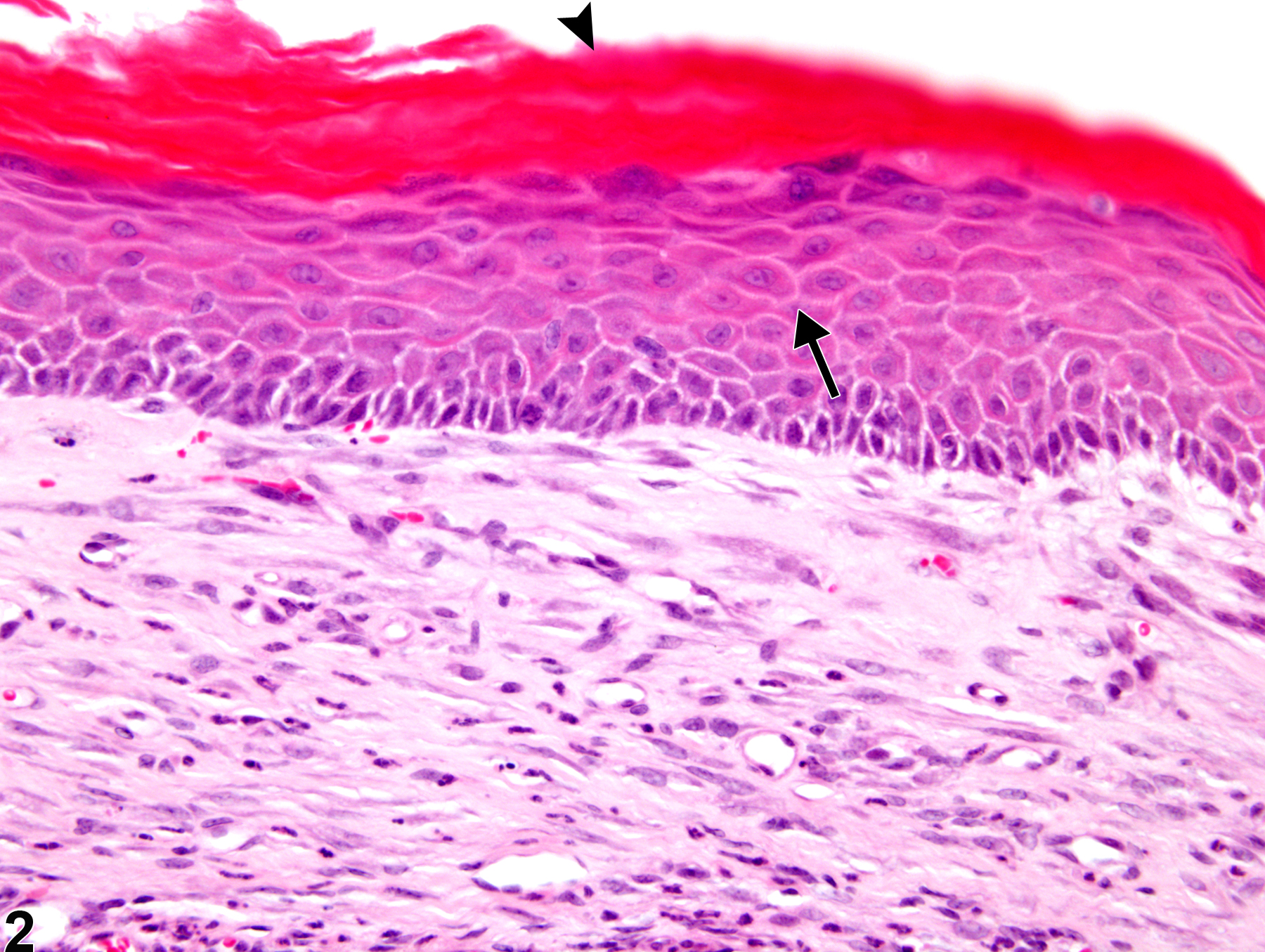
Eye, Cornea - Hyperplasia, Squamous in a male B6C3F1 mouse from a chronic study (higher magnification of Figure 1). The corneal squamous epithelium is thickened (arrow) by increased layers of cells and hyperkeratosis (arrowhead).

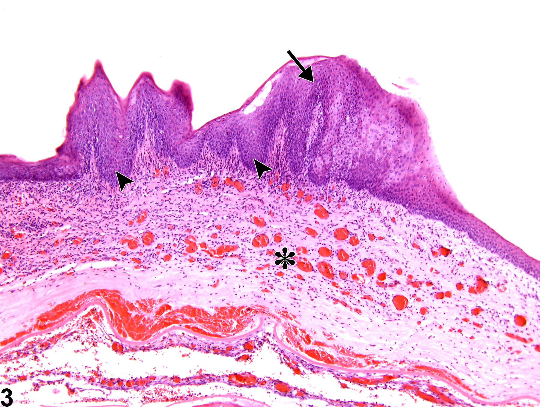
Eye, Cornea - Hyperplasia, Squamous in a male F344/N rat from a chronic study. There are increased numbers of cell layers with exophytic and endophytic

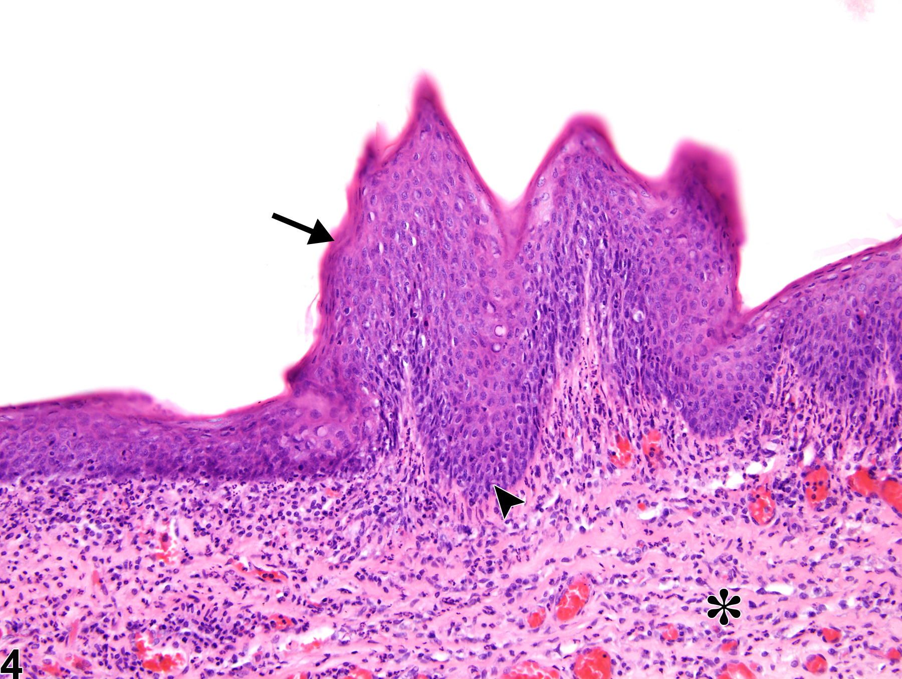
Eye, Cornea - Hyperplasia, Squamous in a male F344/N rat from a chronic study (higher magnification of Figure 3). This higher magnification image shows the marked hyperplasia in greater detail.