

Special Senses System
Eye, Cornea - Metaplasia, Goblet Cell
Narrative
Geiss V, Yoshitomi K. 1991. Eyes. In: Pathology of the Mouse: Reference and Atlas (Maronpot RR, Boorman GA, Gaul BW, eds). Cache River Press, Vienna, IL, 471-489.
Greaves P. 2007. Nervous system and special sense organs. In: Histopathology of Preclinical Toxicity Studies: Interpretation and Relevance in Drug Safety Evaluation, 3rd ed. Academic Press, San Diego, CA, 861-933.
Abstract: http://www.sciencedirect.com/science/book/9780444527714Joussen AM, Poulaki, V, Mitisiades N, Stechschulte SU, Kirchof B, Dartt DA, Fong G-H, Rudge J, Wiegans SJ, Yancopoulos GD, Adamis AP. 2003. VEGF-dependent conjunctivalization of the corneal surface. Invest Ophthalmol Vis Sci 44:117-123.
Full Text: https://doi.org/10.1167/iovs.01-1277Maurer JK, Parker RD. 1996. Light microscopic comparison of surfactant-induced eye irritation in rabbits and rats at three hours and recovery/day 35. Toxicol Pathol 24:403-411.
Abstract: http://tpx.sagepub.com/content/24/4/403.shortNational Toxicology Program. 1992. NTP TR-410. Toxicology and Carcinogenesis Studies of Naphthalene (CAS No. 91-20-3) in B6C3F1 Mice (Inhalation Studies). NTP, Research Triangle Park, NC.
Abstract: https://ntp.niehs.nih.gov/go/7700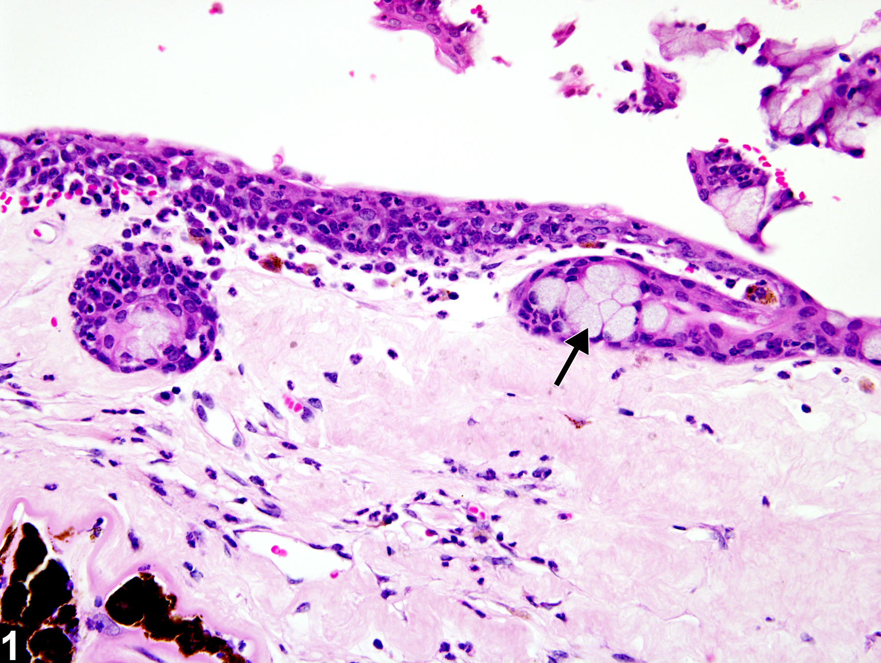
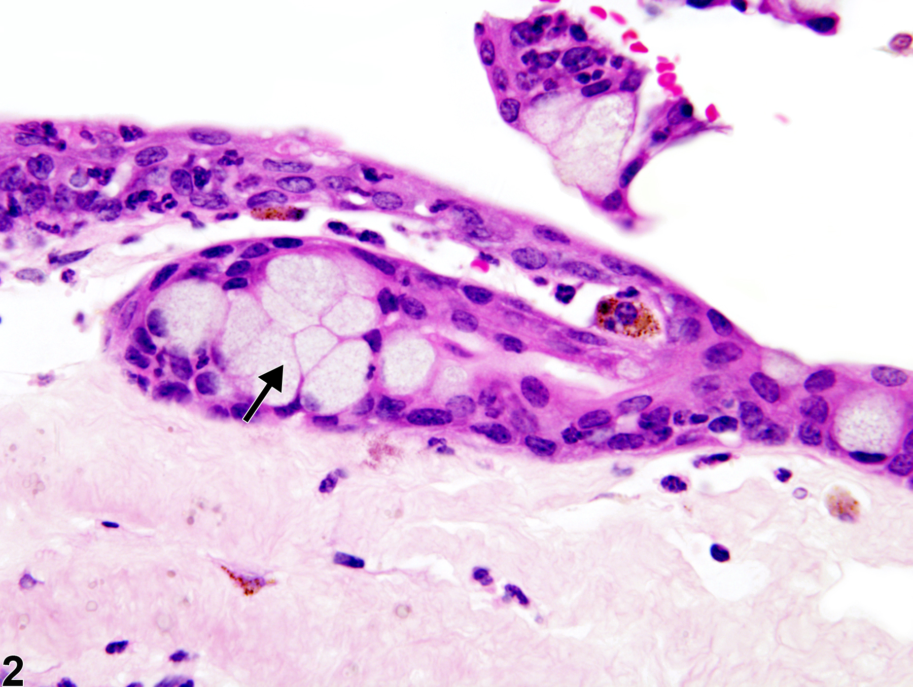
Eye, Cornea - Metaplasia, Goblet cell in a female B6C3F1 mouse from a chronic study. There are clusters of cells similar to normal conjunctival goblet cells in the corneal epithelium (arrow).
All Images

Eye, Cornea - Metaplasia, Goblet cell in a female B6C3F1 mouse from a chronic study. There are clusters of cells similar to normal conjunctival goblet cells in the corneal epithelium (arrow).

Eye, Cornea - Metaplasia, Goblet cell in a female B6C3F1 mouse from a chronic study (higher magnification of Figure 1). There are large goblet cells within the corneal epithelium (arrow).