

Special Senses System
Eye, Cornea - Mineralization
Narrative
Bruner H, Keller WF, Stitzel KA, Sauers LJ, Reer PJ, Long PH, Bruce RD, Alden CL. 1992. Spontaneous corneal dystrophy and generalized basement membrane changes in Fischer-344 rats. Toxicol Pathol 20:357366.
Abstract: https://www.ncbi.nlm.nih.gov/pubmed/1295066Frame SR, Slone TW. 1966. Nonneoplastic and neoplastic changes in the eye. In: Pathobiology of the Aging Mouse, Vol 2 (Mohr U, Dungworth DL, Capen CC, Carlton WW, Sundberg JP, Ward JM, eds). ILSI Press, Washington, DC, 97-103.
Geiss V, Yoshitomi K. 1991. Eyes. In: Pathology of the Mouse: Reference and Atlas (Maronpot RR, Boorman GA, Gaul BW, eds). Cache River Press, Vienna, IL, 471-489.
Greaves P. 2007. Nervous system and special sense organs. In: Histopathology of Preclinical Toxicity Studies: Interpretation and Relevance in Drug Safety Evaluation, 3rd ed. Academic Press, San Diego, CA, 861-933.
Abstract: http://www.sciencedirect.com/science/book/9780444527714Guillet R, Wyatt J, Baggs RB, Kellogg CK. 1988. Anesthetic-induced corneal lesions in developmentally sensitive rats. Invest Ophthalmol Vis Sci 29:949-954.
Abstract: https://pubmed.ncbi.nlm.nih.gov/3372167/Kuno H, Usui T, Eydelloth RS, Wolf ED. 1991. Spontaneous ophthalmic lesions in young Sprague-Dawley rats. J Vet Med Sci 53:607-614.
Abstract: https://www.ncbi.nlm.nih.gov/pubmed/10845604Losco PE, Troup CM. 1988. Corneal dystrophy in Fischer 344 rats. Lab Anim Sci 38:702-710.
Abstract: https://www.ncbi.nlm.nih.gov/pubmed/3265461Meador VP, Tyler RD, Plunkett ML. 1992. Epicardial and corneal mineralization in clinically normal severe combined immunodeficiency (SCID) mice. Vet Pathol 29:247-249.
Abstract: http://vet.sagepub.com/content/29/3/247.shortNational Toxicology Program. 2004. NTP TR-515. Toxicology and Carcinogenesis Studies of Propylene Glycol Mono-t-butyl Ether (CAS No. 57018-52-7) in F344/N Rats and B6C3F1 Mice and a Toxicology Study of Propylene Glycol Mono-t-butyl Ether In Male NBR Rats (Inhalation Studies). NTP, Research Triangle Park, NC.
Abstract: https://ntp.niehs.nih.gov/go/7724Smith RS, Sundberg JP, John SWM. 2002. The anterior segment. In: Systematic Evaluation of the Mouse Eye: Anatomy, Pathology, and Biomethods (Smith RS, John SWM, Nishina PM, Sundberg JP, eds). CRC Press, Boca Raton, FL, 111-159.
Taradach C, Greaves P, Rubin LF. 1984. Spontaneous eye lesions in laboratory animals: Incidence in relation to age. Crit Rev Toxicol 12:121-147.
Abstract: https://www.ncbi.nlm.nih.gov/pubmed/6368130Van Winkle TJ. 1991. Corneal opacities, spontaneous, mouse. In: International Life Sciences Institute Monographs on the Pathology of Laboratory Animals, Vol 10, Eye and Ear (Jones TC, Mohr U, Hunt RD, eds). Springer, Berlin, 21-25.
Williams DL. 2005. Ocular disease in rats: A review. Vet Ophthalmol 5:183-191.
Abstract: https://www.ncbi.nlm.nih.gov/pubmed/12236869Yamate J, Tajima M, Maruyama Y, Kudow S. 1987. Observations on soft tissue calcification in DBA/2NCrj mice in comparison with CRJ:CD-1 mice. Lab Anim 21:289-298.
Abstract: https://www.ncbi.nlm.nih.gov/pubmed/3695386Yoshitomi K, Boorman GA. 1990. Eye and associated glands. In: Pathology of the Fischer Rat: Reference and Atlas (Boorman GA, Eustis SL, Elwell MR, Montgomery CA, MacKenzie WF, eds). Academic Press, San Diego, CA, 239-260.
Abstract: https://www.ncbi.nlm.nih.gov/nlmcatalog/9002563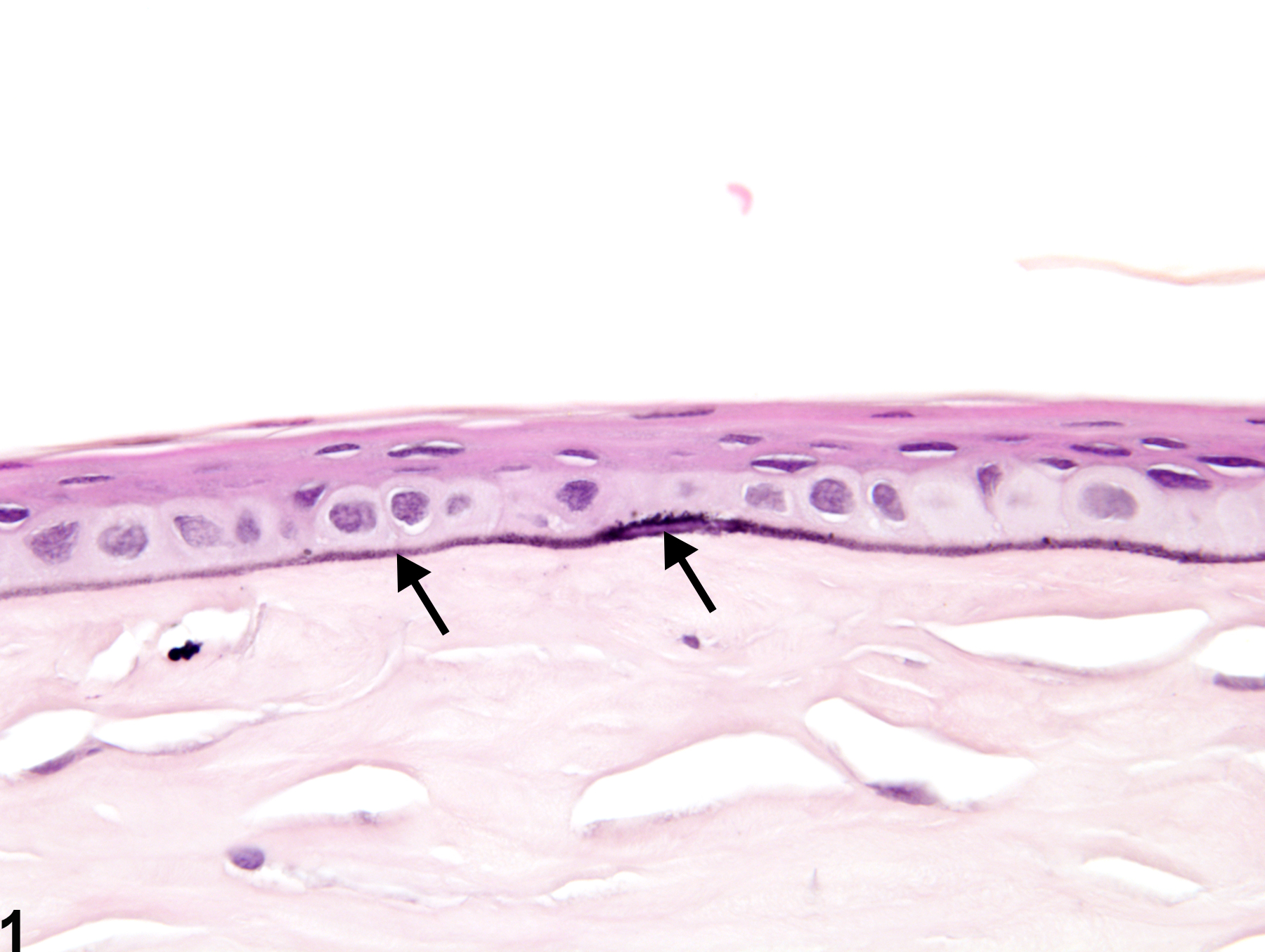
Eye, Cornea - Mineralization in a female F344/N rat from a chronic study. There are basophilic linear deposits within and subjacent to the corneal epithelial basement membrane (arrow).
All Images

Eye, Cornea - Mineralization in a female F344/N rat from a chronic study. There are basophilic linear deposits within and subjacent to the corneal epithelial basement membrane (arrow).

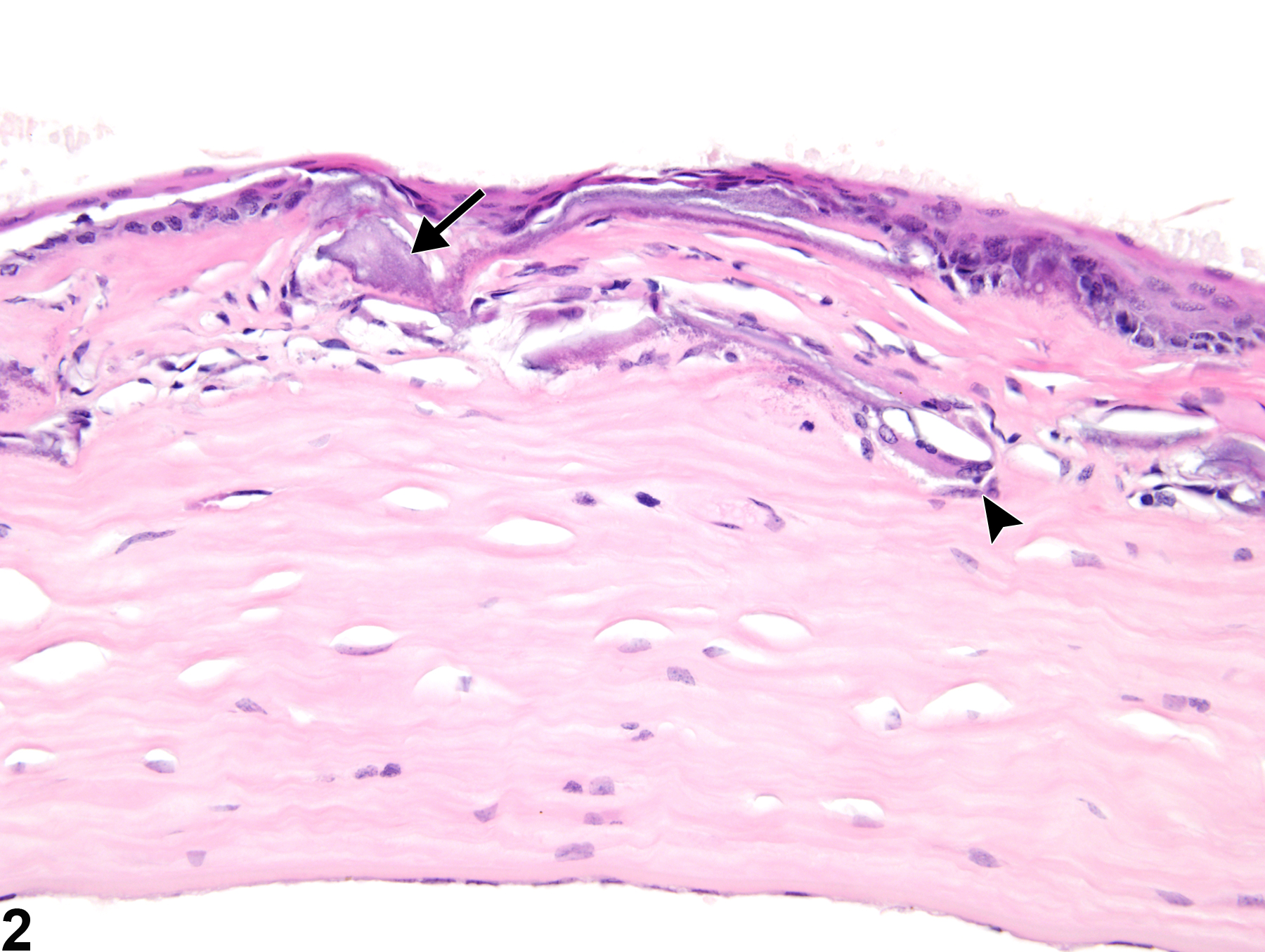
Eye, Cornea - Mineralization in a female B6C3F1 mouse from a chronic study. There are irregular basophilic deposits (arrow) in the stroma with concurrent stromal granulomatous inflammation (arrowhead).

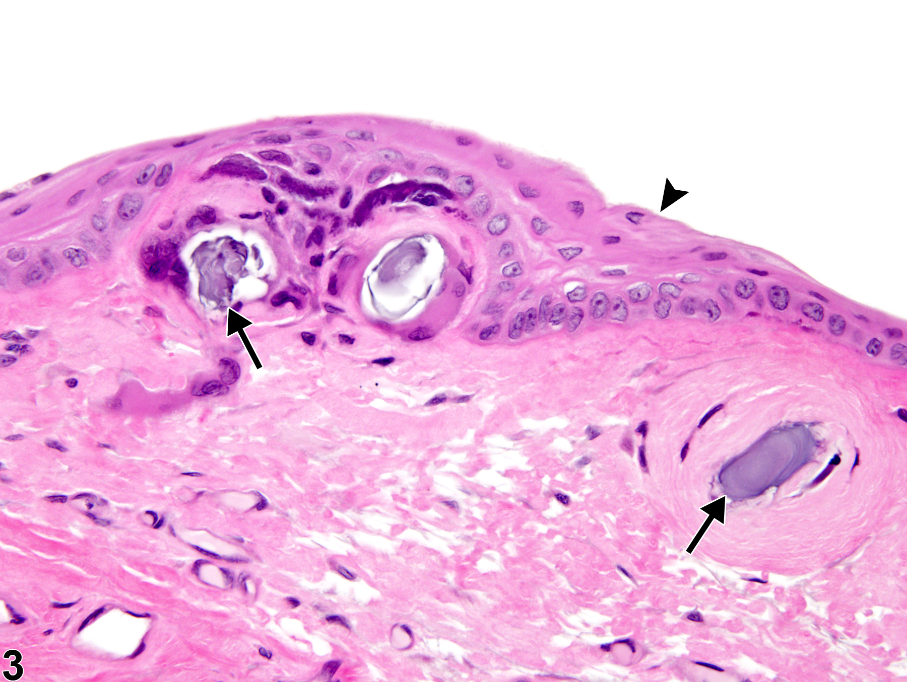
Eye, Cornea - Mineralization in a male F344/N rat from a subchronic study. There are pale to dark basophilic foci of mineralization (arrows) at the corneal limbus in the eye and concurrent corneal epithelial hyperplasia (arrowhead).