

Special Senses System
Eye, Cornea - Neovascularization
Narrative
Ecoiffier T, Yuen D, Chen L. 2010. Differential distribution of blood and lymphatic vessels in the murine cornea. Invest Ophthalmol Vis Sci 51:2436-2440.
Abstract: https://www.ncbi.nlm.nih.gov/pubmed/20019372Frame SR, Slone TW. 1966. Nonneoplastic and neoplastic changes in the eye. In: Pathobiology of the Aging Mouse, Vol 2 (Mohr U, Dungworth DL, Capen CC, Carlton WW, Sundberg JP, Ward JM, eds). ILSI Press, Washington, DC, 97-103.
Geiss V, Yoshitomi K. 1991. Eyes. In: Pathology of the Mouse: Reference and Atlas (Maronpot RR, Boorman GA, Gaul BW, eds). Cache River Press, Vienna, IL, 471-489.
Greaves P. 2007. Nervous system and special sense organs. In: Histopathology of Preclinical Toxicity Studies: Interpretation and Relevance in Drug Safety Evaluation, 3rd ed. Academic Press, San Diego, CA, 861-933.
Abstract: http://www.sciencedirect.com/science/book/9780444527714Hoffart L, Matonti F, Conrath J, Daniel L, Ridings B, Masson GS, Chavane F. 2010. Inhibition of corneal neovascularization after alkali burn: Comparison of different doses of bevacizumab in monotherapy or associated with dexamethasone. Clin Exp Ophthalmol 38:346-353.
Full Text: http://onlinelibrary.wiley.com/doi/10.1111/j.1442-9071.2010.02252.x/fullNational Toxicology Program. 1988. NTP TR-331. Toxicology and Carcinogenesis Studies of Malonaldehyde, Sodium Salt (3-Hydroxy-2-propenal, Sodium Salt) (CAS No. 24382-04-5) in F344/N Rats and B6C3F1 Mice (Gavage Studies). NTP, Research Triangle Park, NC.
Abstract: https://ntp.niehs.nih.gov/go/8898Pauly A, Brignole-Baudouin, Labbé A, Liang H, Warnet J-M, Baudouin C. 2007. New tools for the evaluation of toxic ocular surface changes in the rat. Invest Ophthalmol Vis Sci 48:5473–5463.
Full Text: https://doi.org/10.1167/iovs.06-0728Smith RS, Hawes NL, Chang B, Nishina PM. 2002. Retina. In: Systematic Evaluation of the Mouse Eye: Anatomy, Pathology, and Biomethods (Smith RS, John SWM, Nishina PM, Sundberg JP, eds). CRC Press, Boca Raton, FL, 195-225.
Taradach C, Greaves P, Rubin LF. 1984. Spontaneous eye lesions in laboratory animals: Incidence in relation to age. Crit Rev Toxicol 12:121-147.
Abstract: https://www.ncbi.nlm.nih.gov/pubmed/6368130Yoshitomi K, Boorman GA. 1990. Eye and associated glands. In: Pathology of the Fischer Rat: Reference and Atlas (Boorman GA, Eustis SL, Elwell MR, Montgomery CA, MacKenzie WF, eds). Academic Press, San Diego, CA, 239-260.
Abstract: https://www.ncbi.nlm.nih.gov/nlmcatalog/9002563Yudkin AM, Lambert RM. 1923. Pathogenesis of the ocular lesions produced by a deficiency of vitamin A. J Exp Med 38:17-24.
Full Text: https://www.ncbi.nlm.nih.gov/pmc/articles/PMC2128416/pdf/17.pdf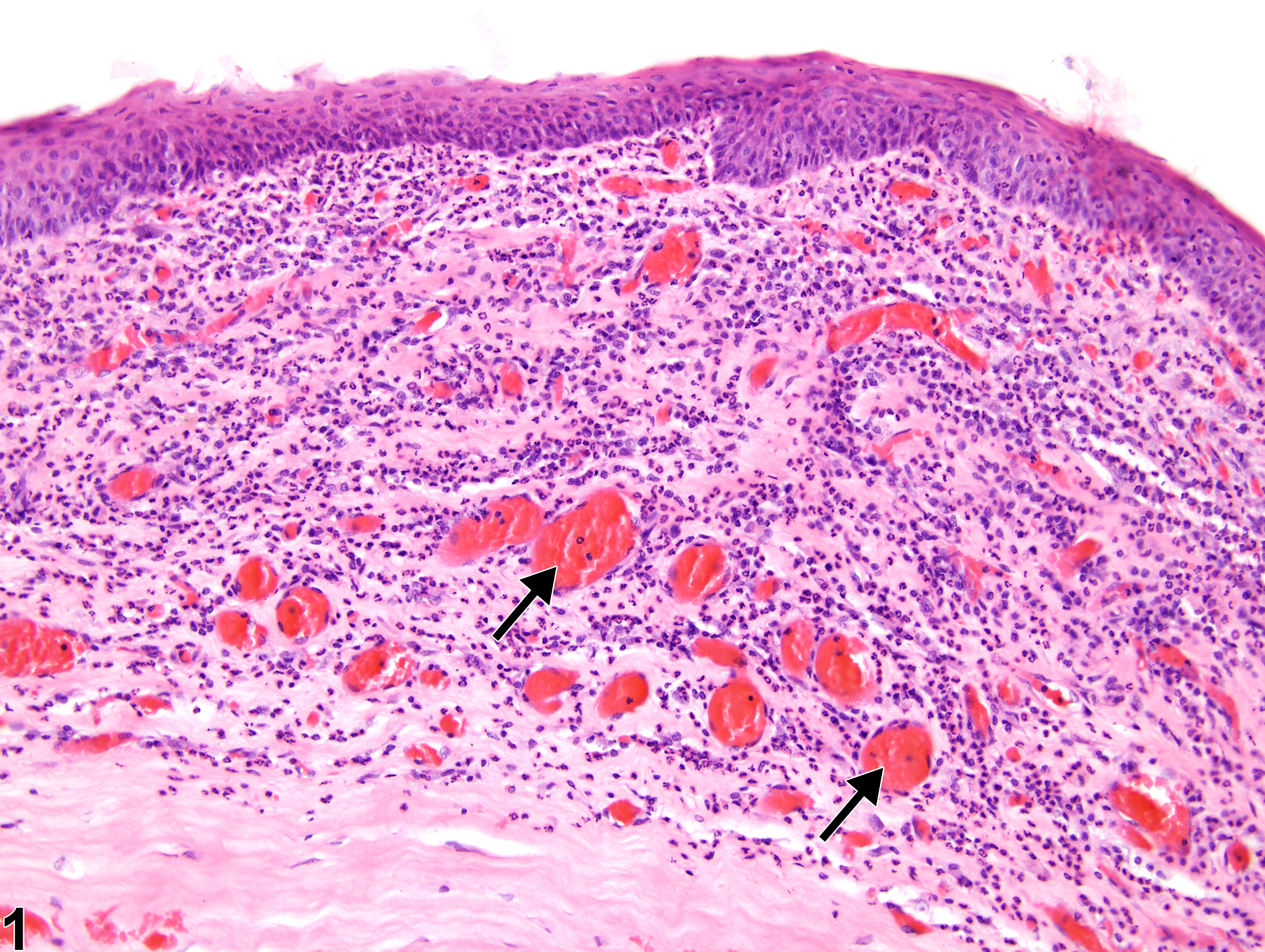
Eye, Cornea - Neovascularization in a male F344/N rat from a chronic study. There are multiple small blood vessels in the stroma (arrows) with concurrent inflammation and epithelial hyperplasia.
All Images

Eye, Cornea - Neovascularization in a male F344/N rat from a chronic study. There are multiple small blood vessels in the stroma (arrows) with concurrent inflammation and epithelial hyperplasia.

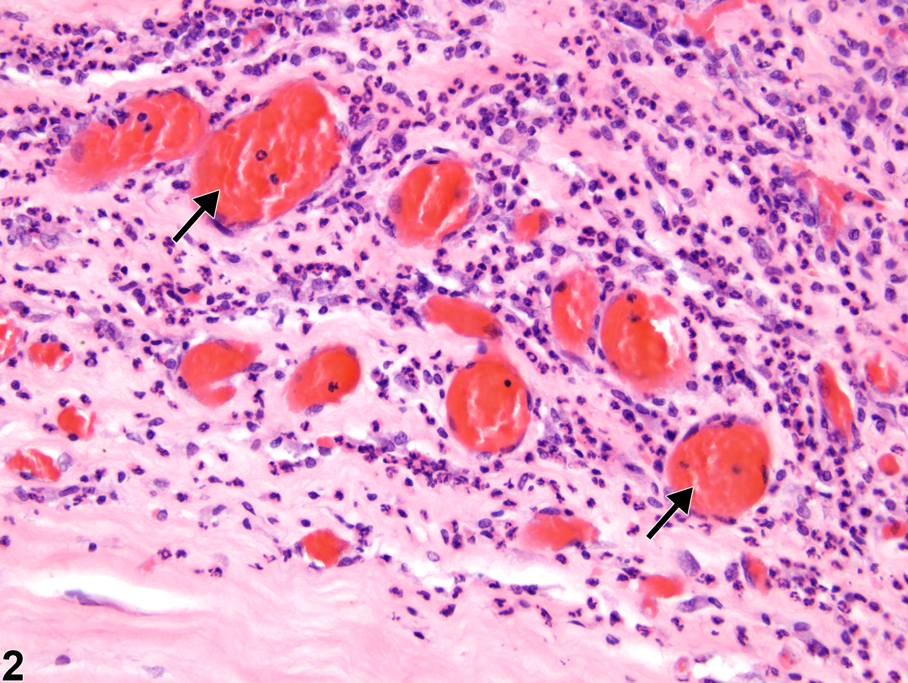
Eye, Cornea - Neovascularization in a male F344/N rat from a chronic study (higher magnification of Figure 1). Multiple small blood vessels are proliferating in the stroma (arrows); there is concurrent stromal inflammation.