

Special Senses System
Eye, Iris - Synechia
Narrative
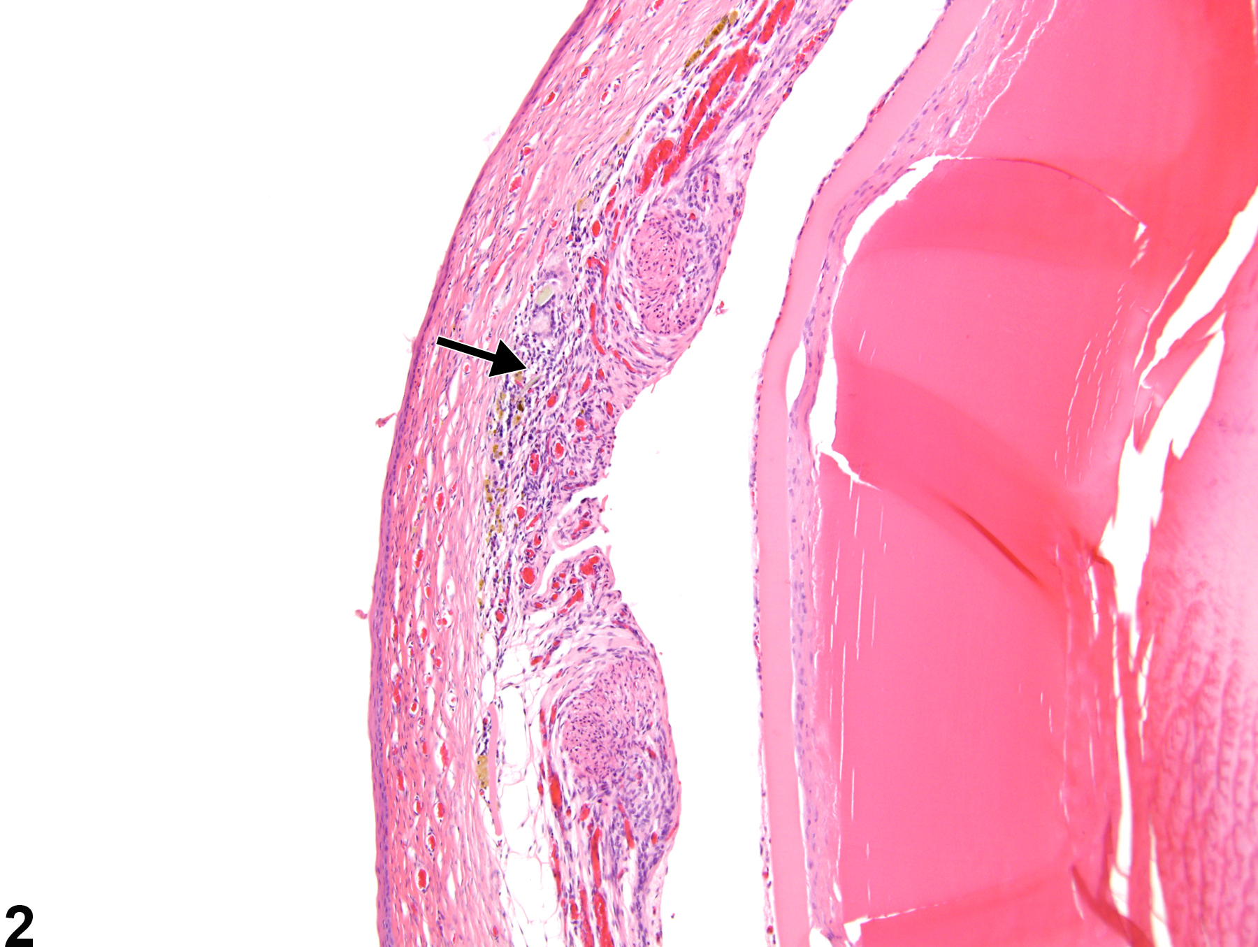
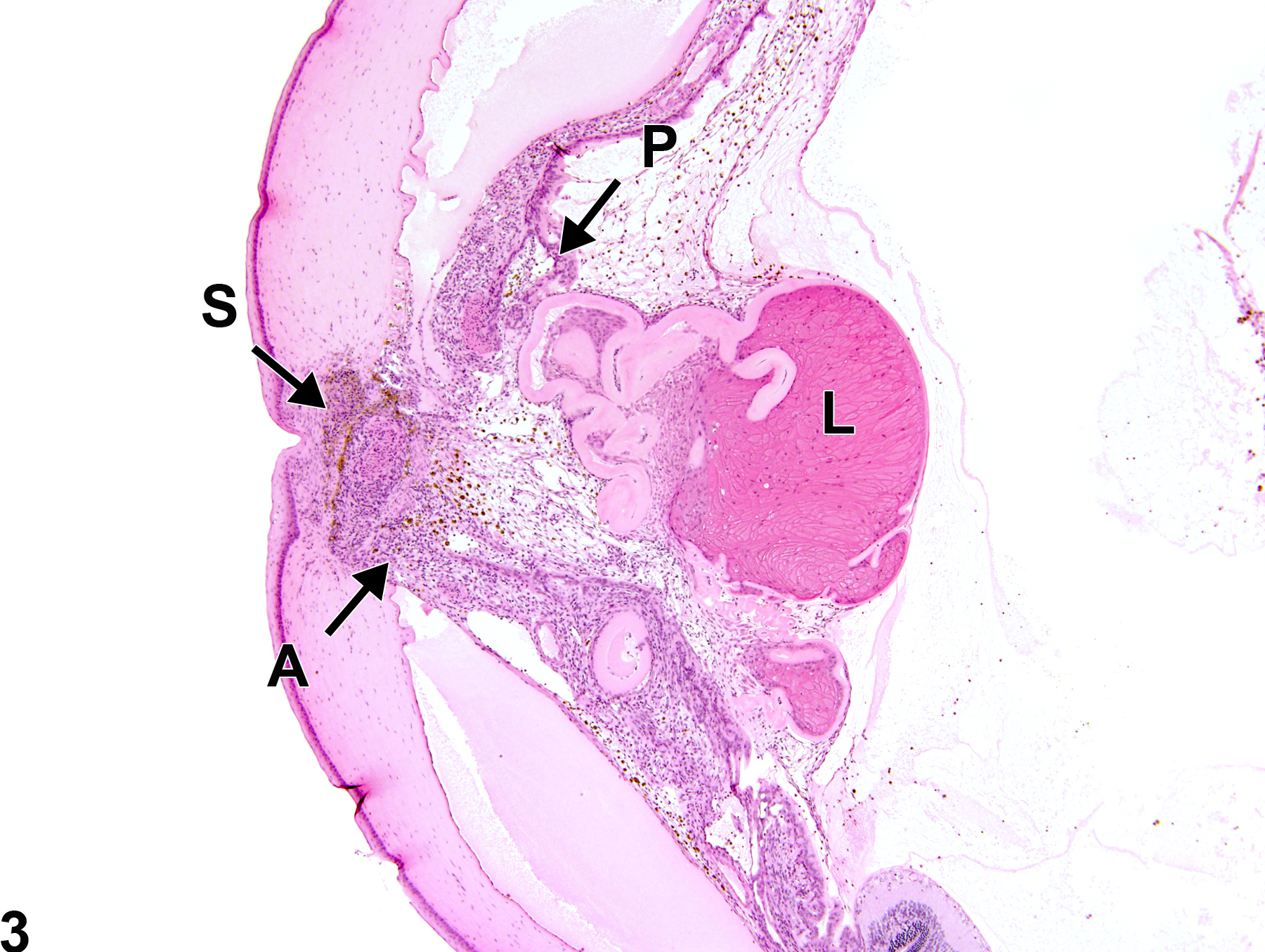
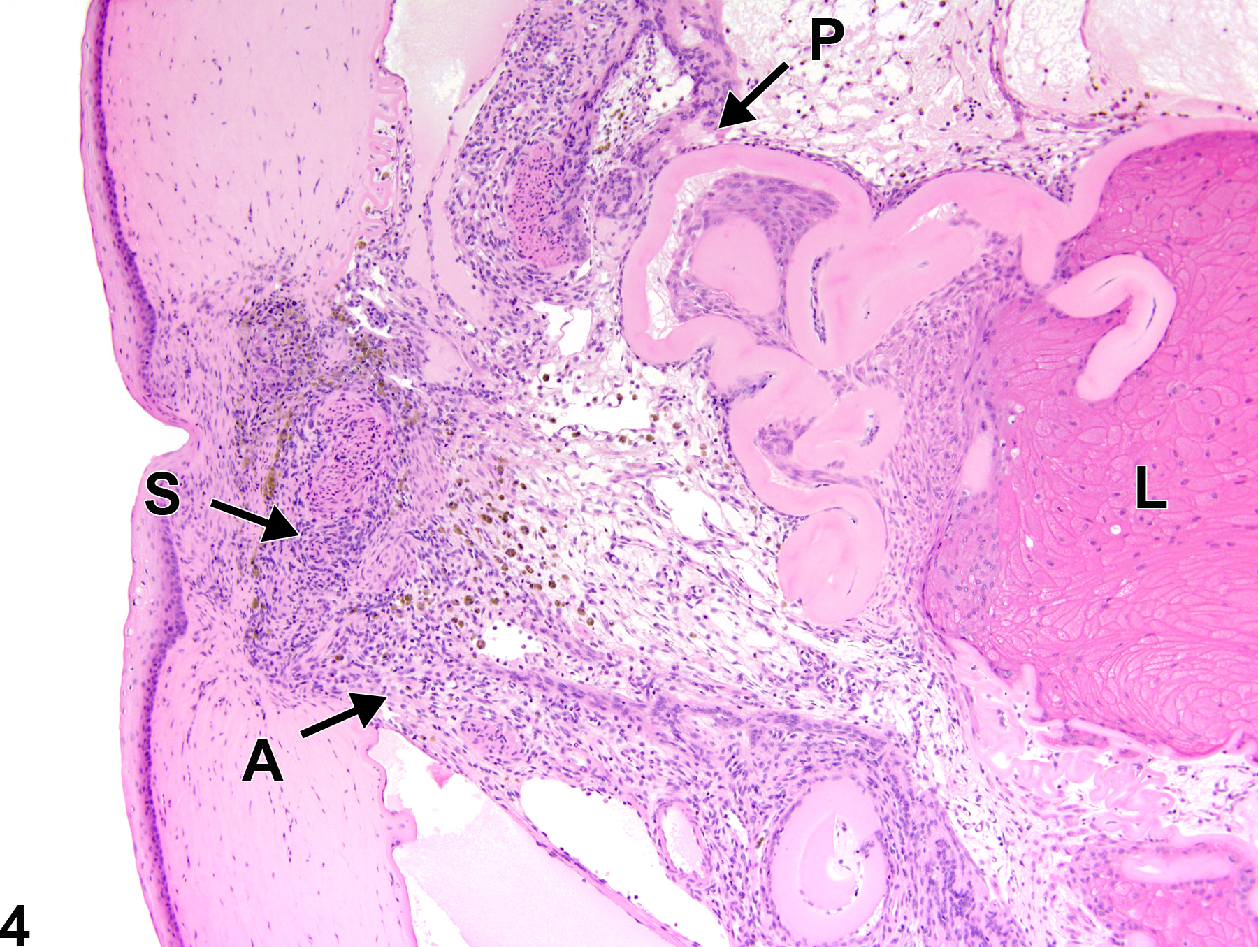
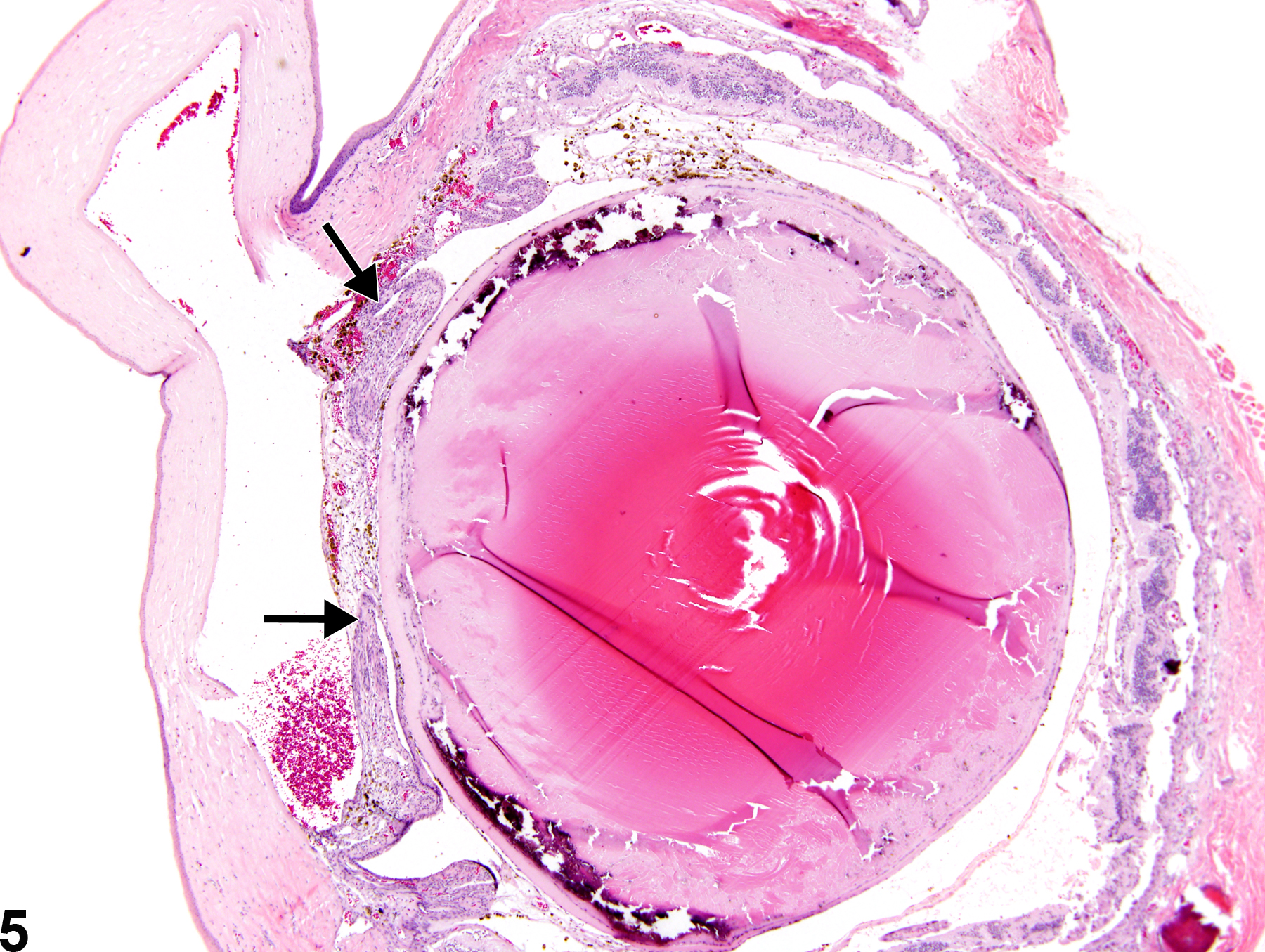
Anterior synechia (Figure 1, Figure 2, Figure 3, and Figure 4) is an adhesion of the iris to the posterior cornea due to abnormal fibrovascular tissue formation. Posterior synechia (Figure 3, Figure 4, Figure 5, and Figure 6) is an adhesion of the iris to the anterior lens capsule and/or vitreous due to abnormal fibrovascular tissue formation. There can also be concurrent anterior and posterior synechiae (Figure 3 and Figure 4). Associated lesions include staphyloma (partial protrusion of the iris into the corneal stroma), entropion uveae (posterior inversion of the pupillary margin of the iris), occlusion of the pupil by an abnormal fibrovascular membrane, and inflammation, among others.
Frame SR, Slone TW. 1966. Nonneoplastic and neoplastic changes in the eye. In: Pathobiology of the Aging Mouse, Vol 2 (Mohr U, Dungworth DL, Capen CC, Carlton WW, Sundberg JP, Ward JM, eds). ILSI Press, Washington, DC, 97-103.
Geiss V, Yoshitomi K. 1991. Eyes. In: Pathology of the Mouse: Reference and Atlas (Maronpot RR, Boorman GA, Gaul BW, eds). Cache River Press, Vienna, IL, 471-489.
John SW, Smith RS, Savinova OV, Hawes NL, Chang B, Turnbull D, Davisson M, Roderick TH, Heckenlively JR. 1998. Essential iris atrophy, pigment dispersion, and glaucoma in DBA/2J mice. Invest Ophthalmol Vis Sci 39:951-962.
Abstract: https://pubmed.ncbi.nlm.nih.gov/9579474/Kakehashi A, Saito Y, Mori K, Sugi N, Ono R, Yamagami H, Shinohara M, Tamemoto H, Ishikawa SE, Kawakami M, Kanazawa Y. 2006. Characteristics of diabetic retinopathy in SDT rats. Diabetes Metab Res Rev 22:455-461.
Full Text: http://onlinelibrary.wiley.com/doi/10.1002/dmrr.638/fullLai Y-L, Jacoby RO, Bhatt PN, Jonas AM. 1976. Keratoconjunctivitis associated with sialodacryoadenitis Invest Ophthalmol Vis Sci 15:538-541.
Abstract: https://pubmed.ncbi.nlm.nih.gov/931700/National Toxicology Program. 1992. NTP TR-415. Toxicology and Carcinogenesis Studies of Polysorbate 80 (CAS No. 9005-65-6) in F344/N Rats and B6C3F1 Mice (Feed Studies). NTP, Research Triangle Park, NC.
Abstract: https://ntp.niehs.nih.gov/go/7710National Toxicology Program. 2001. NTP TR-479. Toxicology and Carcinogenesis Studies of Coconut Oil Acid Diethanolamine Condensate (CAS No. 68603-42-9) in F344/N Rats And B6C3F1 Mice (Dermal Studies). NTP, Research Triangle Park, NC.
Abstract: https://ntp.niehs.nih.gov/go/9760Smith RS, Sundberg JP, John SWM. 2002. The anterior segment. In: Systematic Evaluation of the Mouse Eye: Anatomy, Pathology, and Biomethods (Smith RS, John SWM, Nishina PM, Sundberg JP, eds). CRC Press, Boca Raton, FL, 111-159.
Taradach C, Greaves P, Rubin LF. 1984. Spontaneous eye lesions in laboratory animals: Incidence in relation to age. Crit Rev Toxicol 12:121-147.
Abstract: https://www.ncbi.nlm.nih.gov/pubmed/6368130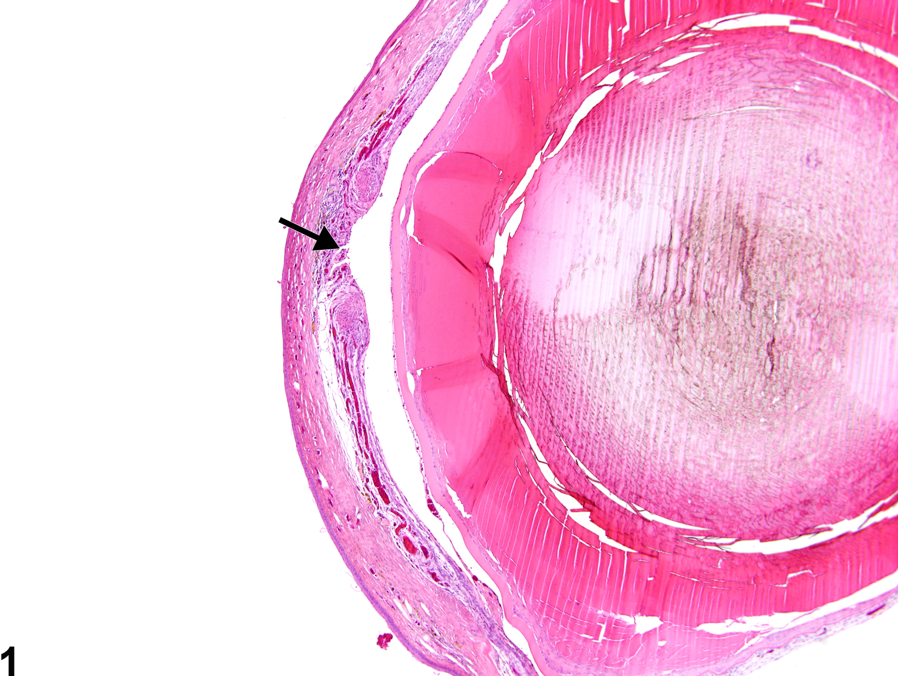
Eye, Iris - Synechia, Anterior in a female F344/N rat from a chronic study. There is adhesion of the iris to the posterior cornea (arrow).
All Images

Eye, Iris - Synechia, Anterior in a female F344/N rat from a chronic study. There is adhesion of the iris to the posterior cornea (arrow).

Eye, Iris - Synechia, Anterior in a female F344/N rat from a chronic study (higher magnification of Figure 1). There is adhesion of the iris to the posterior cornea (arrow) due to abnormal fibrovascular tissue formation.

Eye, Iris - Synechia in a male F344/NTac rat from a subchronic study. There are concurrent anterior (A) and posterior (P) iridial synechiae, partial protrusion of the iris into the corneal stroma (staphyloma) (S), and a cataractous lens (L).

Eye, Iris - Synechia in a male F344/NTac rat from a subchronic study (higher magnification of Figure 3). There is concurrent anterior (A) and posterior (P) iridial synechiae, as well as partial protrusion of the iris in the corneal stroma (staphyloma) (S), and a cataractous lens (L).

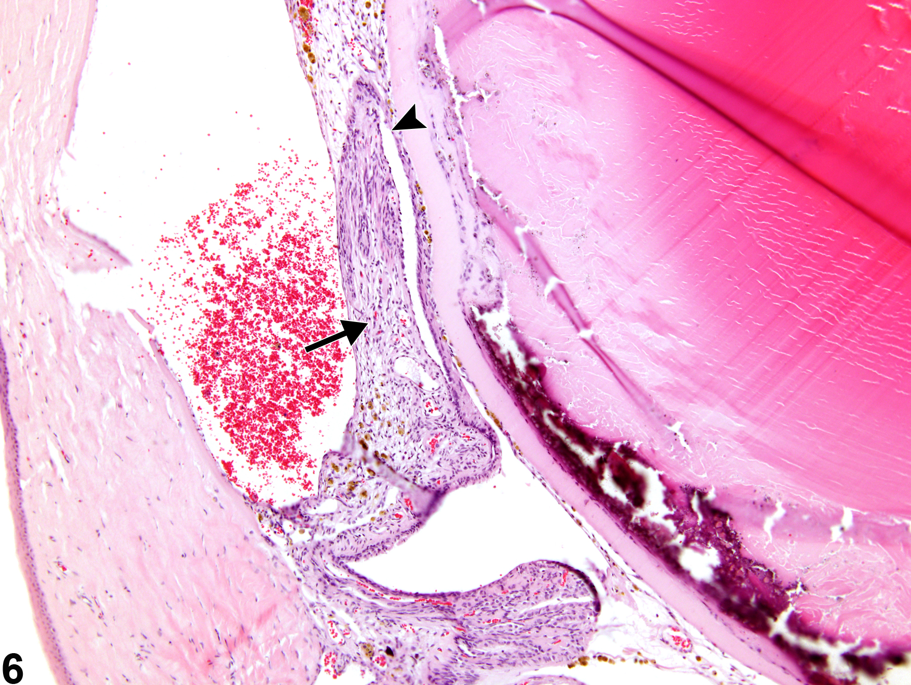
Eye, Iris - Synechia, Posterior in a female F344/N rat from a chronic study. There is adhesion of the iris to the lens capsule (arrow).

Eye, Iris - Synechia, Posterior in a female F344/N rat from a chronic study (higher magnification of Figure 5). There is adhesion of the iris to the lens capsule (arrow) due to abnormal fibrovascular tissue formation is present in the eye, as well as entropion uveae (arrowhead).