

Special Senses System
Eye, Lens - Cataract
Narrative
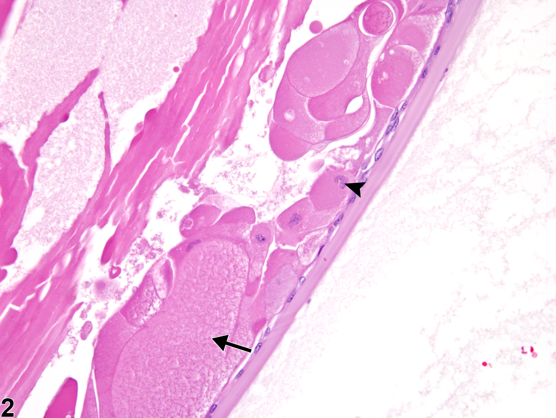
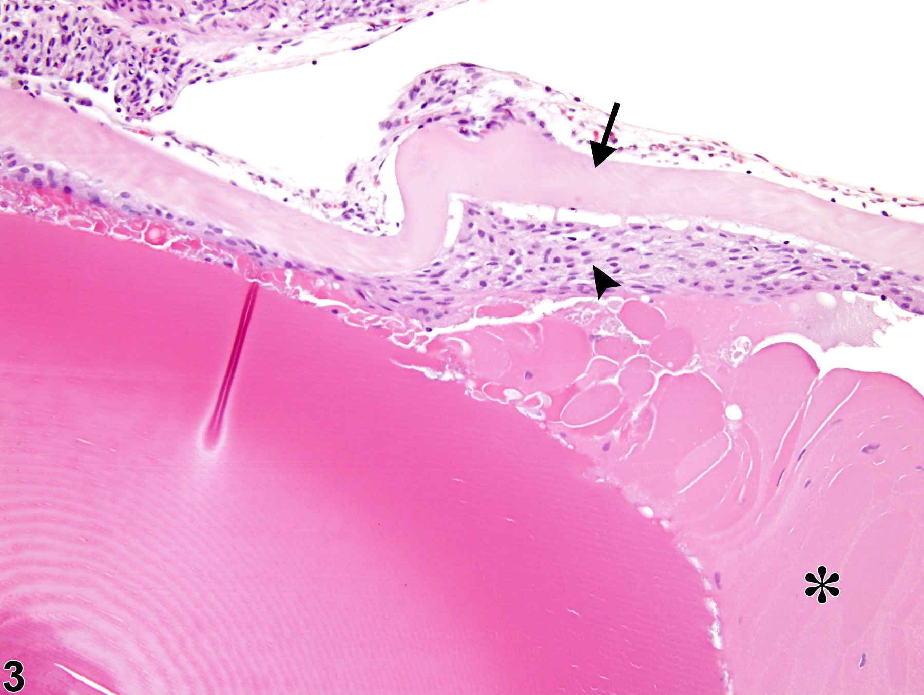
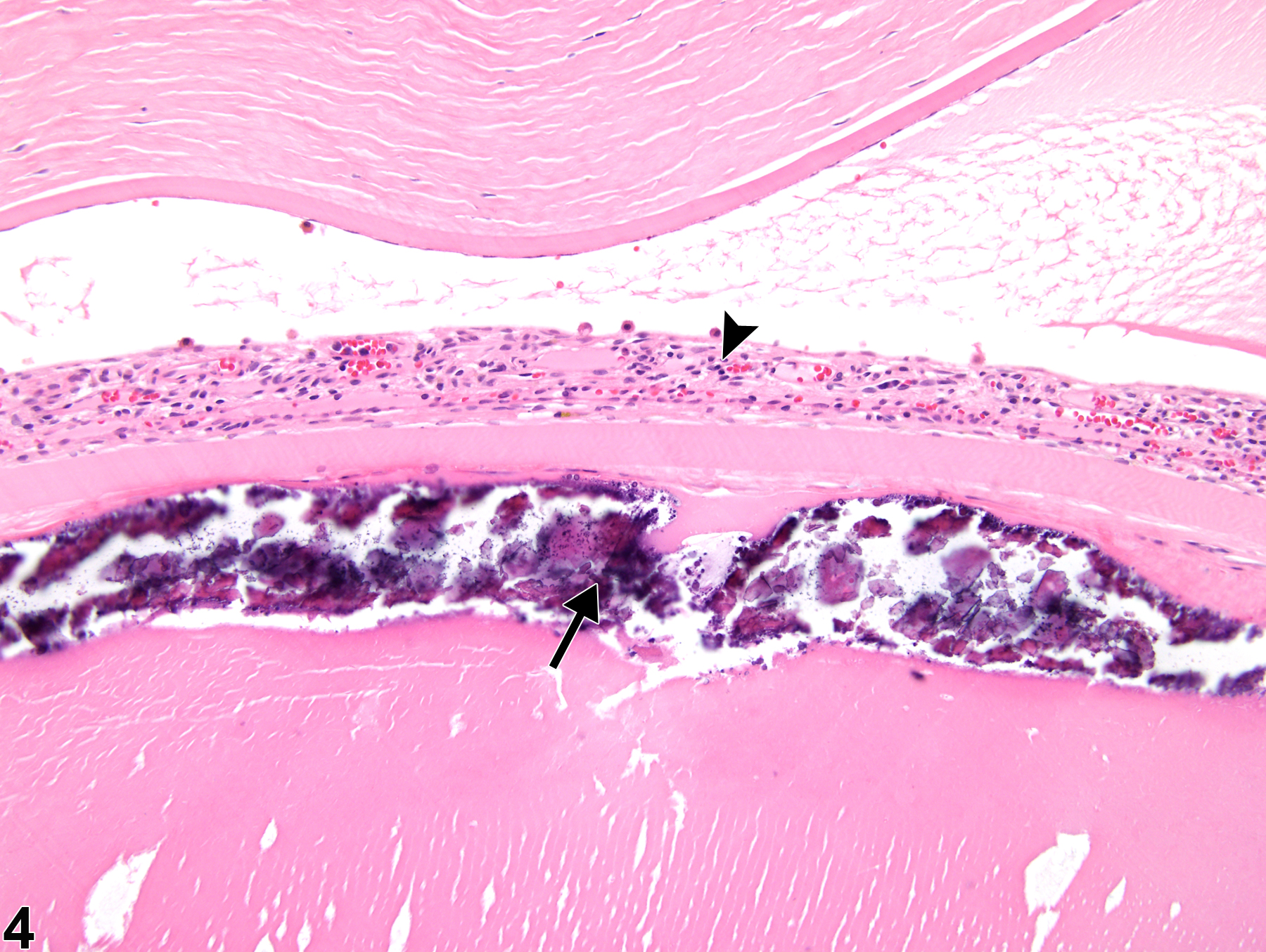
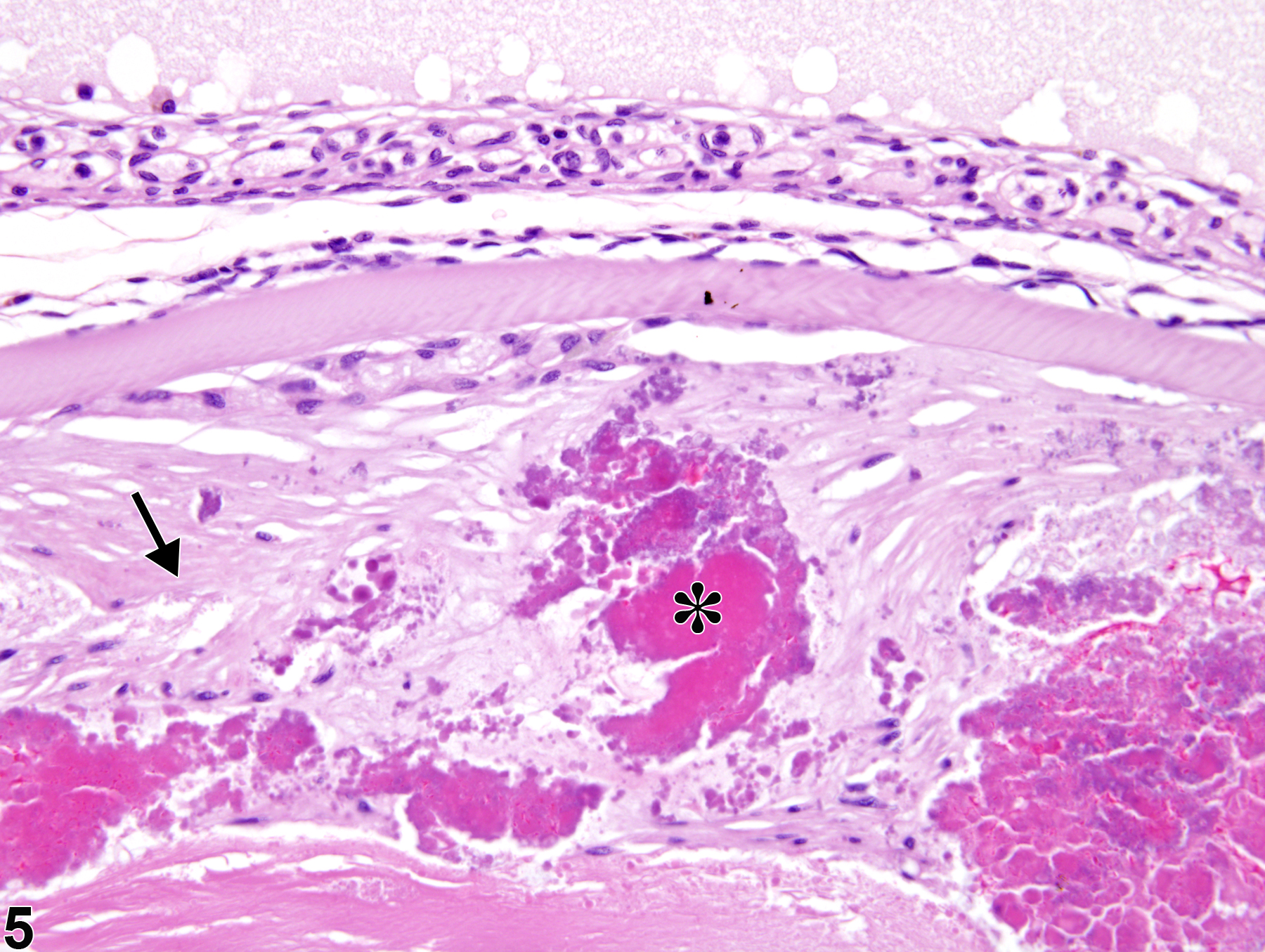
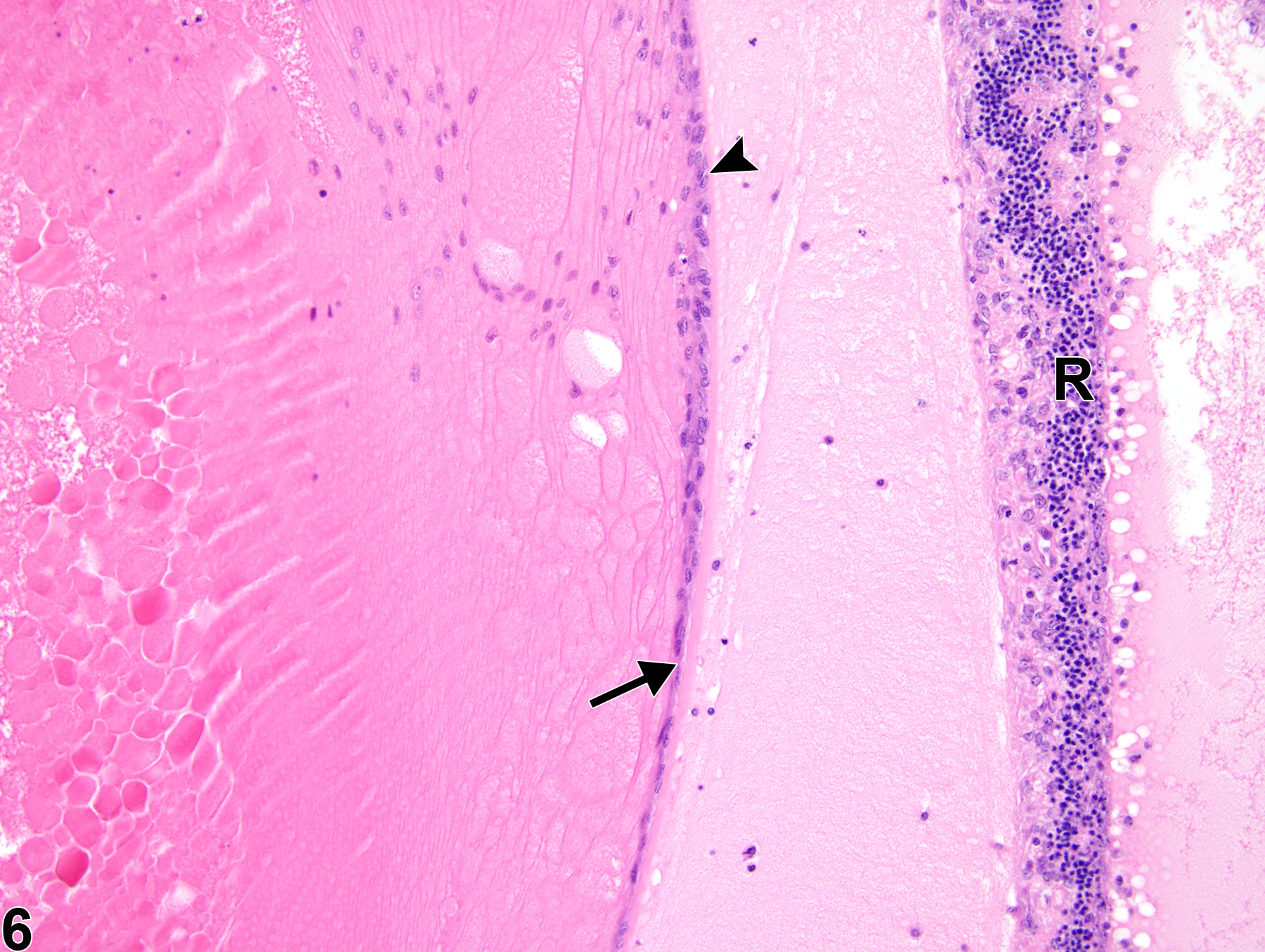
Cataracts are characterized by lens fibers with various abnormal features, including separation, swelling, granularity, condensation, fragmentation, and disruption of the normally orderly configuration, as well as abnormal retention of lens fiber nuclei in several swollen lens fibers (Figure 1 and Figure 2). Swollen lens fibers with abnormally retained nuclei are sometimes referred to as "bladder" or "balloon" cells. Cataracts can also exhibit additional features, such as capsular thickening, wrinkling, and/or rupture (Figure 3) or lens fiber or capsular mineralization (Figure 4). Anterior epithelial cells of cataractous lens can also undergo proliferation and fibrous metaplasia (epithelial-to-mesenchymal transition) in response to injury. There may also be anterior subcapsular fibrosis (Figure 5) in a cataractous lens (note the intermixed dark eosinophilic, fragmented lens fiber material), as well as hyperplasia of the anterior epithelium of (Figure 6). The proliferative anterior epithelium may also extend posterior to the lens bow (posterior migration of lens epithelium) (Figure 6).
Frame SR, Slone TW. 1966. Nonneoplastic and neoplastic changes in the eye. In: Pathobiology of the Aging Mouse, Vol 2 (Mohr U, Dungworth DL, Capen CC, Carlton WW, Sundberg JP, Ward JM, eds). ILSI Press, Washington, DC, 97-103.
Geiss V, Yoshitomi K. 1991. Eyes. In: Pathology of the Mouse: Reference and Atlas (Maronpot RR, Boorman GA, Gaul BW, eds). Cache River Press, Vienna, IL, 471-489.
Graw J. 2009. Mouse models of cataract. J Genetics 88:469-486.
Abstract: https://www.ncbi.nlm.nih.gov/pubmed/20090208Greaves P. 2007. Nervous system and special sense organs. In: Histopathology of Preclinical Toxicity Studies: Interpretation and Relevance in Drug Safety Evaluation, 3rd ed. Academic Press, San Diego, CA, 861-933.
Abstract: http://www.sciencedirect.com/science/book/9780444527714Hara A, Matsumoto M, Uga S. 1999. Morphological study on cataractogenesis of the Nakano mouse lens. Graefes Arch Clin Exp Ophthalmol 237:249-255.
Abstract: https://www.ncbi.nlm.nih.gov/pubmed/10090589Kuno H, Usui T, Eydelloth RS, Wolf ED. 1991. Spontaneous ophthalmic lesions in young Sprague-Dawley rats. J Vet Med Sci 53:607-614.
Abstract: https://www.ncbi.nlm.nih.gov/pubmed/10845604Newkirk KM, Chandler HL, Parent AE, Young DC, Colitz CMH, Wilkie DA, Kusewitt DF. 2007. Ultraviolet radiation-induced corneal degeneration in 129 mice. Toxicol Pathol 35:817-824.
Full Text: http://tpx.sagepub.com/content/35/6/817.fullNational Toxicology Program. 1996. NTP TR-452. Toxicology and Carcinogenesis Studies of 2,2-Bis(Bromomethyl)-1,3-Propanediol (FR-1138®) (CAS No. 3296-90-0) in F344 Rats and B6C3F1 Mice (Feed Studies). NTP, Research Triangle Park, NC.
Abstract: https://ntp.niehs.nih.gov/go/6048National Toxicology Program. 1997. NTP TR-450. Toxicology and Carcinogenesis Studies of Tetrafluoroethylene (CAS No. 116-14-3) in F344 Rats and B6C3F1 Mice (Inhalation Studies). NTP, Research Triangle Park, NC.
Abstract: https://ntp.niehs.nih.gov/go/6044National Toxicology Program. 2012. NTP TR-572. Toxicology and Carcinogenesis Studies of Methyl trans-Styryl Ketone (CAS No. 1896-62-4) in F344/N Rats and B6C3F1 Mice (Feed and Dermal Studies). NTP, Research Triangle Park, NC.
Abstract: https://ntp.niehs.nih.gov/go/36154National Toxicology Program. 2012. NTP TR-579. Toxicology and Carcinogenesis Studies of N, N-Dimethyl-p-Toluidine (CAS No. 99-97-8) in F344/N Rats and B6C3F1/N Mice (Gavage Studies). NTP, Research Triangle Park, NC.
Abstract: https://ntp.niehs.nih.gov/go/37162Okano T, Uga S, Ishikawa S, Shumiya S. 1993. Histopathological study of hereditary cataractous lenses in the SCR strain rat. Exp Eye Res 57:567-576.
Abstract: https://www.ncbi.nlm.nih.gov/pubmed/8282043Rao GN. 1991. Light intensity-associated eye lesions of Fischer 344 rats in long-term studies. Toxicol Pathol 19:148-155.
Abstract: http://tpx.sagepub.com/content/19/2/148.shortSaika S, Kono-Saika S, Ohnishi Y, Sato M, Muragaki Y, Ooshima A, Flanders KC, Yoo J, Anzano M, Liu C-Y, Kao W W-Y, Roberts AB. 2004. Smad3 signaling is required for epithelial-mesenchymal transition of lens epithelium after injury. Am J Pathol 164:651-663.
Abstract: https://www.ncbi.nlm.nih.gov/pubmed/14742269Shin EHH, Basson MA, Robinson ML, McAvoy JW, Lovicu FJ. 2012. Sprouty is a negative regulator of transforming growth factor β-induced epithelial-to-mesenchymal transition and cataract. Mol Med 18:861-873.
Abstract: https://www.ncbi.nlm.nih.gov/pubmed/22517312Shinohara M, Masuyama T, Shoda T, Takahashi T, Katsuda Y, Komeda K, Kuroki M, Kakehashi A, Kanazawa Y. 2000. A new spontaneously diabetic non-obese Torii rat strain with severe ocular complications. Int J Exp Diabetes Res 1:89-100.
Abstract: http://www.hindawi.com/journals/jdr/2000/371689/abs/Smith RS. 2002. Choroid, lens, and vitreous. In: Systematic Evaluation of the Mouse Eye: Anatomy, Pathology, and Biomethods (Smith RS, John SWM, Nishina PM, Sundberg JP, eds). CRC Press Boca Raton, FL, 161-193.
Taradach C, Greaves P, Rubin LF. 1984. Spontaneous eye lesions in laboratory animals: Incidence in relation to age. Crit Rev Toxicol 12:121-147.
Abstract: https://www.ncbi.nlm.nih.gov/pubmed/6368130Uga S, Tsuchiya K, Ishikawa S. 1988. Histopathological study of Emory mouse cataract. Graefes Arch Clin Exp Ophthalmol 226:15-21.
Abstract: https://www.ncbi.nlm.nih.gov/pubmed/3342971Whiteley HL, Peiffer RL. 2002. The eye. In: Handbook of Toxicologic Pathology, 2nd ed, Vol 2 (Haschek WM, Rousseaux, CG, Wallig MA, eds). Academic Press, San Diego, CA, 539-584.
Williams DL. 2005. Ocular disease in rats: A review. Vet Ophthalmol 5:183-191.
Abstract: https://www.ncbi.nlm.nih.gov/pubmed/12236869Yoshitomi K, Boorman GA. 1990. Eye and associated glands. In: Pathology of the Fischer Rat: Reference and Atlas (Boorman GA, Eustis SL, Elwell MR, Montgomery CA, MacKenzie WF, eds). Academic Press, San Diego, CA, 239-260.
Abstract: https://www.ncbi.nlm.nih.gov/nlmcatalog/9002563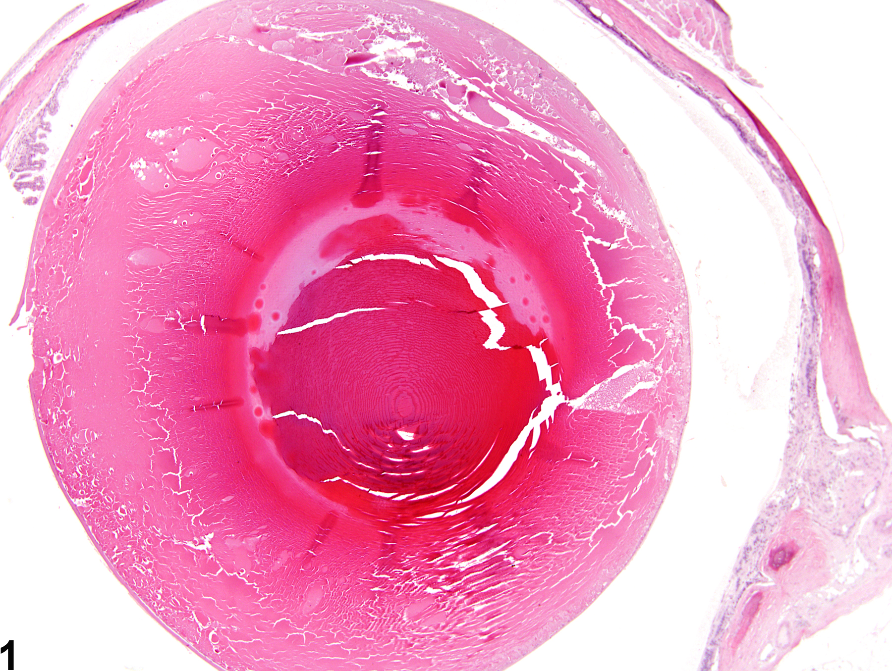
Eye, Lens - Cataract in a female F344/N rat from a chronic study. There are lens fibers with separation, swelling, granularity, condensation, fragmentation, and disruption of the normally orderly configuration.
All Images

Eye, Lens - Cataract in a female F344/N rat from a chronic study. There are lens fibers with separation, swelling, granularity, condensation, fragmentation, and disruption of the normally orderly configuration.

Eye, Lens - Cataract in a male F344/N rat from a chronic study. There are swollen, disrupted lens fibers (arrow) with abnormal retention of lens fiber nuclei (arrowhead).

Eye, Lens - Cataract in a male F344/N rat from a chronic study. There is capsular thickening and wrinkling (arrow), subcapsular anterior epithelial hyperplasia (arrowhead), and swollen, disrupted lens fibers (asterisk).

Eye, Lens - Cataract in a female F344/N rat from a chronic study. There is subcapsular mineralization (long arrow) of the lens; posterior synechia of the iris (short arrow) is also present.

Eye, Lens - Cataract in a male F344/N rat from a chronic study. Anterior subcapsular fibrosis (arrow) is intermixed with dark eosinophilic, fragmented lens fiber material (asterisk).

Eye, Lens - Cataract in a male F344/N rat from a subchronic study. There is hyperplasia (arrowhead) and posterior migration (behind the lens bow) (arrow) of the anterior lens epithelium; there is also retinal degeneration and detachment (R).