

Special Senses System
Eye, Retina - Dysplasia
Narrative
Frame SR, Slone TW. 1966. Nonneoplastic and neoplastic changes in the eye. In: Pathobiology of the Aging Mouse, Vol 2 (Mohr U, Dungworth DL, Capen CC, Carlton WW, Sundberg JP, Ward JM, eds). ILSI Press, Washington, DC, 97-103.
Gottschall-Pass KT, Grahn BH, Gorecki DKJ, Paterson PG. 1997. Oscillatory potentials and light microscopic changes demonstrate an interaction between zinc and taurine in the developing rat retina. J Nutr 127:1206-1213.
Abstract: http://jn.nutrition.org/content/127/6/1206.fullHaider NB, Naggart JK, Nishina PM. 2001. Excess cone cell proliferation due to lack of a functional NR2E3 causes retinal dysplasia in rd7/rd7 mice. Hum Mol Gen 10:1619-1626.
Full Text: http://hmg.oxfordjournals.org/content/10/16/1619.fullNational Toxicology Program. 2012. NTP TR-571. Toxicology and Carcinogenesis Studies of Kava Kava Extract (CAS No. 9000-38-8) in F344/N Rats and B6C3F1 Mice (Gavage Studies). NTP, Research Triangle Park, NC.
Abstract: https://ntp.niehs.nih.gov/go/36127Poulson R, Hayes B. 1988. Congenital retinal folds in Sheffield-Wistar rats. Graefes Arch Clin Exp Ophthalmol 226:31-33.
Full Text: https://www.ncbi.nlm.nih.gov/pubmed/3342972Smith RS, Hawes NL, Chang B, Nishina PM. 2002. Retina. In: Systematic Evaluation of the Mouse Eye: Anatomy, Pathology, and Biomethods (Smith RS, John SWM, Nishina PM, Sundberg JP, eds). CRC Press, Boca Raton, FL, 195-225.
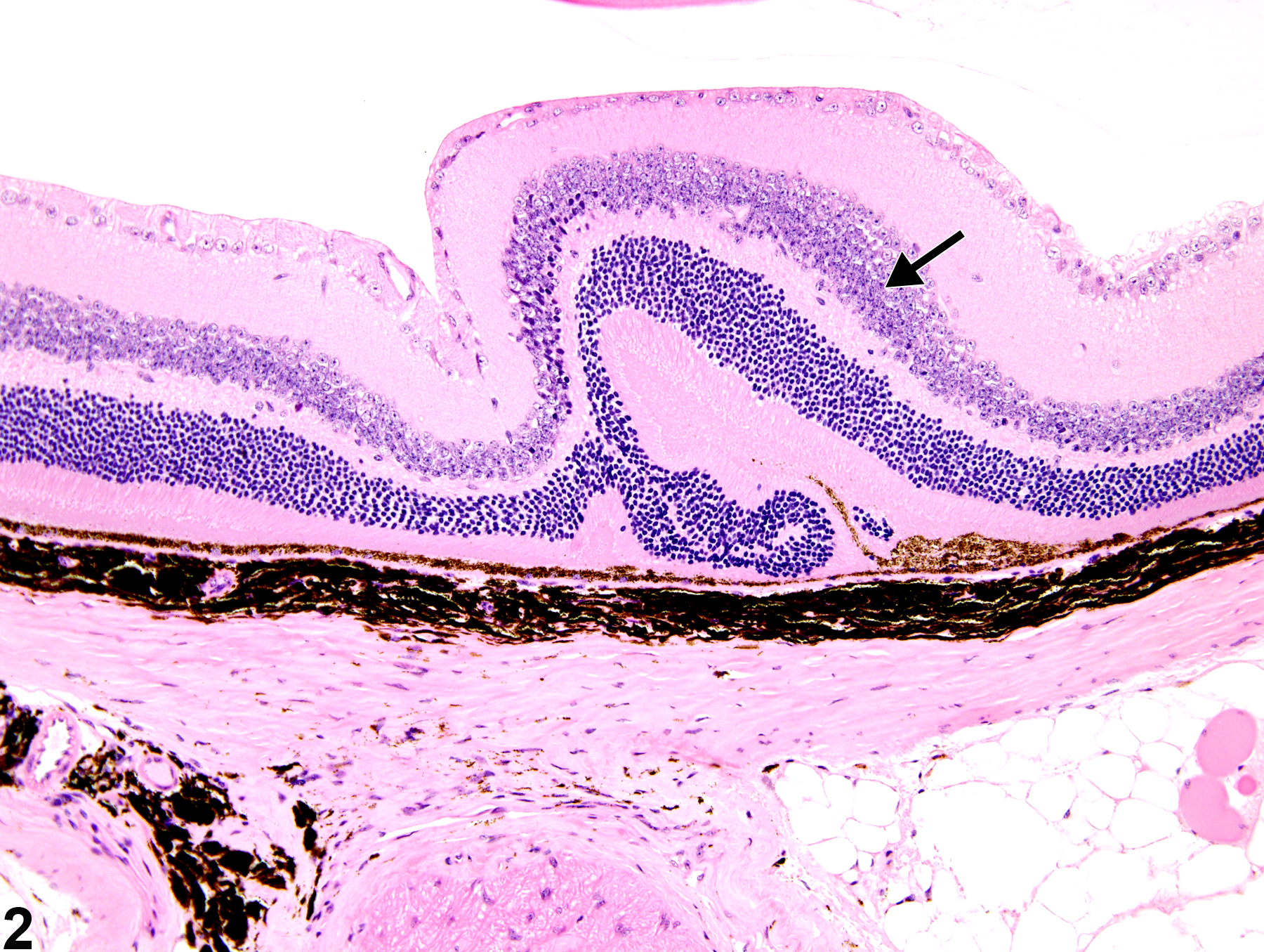
Eye, Retina - Dysplasia in a female B6C3F1 mouse from a chronic study. There are focal infoldings or rosette-like formations of the retinal layers (arrow).
All Images

Eye, Retina - Dysplasia in a female B6C3F1 mouse from a chronic study. There are focal infoldings or rosette-like formations of the retinal layers (arrow).

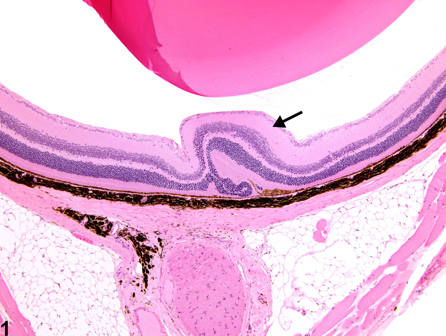
Eye, Retina - Dysplasia in a female B6C3F1 mouse from a chronic study (higher magnification of Figure 1). There is retinal dysplasia (arrow) with little, if any, concurrent degeneration or necrosis.