

Special Senses System
Eye, Retina - Gliosis
Narrative
Adams ET, Auerbach S, Blackshear PE, Bradley A, Gruebbel MM, Little PB, Malarkey D, Maronpot R, McKay JS, Miller RA, Moore RR, Morrison JP, Nyska A, Ramot Y, Rao D, Suttie A, Wells MY, Willson GA, Elmore SA. 2011. Proceedings of the 2010 National Toxicology Program Satellite Symposium. Toxicol Pathol 39:240-266.
Abstract: https://www.ncbi.nlm.nih.gov/pubmed/21177527Brooks DE, Komàromy AM, Källberg ME. 1999. Comparative retinal ganglion cell and optic nerve morphology. Vet Ophthalmol 2:3-11.
Abstract: https://www.ncbi.nlm.nih.gov/pubmed/11397238Ho G, Kumar S, Min XS, Kng YL, Loh MY, Gao S, Zhuo L. 2009. Molecular imaging of retinal gliosis in transgenic mice induced by kainic acid neurotoxicity. Invest Ophthalmol Vis Sci 50:2459-2464.
Full Text: https://doi.org/10.1167/iovs.08-2133Jung HJ, Raine CS, Suzuki K. 1978. Schwann cells and peripheral nervous system myelin in the rat retina. Acta Neuropathol 44:245-247.
Abstract: http://link.springer.com/article/10.1007/BF00691075Langmann T. 2007. Microglia activation in retinal degeneration. J Leukocyte Biol 81:1345-1351.
Full Text: http://www.jleukbio.org/content/81/6/1345.fullNational Toxicology Program. 2012. NTP TR-571. Toxicology and Carcinogenesis Studies of Kava Kava Extract (CAS No. 9000-38-8) in F344/N Rats and B6C3F1 Mice (Gavage Studies). NTP, Research Triangle Park, NC.
Abstract: https://ntp.niehs.nih.gov/go/36127Seoane A, Espejo M, Pallàs M, Rodriguez-Farre E, Ambrosio S, Llorens J. 1999. Degeneration and gliosis in rat retina and central nervous system following 3,3'-iminodipropionitrile exposure. Brain Res 833:258-271.
Full Text: http://toxsci.oxfordjournals.org/content/88/2/456.full.pdfYanoff K, Zimmerman LE, Davis R. 1971. Massive gliosis of the retina. Int Ophthalmol Clin 11:211-229.
Yoshitomi K, Boorman GA. 1990. Eye and associated glands. In: Pathology of the Fischer Rat: Reference and Atlas (Boorman GA, Eustis SL, Elwell MR, Montgomery CA, MacKenzie WF, eds). Academic Press, San Diego, CA, 239-260.
Abstract: https://www.ncbi.nlm.nih.gov/nlmcatalog/9002563Zeng H, Zhu X, Zhang C, Yang L-P, Wu L, Tso MOM. 2005. Identification of sequential events and factors associated with microglial activation, migration, and cytotoxicity in retinal degeneration in rd mice. Invest Ophthalmol Vis Sci 46:2992-2999.
Full Text: https://doi.org/10.1167/iovs.05-0118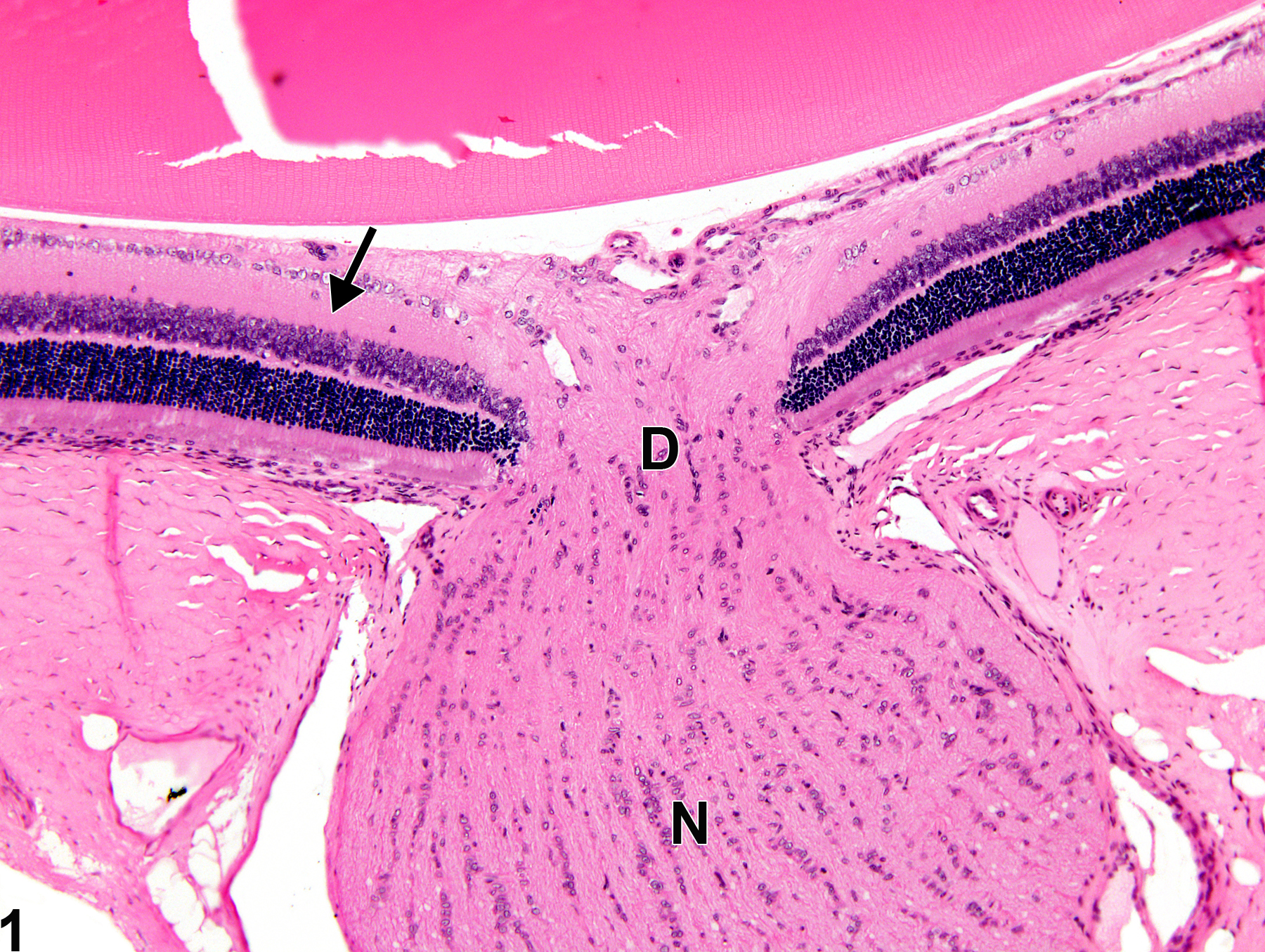
Eye, Retina - Normal in a female F344/N rat from a chronic study. Normal retina (arrow), optic disc (D), and optic nerve (N) for comparison to Figure 2.
All Images

Eye, Retina - Normal in a female F344/N rat from a chronic study. Normal retina (arrow), optic disc (D), and optic nerve (N) for comparison to Figure 2.

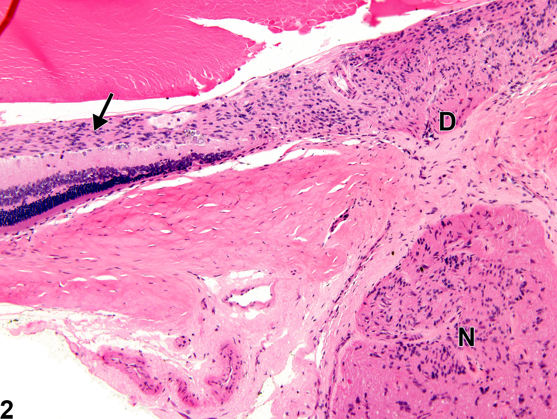
Eye, Retina - Gliosis in a female F344/N rat from a chronic study. There are increased numbers of glial cells in the nerve fiber layer (arrow), optic disc (D), and optic nerve (N).