

Special Senses System
Eye, Retina - Hemorrhage
Narrative
Kakehashi A, Saito Y, Mori K, Sugi N, Ono R, Yamagami H, Shinohara M, Tamemoto H, Ishikawa SE, Kawakami M, Kanazawa Y. 2006. Characteristics of diabetic retinopathy in SDT rats. Diabetes Metab Res Rev 22:455-461.
Full Text: http://onlinelibrary.wiley.com/doi/10.1002/dmrr.638/fullKuno H, Usui T, Eydelloth RS, Wolf ED. 1991. Spontaneous ophthalmic lesions in young Sprague-Dawley rats. J Vet Med Sci 53:607-614.
Abstract: https://www.ncbi.nlm.nih.gov/pubmed/10845604National Toxicology Program. 1997. NTP TR-450. Toxicology and Carcinogenesis Studies of Tetrafluoroethylene (CAS No. 116-14-3) in F344 Rats and B6C3F1 Mice (Inhalation Studies). NTP, Research Triangle Park, NC.
Abstract: https://ntp.niehs.nih.gov/go/6044Nyska A, Maronpot RR, Ghanayem BI. 1999. Ocular thrombosis and retinal degeneration induced in female F344 rats by 2-butoxyethanol. Hum Exp Toxicol 18:577-582.
Abstract: http://het.sagepub.com/content/18/9/577.abstractRamos M, Reilly CM, Bolon B. 2011. Toxicological pathology of the retina and optic nerve. In: Fundamental Neuropathology for Pathologists and Toxicologists (Bolon B, Butt MT, eds). Wiley, Hoboken, NJ, 385-412.
Abstract: http://onlinelibrary.wiley.com/doi/10.1002/9780470939956.ch24/summarySilva-Araújo AL, Tavares MA, Patacao MH, Carolino RM. 1996. Retinal hemorrhages associated with in utero exposure to cocaine: Experimental and clinical findings. Retina 16:411-418.
Abstract: https://www.ncbi.nlm.nih.gov/pubmed/8912968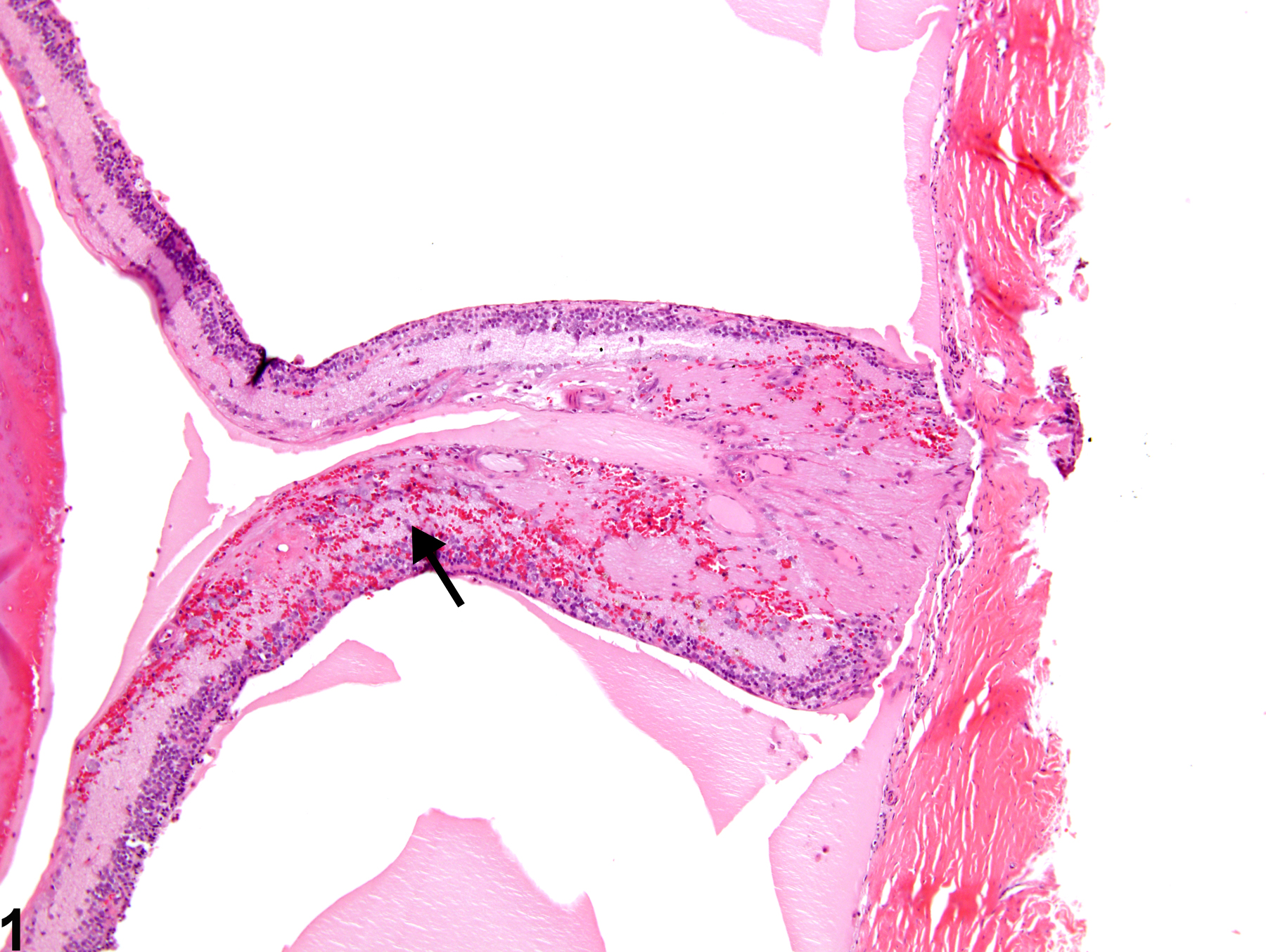
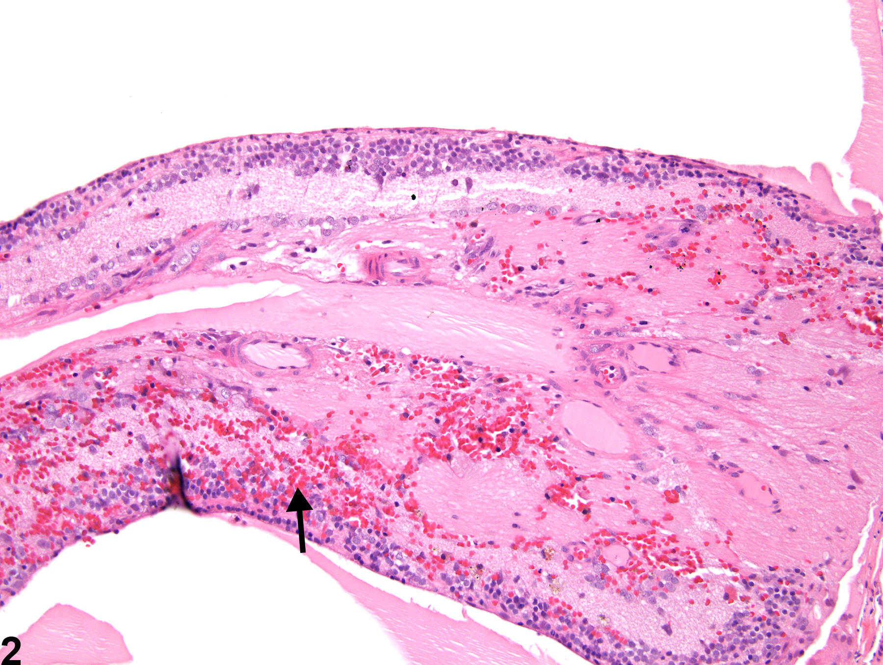
Eye, Retina - Hemorrhage in a female F344/N rat from a chronic study. There are extravasated blood cells (arrow) in a detached and degenerate retina.
All Images

Eye, Retina - Hemorrhage in a female F344/N rat from a chronic study. There are extravasated blood cells (arrow) in a detached and degenerate retina.

Eye, Retina - Hemorrhage in a female F344/N rat from a chronic study (higher magnification of Figure 1). This detached and degenerate retina contains extravasated blood cells (arrow).

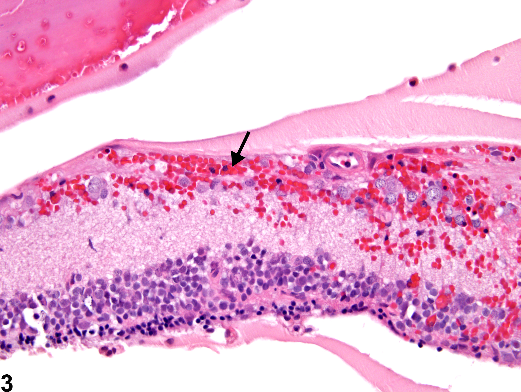
Eye, Retina - Hemorrhage in a female F344/N rat from a chronic study (higher magnification of Figure 1). The hemorrhage (arrow) in this detached and degenerate retina is mainly in the inner layers.