

Special Senses System
Eye, Vitreous - Fibrosis
Narrative
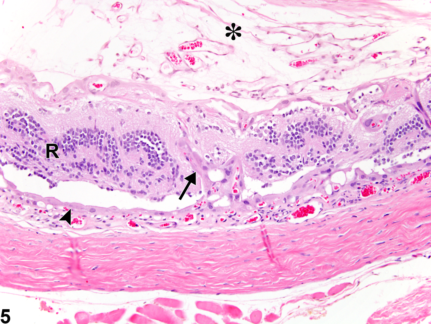
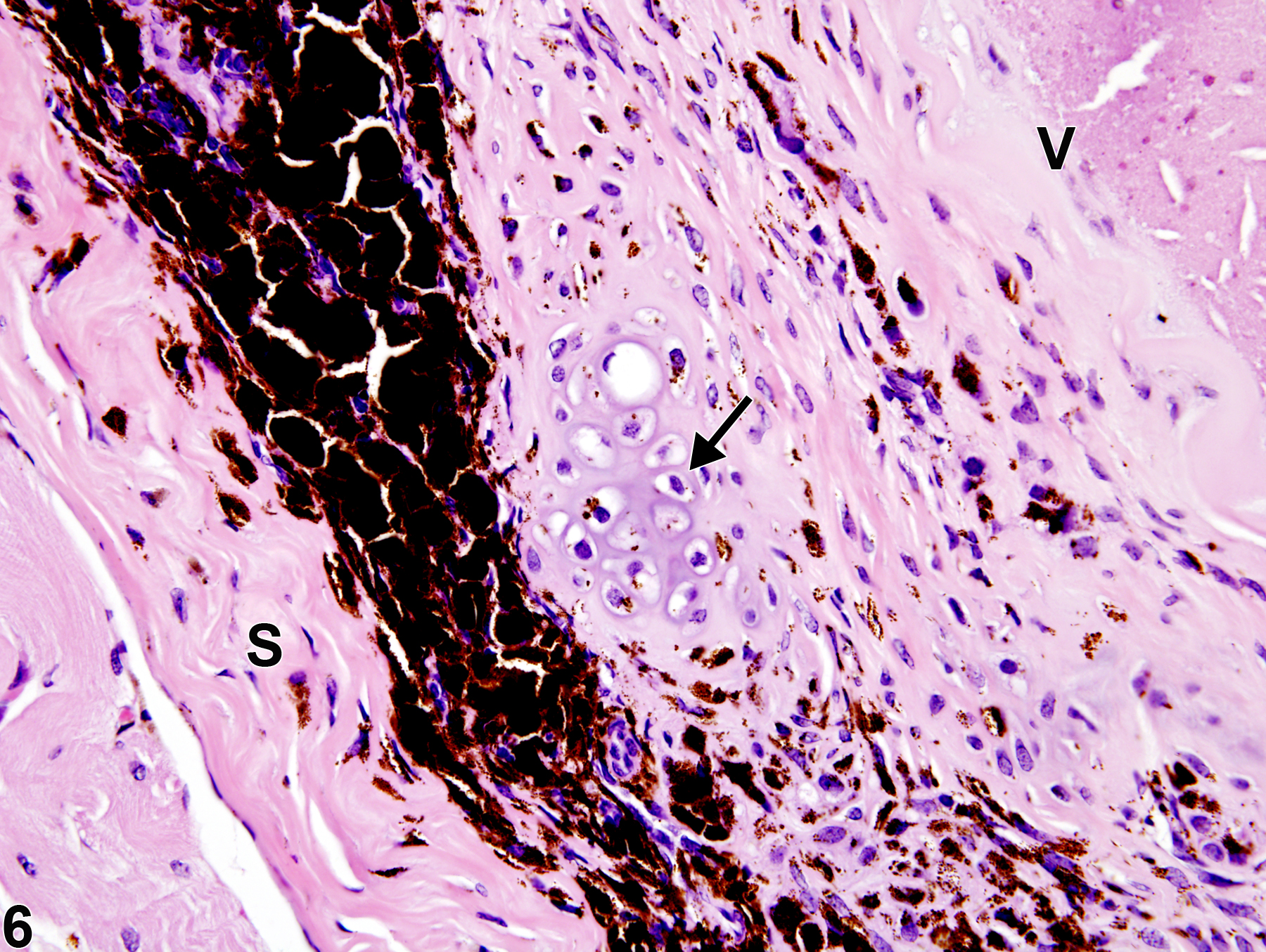
Reactive retinal pigment epithelium (RPE) cells may migrate transretinally into the vitreous (Figure 5) and spreading along the inner surface of the retina. Such migrant RPE cells can undergo fibrous metaplasia (epithelial-to-mesenchymal transition) into fibroblast-like cells and participate in the formation of abnormal vitreal fibrous tissue, including fibrous membranes on the surface of the retina and other posterior ocular structures. Other cells that participate in development of vitreal fibrosis include activated resident vitreal hyalocytes, retinal Müller cells and astrocytes, immigrant macrophages, and scleral fibroblasts. Cartilaginous or osseous metaplasia can occasionally form in late-stage vitreal fibrosis (Figure 6).
Bringmann A, Wiedemann, P. 2009. Involvement of Müller glial cells in epiretinal membrane formation. Graefes Arch Clin Exp Ophthalmol 247:865-883.
Abstract: https://www.ncbi.nlm.nih.gov/pubmed/19415318Frame SR, Slone TW. 1966. Nonneoplastic and neoplastic changes in the eye. In: Pathobiology of the Aging Mouse, Vol 2 (Mohr U, Dungworth DL, Capen CC, Carlton WW, Sundberg JP, Ward JM, eds). ILSI Press, Washington, DC, 97-103.
Hogg PA, Grierson I, Hiscott P. 2002. Direct comparison of the migration of three cell types involved in epiretinal membrane formation. Invest Ophthalmol Vis Sci 43:2749-2757.
Full Text: http://tpx.sagepub.com/content/35/2/252.fullMecklenburg L, Schraermeyer U. 2007. An overview on the toxic morphological changes in the retinal pigment epithelium after systemic compound administration. Toxicol Pathol 35:252-267.
Full Text: http://tpx.sagepub.com/content/35/2/252.fullNational Toxicology Program. 1992. NTP TR-407. Toxicology and Carcinogenesis Studies of C.I. Pigment Red 3 (CAS No. 2425-85-6) in F344/N Rats and B6C3F1 Mice (Feed Studies). NTP, Research Triangle Park, NC.
Abstract: https://ntp.niehs.nih.gov/go/7694National Toxicology Program. 1992. NTP TR-415. Toxicology and Carcinogenesis Studies of Polysorbate 80 (CAS No. 9005-65-6) in F344/N Rats and B6C3F1 Mice (Feed Studies). NTP, Research Triangle Park, NC.
Abstract: https://ntp.niehs.nih.gov/go/7710National Toxicology Program. 2001. NTP TR-501. Toxicology and Carcinogenesis Studies of p, p'-Dichlorodiphenyl Sulfone (CAS No. 80-07-9) in F344/N Rats and B6C3F1 Mice (Feed Studies). NTP, Research Triangle Park, NC.
Abstract: https://ntp.niehs.nih.gov/go/14880Saika S, Kono-Saika S, Ohnishi Y, Sato M, Muragaki Y, Ooshima A, Flanders KC, Yoo J, Anzano M, Liu C-Y, Kao W W-Y, Roberts AB. 2004. Smad3 signaling is required for epithelial-mesenchymal transition of lens epithelium after injury. Am J Pathol 164:651-663.
Abstract: https://www.ncbi.nlm.nih.gov/pubmed/14742269Smith RS. 2002. Choroid, lens, and vitreous. In: Systematic Evaluation of the Mouse Eye: Anatomy, Pathology, and Biomethods (Smith RS, John SWM, Nishina PM, Sundberg JP, eds). CRC Press Boca Raton, FL, 161-193.
Smith RS, Hawes NL, Chang B, Nishina PM. 2002. Retina. In: Systematic Evaluation of the Mouse Eye: Anatomy, Pathology, and Biomethods (Smith RS, John SWM, Nishina PM, Sundberg JP, eds). CRC Press, Boca Raton, FL, 195-225.
Yoshitomi K, Boorman GA. 1990. Eye and associated glands. In: Pathology of the Fischer Rat: Reference and Atlas (Boorman GA, Eustis SL, Elwell MR, Montgomery CA, MacKenzie WF, eds). Academic Press, San Diego, CA, 239-260.
Abstract: https://www.ncbi.nlm.nih.gov/nlmcatalog/9002563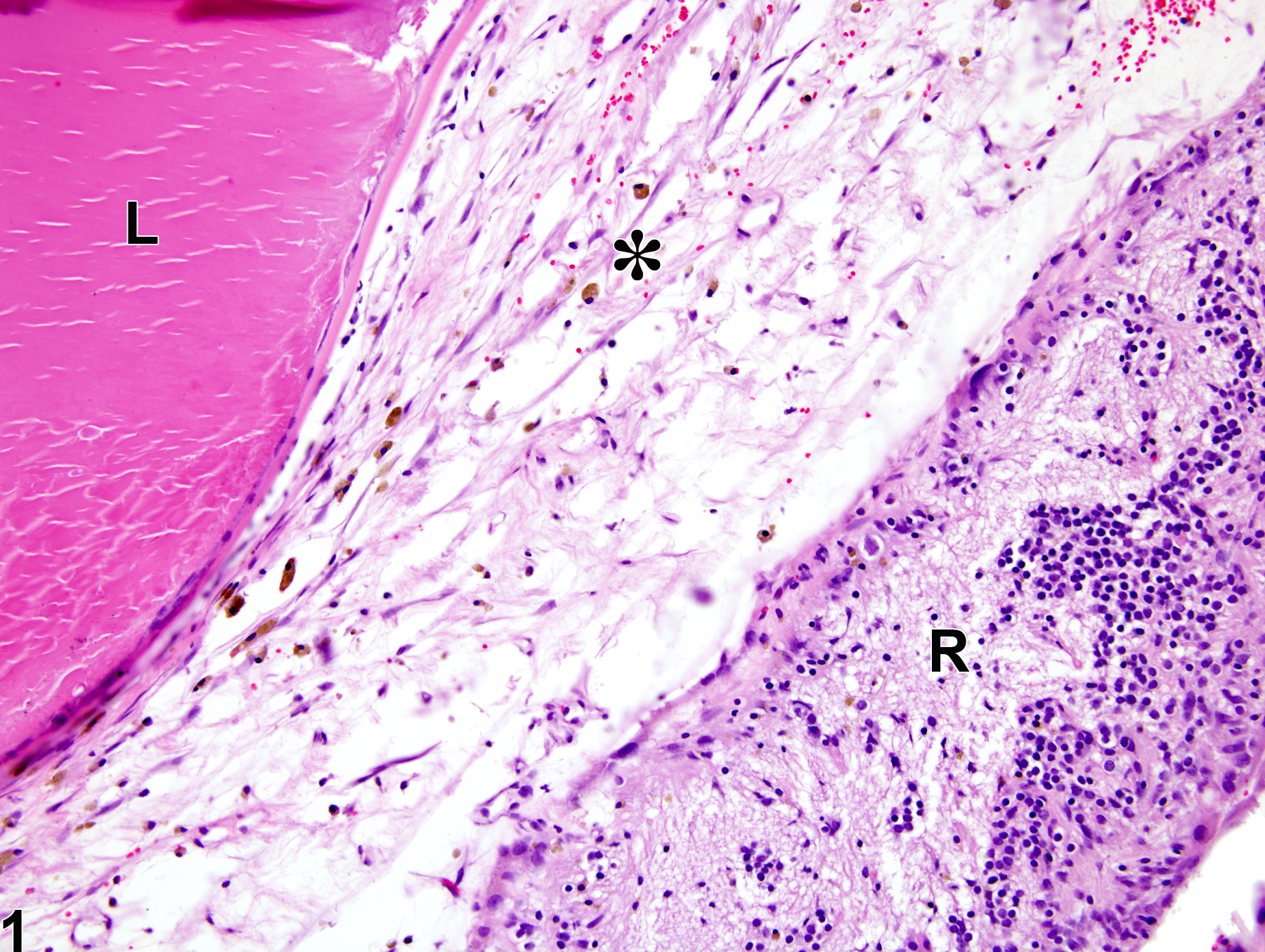
Eye, Vitreous - Fibrosis in a male F344/N rat from a chronic study. There is loose fibrous connective tissue (asterisk) in the vitreous space between a cataractous lens (L) and a detached, degenerate retina (R).
All Images

Eye, Vitreous - Fibrosis in a male F344/N rat from a chronic study. There is loose fibrous connective tissue (asterisk) in the vitreous space between a cataractous lens (L) and a detached, degenerate retina (R).

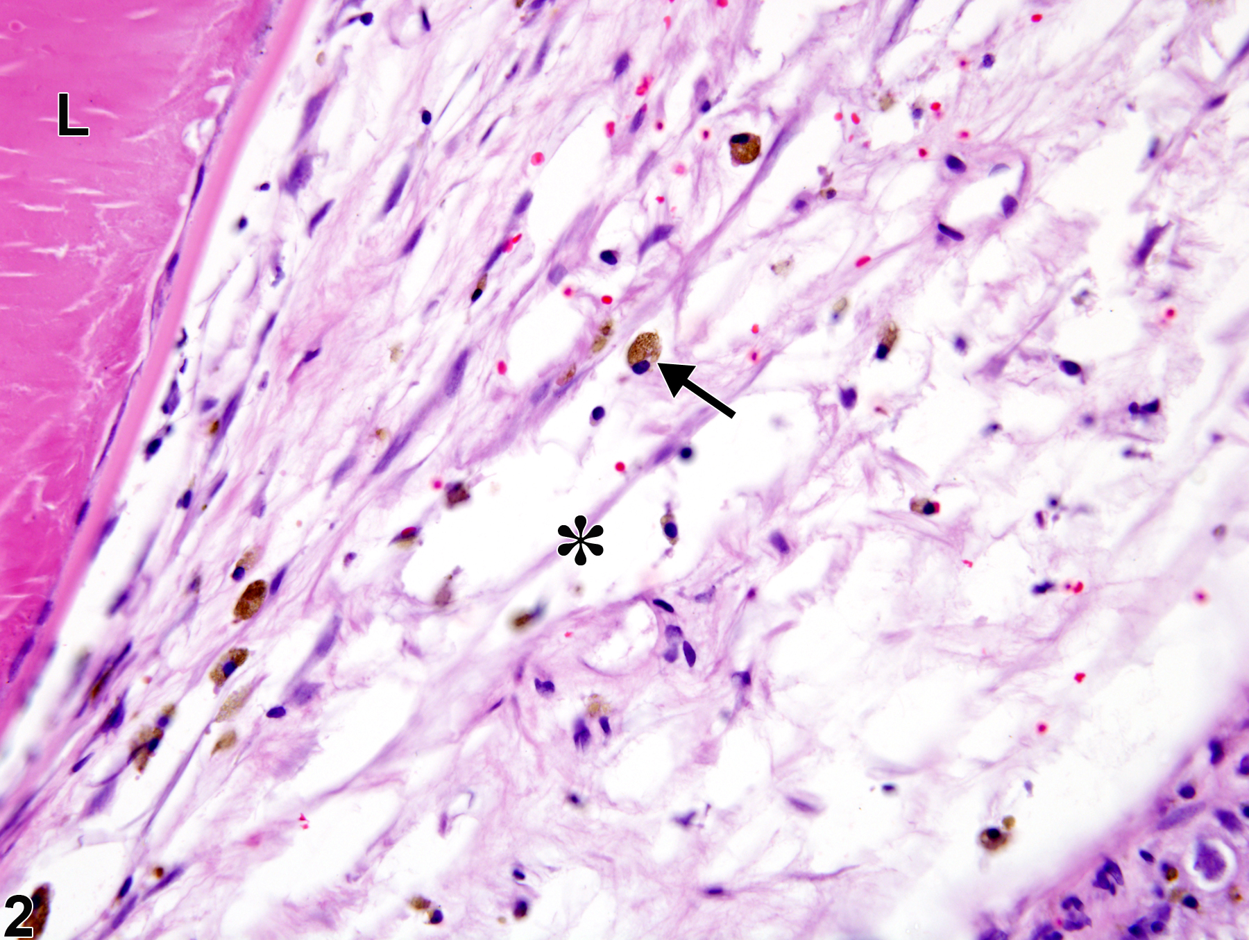
Eye, Vitreous - Fibrosis in a male F344/N rat from a chronic study (higher magnification of Figure 1). There is loose fibrous connective tissue (asterisk) with space scattered pigmented macrophages (arrow) in the vitreous; L = cataractous lens.

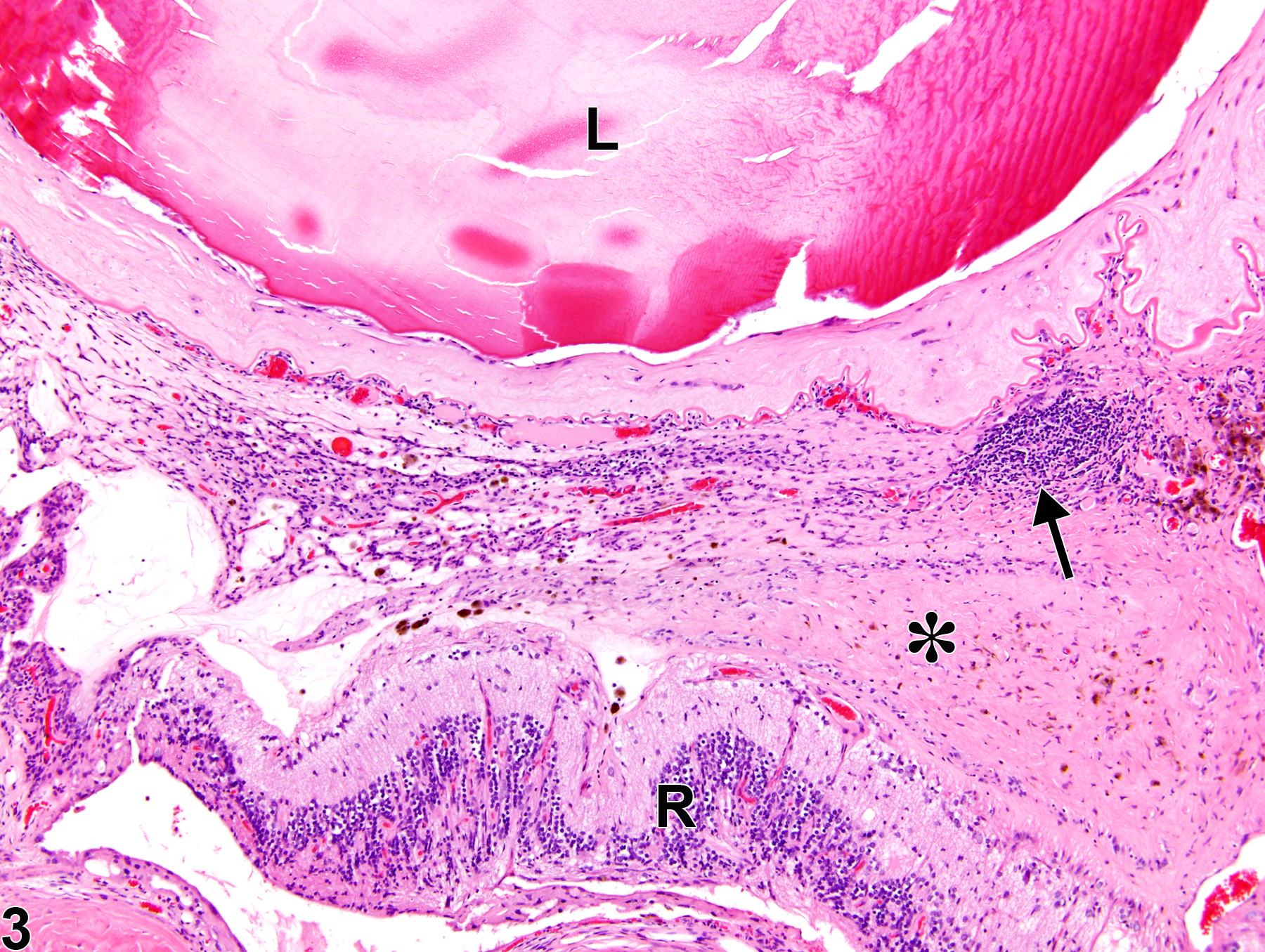
Eye, Vitreous - Fibrosis in a female F344/N rat from a chronic study. There is advanced fibrosis in the vitreous space characterized by dense fibrous connective tissue (asterisk) with chronic inflammation (arrow); L = cataractous lens, R = detached, degenerate retina.

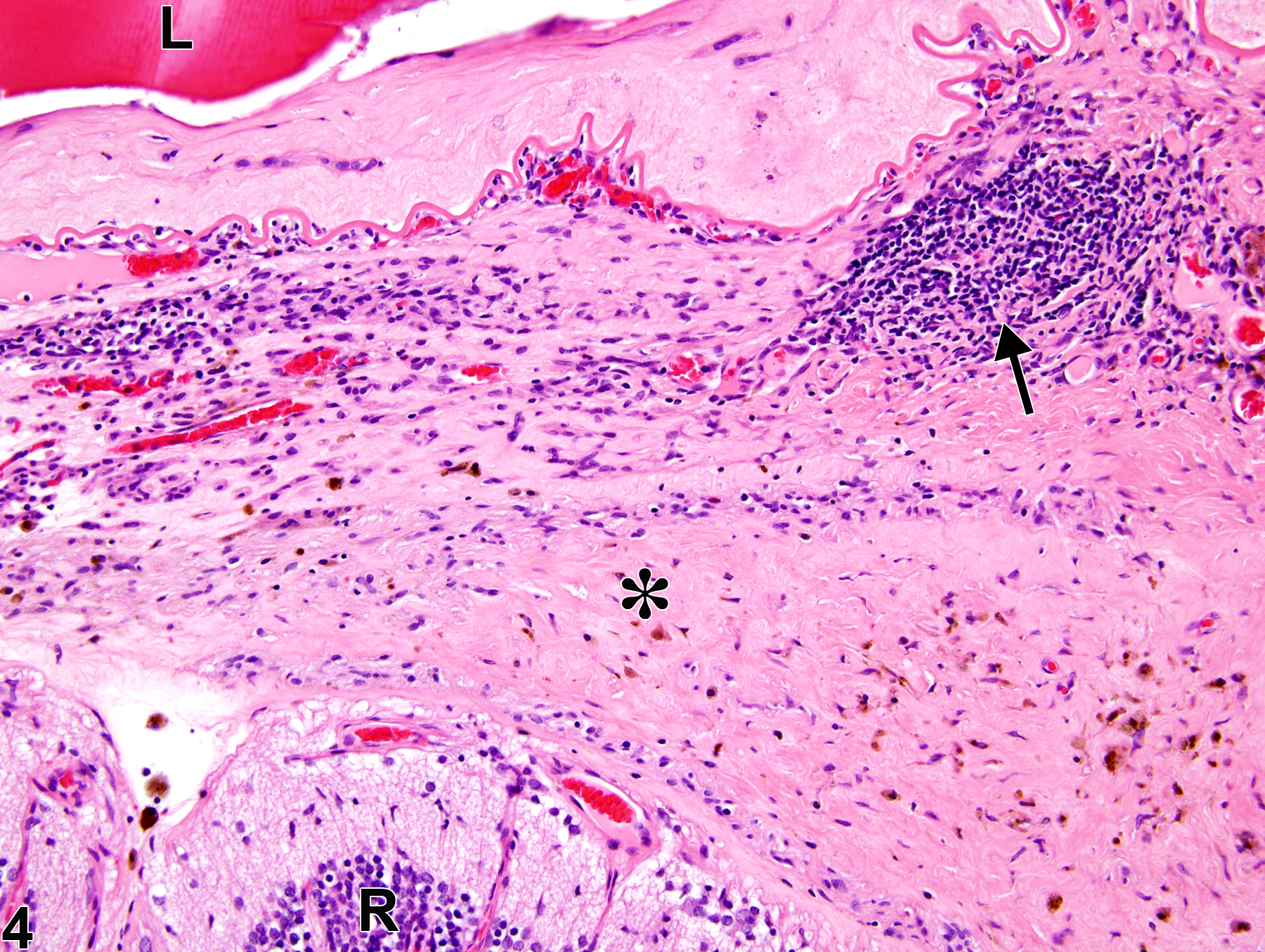
Eye, Vitreous - Fibrosis in a female F344/N rat from a chronic study (higher magnification of Figure 3). The advanced fibrosis is characterized by dense fibrous connective tissue (asterisk) in the vitreous space with chronic inflammation (arrow); L = cataractous lens, R = detached degenerate retina.

Eye, Vitreous - Fibrosis in a female F344/N rat from a chronic study. There is fibrosis (asterisk) and infiltration by migrating reactive epithelial cells (arrow) from the retinal pigment epithelium (arrowhead) in the vitreous space in the eye; R = detached and degenerate retina.

Eye, Vitreous - Fibrosis in a male B6C3F1 mouse from a chronic study. Area of dense, mature fibrous connective tissue in the vitreous (V) and focal cartilaginous metaplasia (arrow); S = sclera.