

Special Senses System
Eye, Vitreous - Hemorrhage
Narrative
Kuno H, Usui T, Eydelloth RS, Wolf ED. 1991. Spontaneous ophthalmic lesions in young Sprague-Dawley rats. J Vet Med Sci 53:607-614.
Abstract: https://www.ncbi.nlm.nih.gov/pubmed/10845604National Toxicology Program. 1992. NTP TR-407. Toxicology and Carcinogenesis Studies of C.I. Pigment Red 3 (CAS No. 2425-85-6) in F344/N Rats and B6C3F1 Mice (Feed Studies). NTP, Research Triangle Park, NC.
Abstract: https://ntp.niehs.nih.gov/go/7694National Toxicology Program. 1997. NTP TR-450. Toxicology and Carcinogenesis Studies of Tetrafluoroethylene (CAS No. 116-14-3) in F344 Rats and B6C3F1 Mice (Inhalation Studies). NTP, Research Triangle Park, NC.
Abstract: https://ntp.niehs.nih.gov/go/6044Smith RS. 2002. Choroid, lens, and vitreous. In: Systematic Evaluation of the Mouse Eye: Anatomy, Pathology, and Biomethods (Smith RS, John SWM, Nishina PM, Sundberg JP, eds). CRC Press Boca Raton, FL, 161-193.
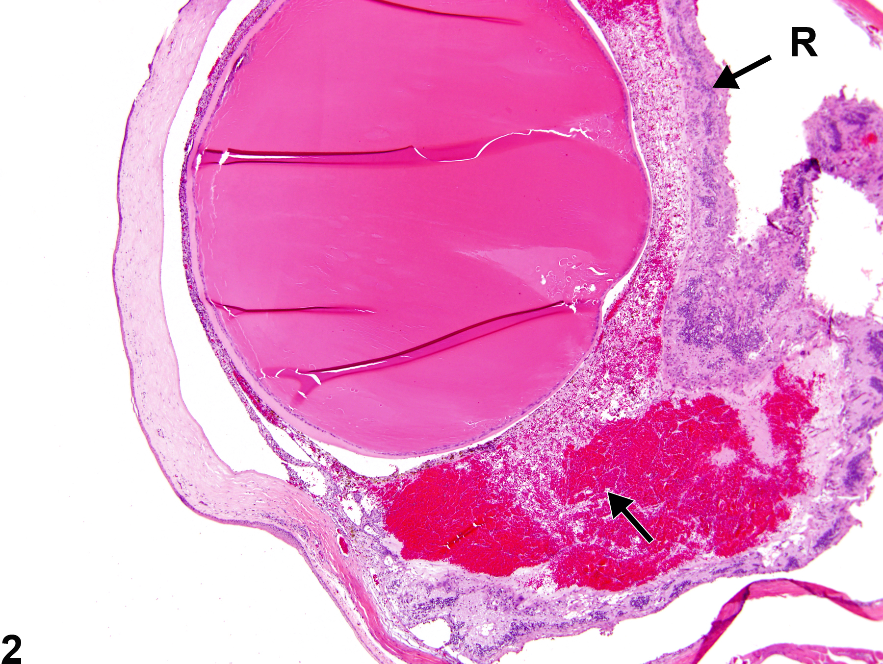
Eye, Vitreous - Hemorrhage in a male F344/N rat from a chronic study. There are intravitreal accumulations of extravasated blood cells (arrow) with retinal detachment and degeneration (R).
All Images

Eye, Vitreous - Hemorrhage in a male F344/N rat from a chronic study. There are intravitreal accumulations of extravasated blood cells (arrow) with retinal detachment and degeneration (R).

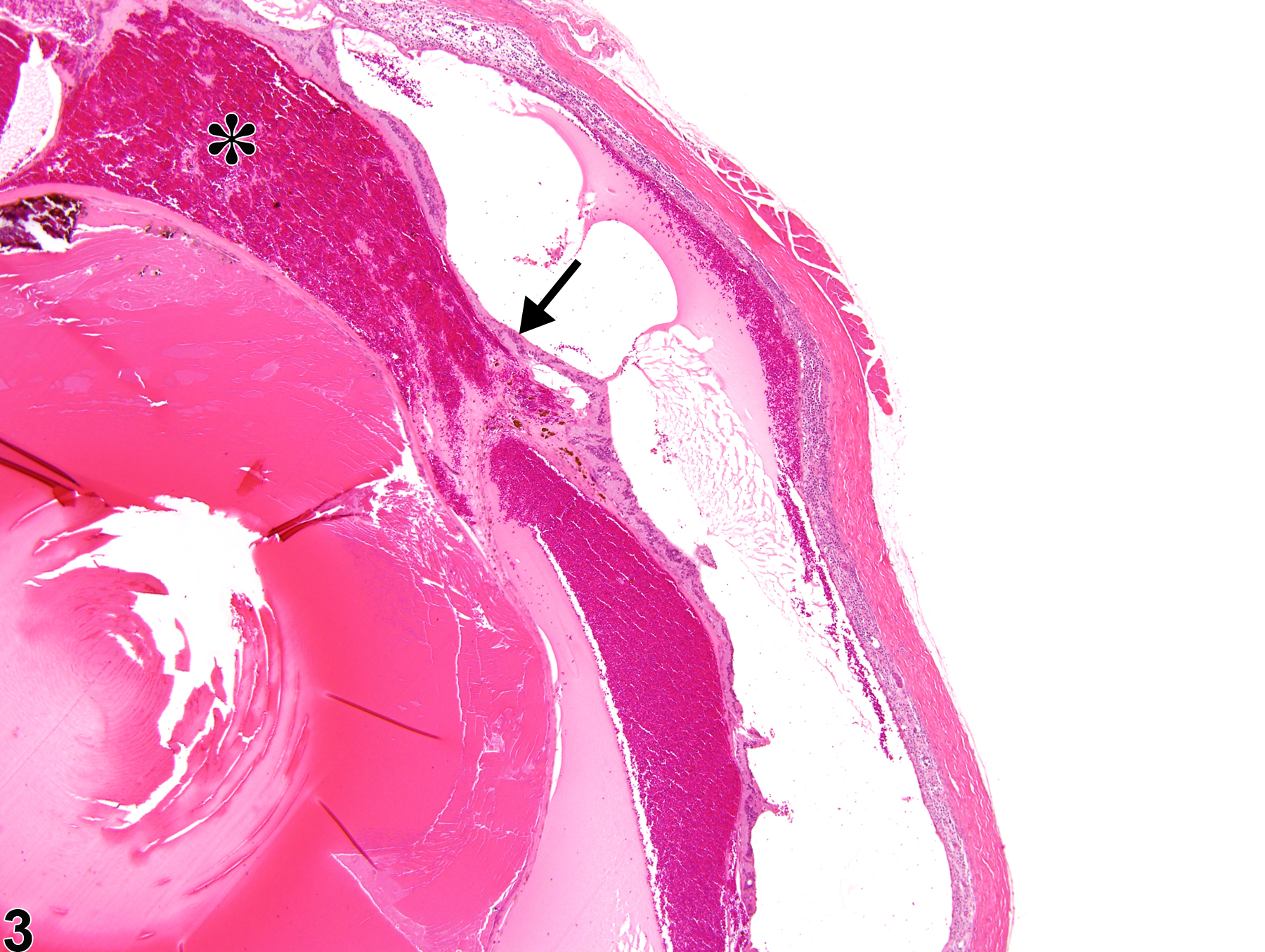
Eye, Vitreous - Hemorrhage in a male F344/N rat from a chronic study (higher magnification of Figure 1). This higher magnification image shows the intravitreal hemorrhage (arrow) and retinal detachment and degeneration (R) in more detail.

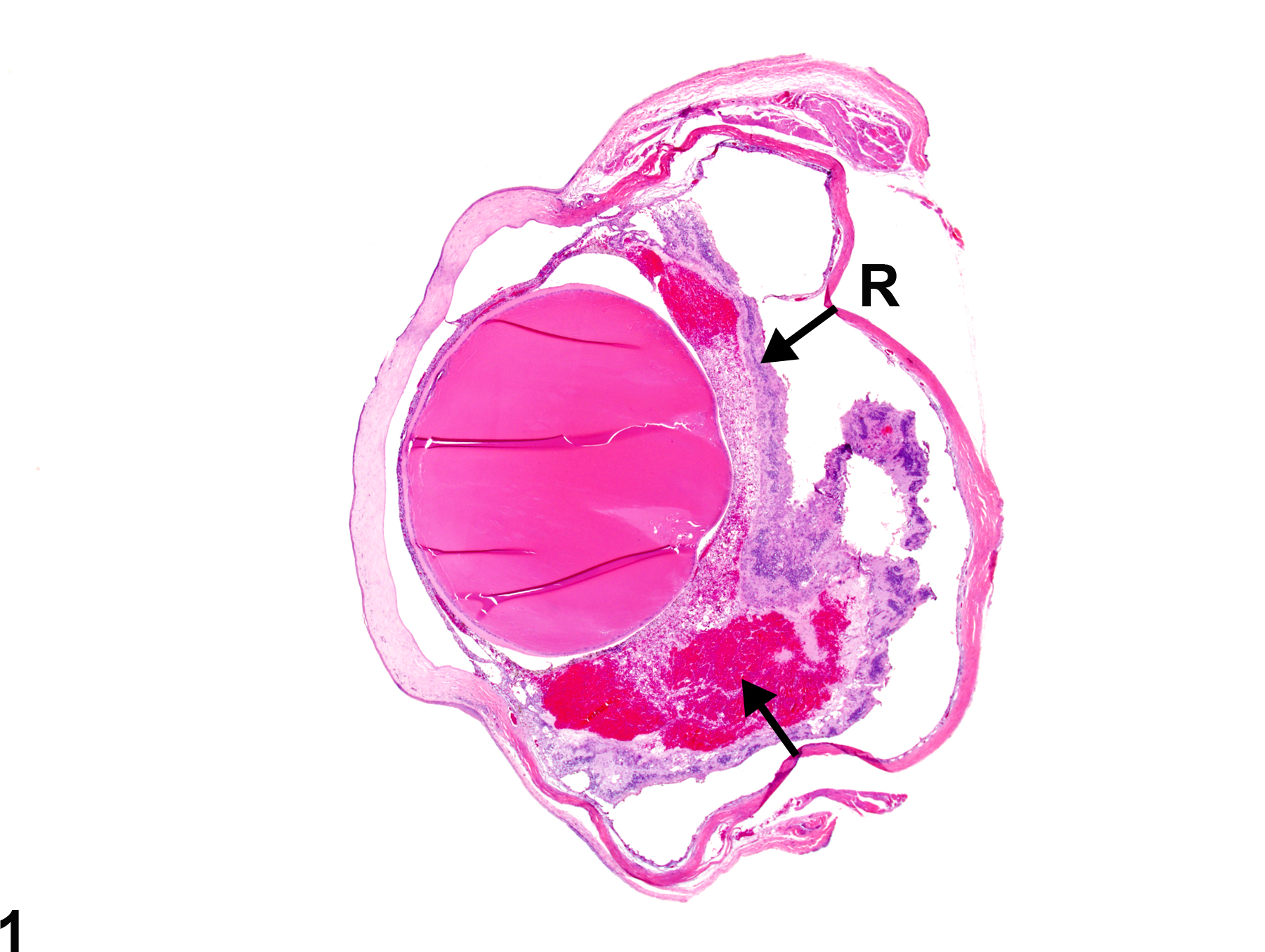
Eye, Vitreous - Hemorrhage in a female F344/N rat from a chronic study. There is vitreal hemorrhage (asterisk) with retinal detachment and degeneration (arrow).

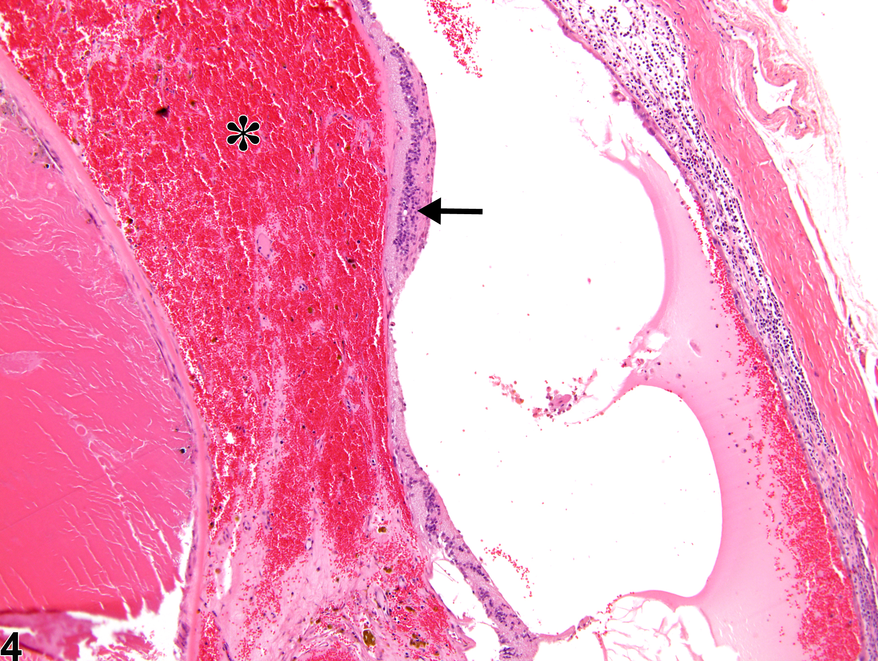
Eye, Vitreous - Hemorrhage in a female F344/N rat from a chronic study. Higher magnification of Figure 3. There is vitreal hemorrhage (asterisk) consisting of intravitreal accumulations of extravasated blood cells with retinal detachment and degeneration (arrow).