

Special Senses System
Zymbal's Gland - Hyperplasia
Narrative
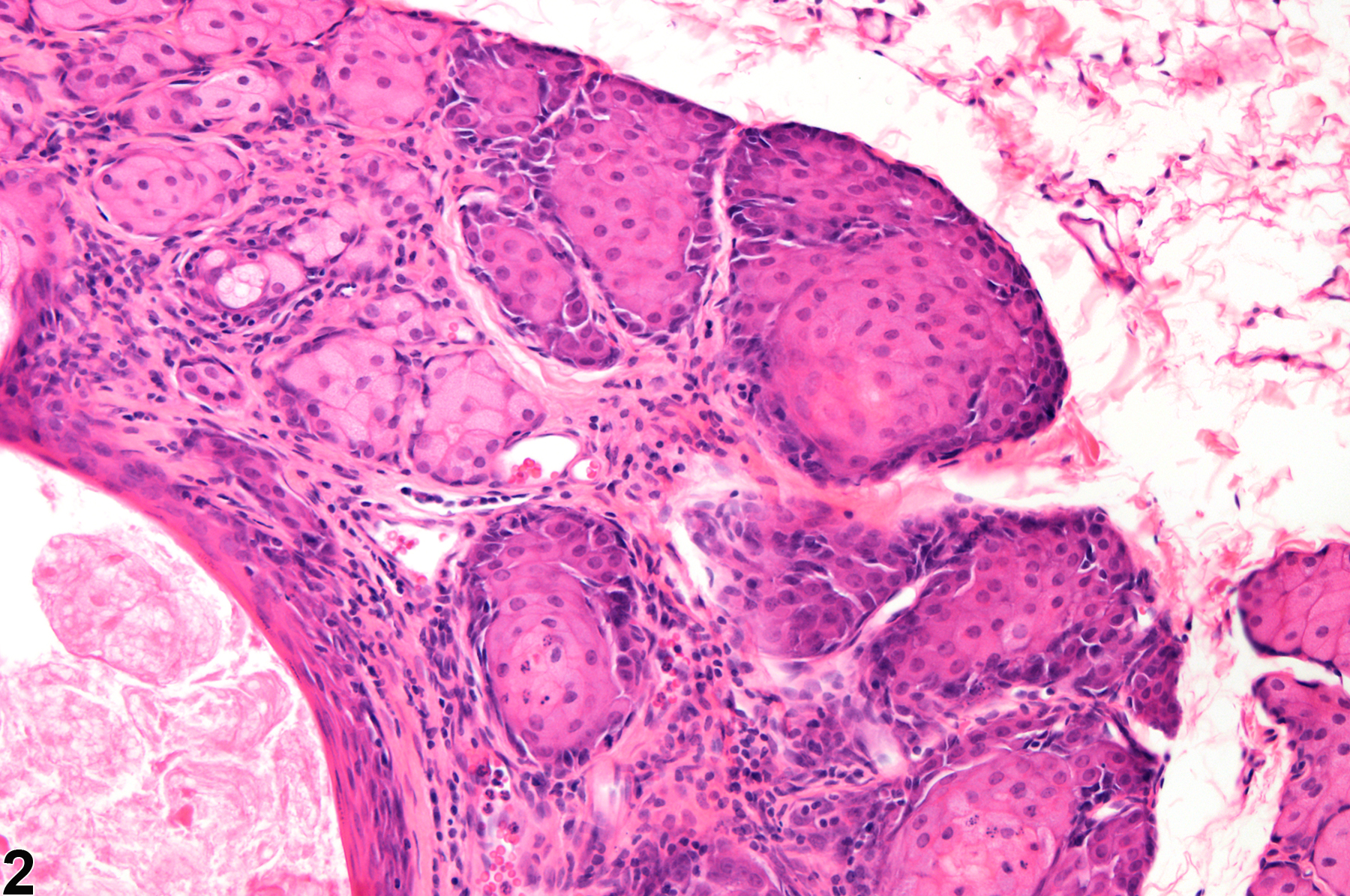
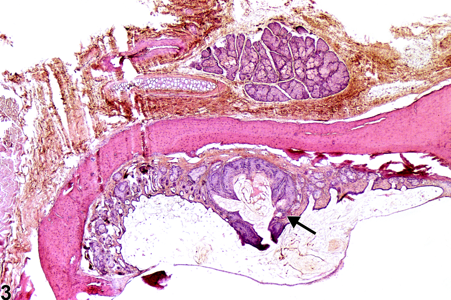
Hyperplastic areas in the Zymbal's gland are characterized by a focal increase in the number and often the size of the sebaceous acini, often with slight distortion of the lobular architecture and slight compression of the adjacent tissue. They are composed of variably sized acini lined by relatively well-differentiated epithelial cells with enlarged nuclei and cytoplasm that is more basophilic and less foamy than in normal cells. The normal maturation sequence of the acinar cells may be obscured or absent. Hyperplasia lacks the pronounced cellular atypia, marked architectural distortion, and invasion of adjacent tissue, which are features of neoplastic lesions. Hyperplasia may be a reactive change secondary to inflammation or degeneration. Hyperplasia occurring in the absence of concurrent degeneration or inflammation is considered a preneoplastic lesion and an early-stage change in the morphologic continuum from hyperplasia to carcinoma.
Copeland-Haines D, Eustis SL. 1990. Specialized sebaceous glands. In: Pathology of the Fischer Rat: Reference and Atlas (Boorman GA, Eustis SL, Elwell MR, Montgomery CA, MacKenzie WF, eds). Academic Press, San Diego, CA, 279-294.
Abstract: https://www.ncbi.nlm.nih.gov/nlmcatalog/9002563Hebel R, Stromberg MW. 1986. Sensory organs. In: Anatomy and Embryology of the Laboratory Rat, 2nd ed. BioMed Verlag, Wörthsee, Germany, 218-224.
Huff JE, Eastin W, Roycroft J, Eustis SL, Haseman JK. 1988. Carcinogenesis studies of benzene, methyl benzene, and dimethylbenzenes. Ann NY Acad Sci 534:427-440.
Abstract: http://onlinelibrary.wiley.com/doi/10.1111/j.1749-6632.1988.tb30132.x/abstractLaws JO, Rudali G, Royer R, Mabile P. 1955. The early changes produced in the auditory sebaceous gland (Zymbal’s gland) of the rat by 2-acetylaminofluorene. Cancer Res 15:139-143.
Abstract: http://cancerres.aacrjournals.org/content/15/3/139National Toxicology Program. 1990. NTP TR-372. Toxicology and Carcinogenesis Studies of 3,3'-Dimethoxybenzidine Dihydrochloride (CAS No. 20325-40-0) in F344/N Rats (Drinking Water Studies). NTP, Research Triangle Park, NC.
Abstract: https://ntp.niehs.nih.gov/go/8884National Toxicology Program. 1993. NTP TR-400. Toxicology and Carcinogenesis Studies of 2,3-Dibromo-1-Propanol (CAS No. 96-13-9) in F344/N Rats and B6C3F1 Mice (Dermal Studies). NTP, Research Triangle Park, NC.
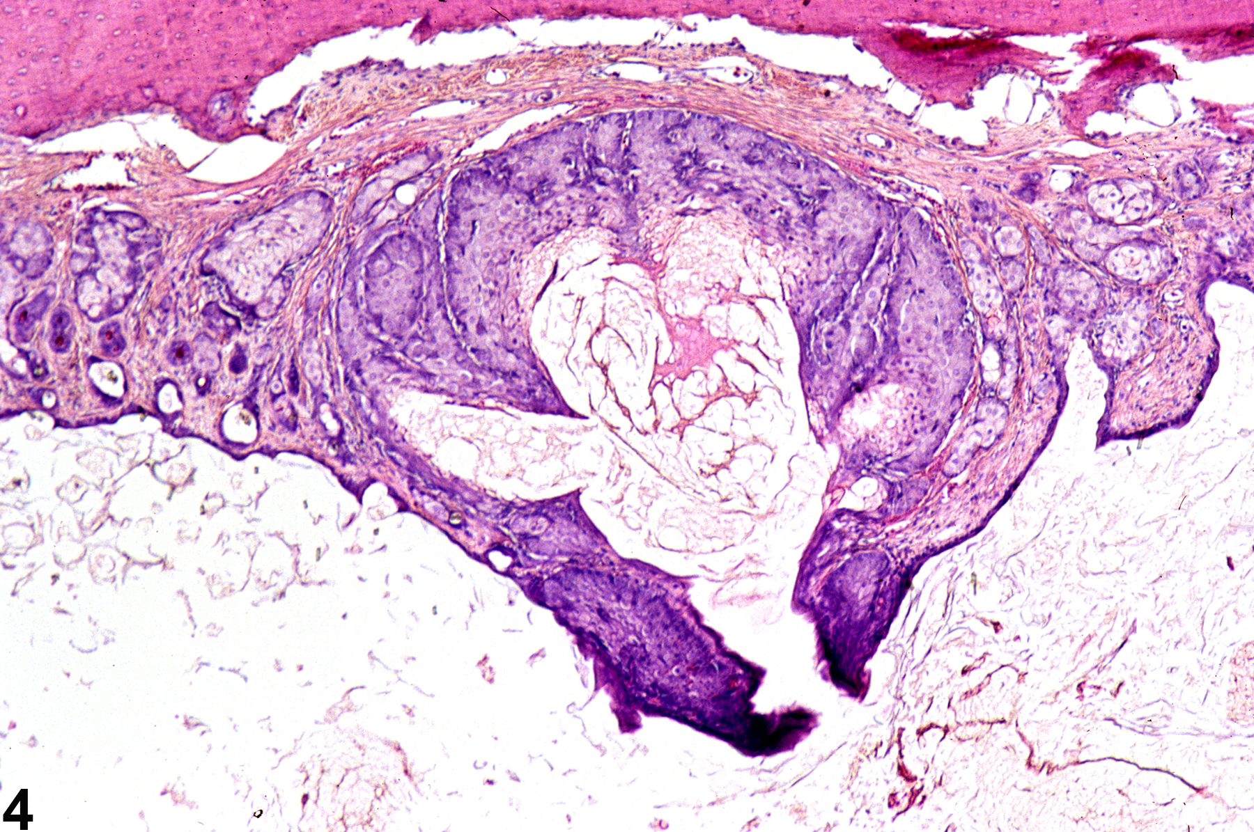
Abstract: https://ntp.niehs.nih.gov/go/12251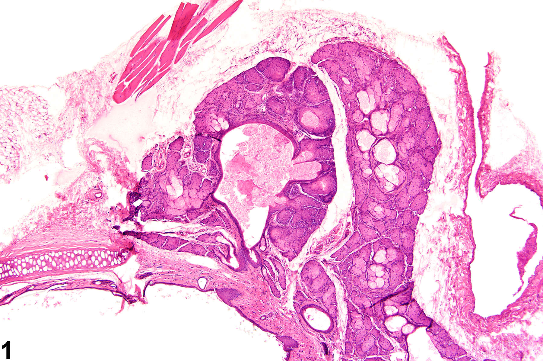 Zymbal's gland - Hyperplasia in a female F344/N rat from a chronic study. There is a focal increase in the number of sebaceous acini (arrow) with slight lobular architectural distortion around a dilated duct.
All Images


