U.S. flag

An official website of the United States government

Dot gov

The .gov means it's official.
Federal government websites often end in .gov or .mil. Before sharing sensitive information, make sure you're on a federal government site.

Https

The site is secure.
The https:// ensures that you are connecting to the official website and that any information you provide is encrypted and transmitted securely.

Skip to Main Navigation
Skip to Page Content
Skip to Atlas Navigation

Kidney - Amyloid

Image of amyloid in the kidney from a female B6C3F1 mouse in a chronic study
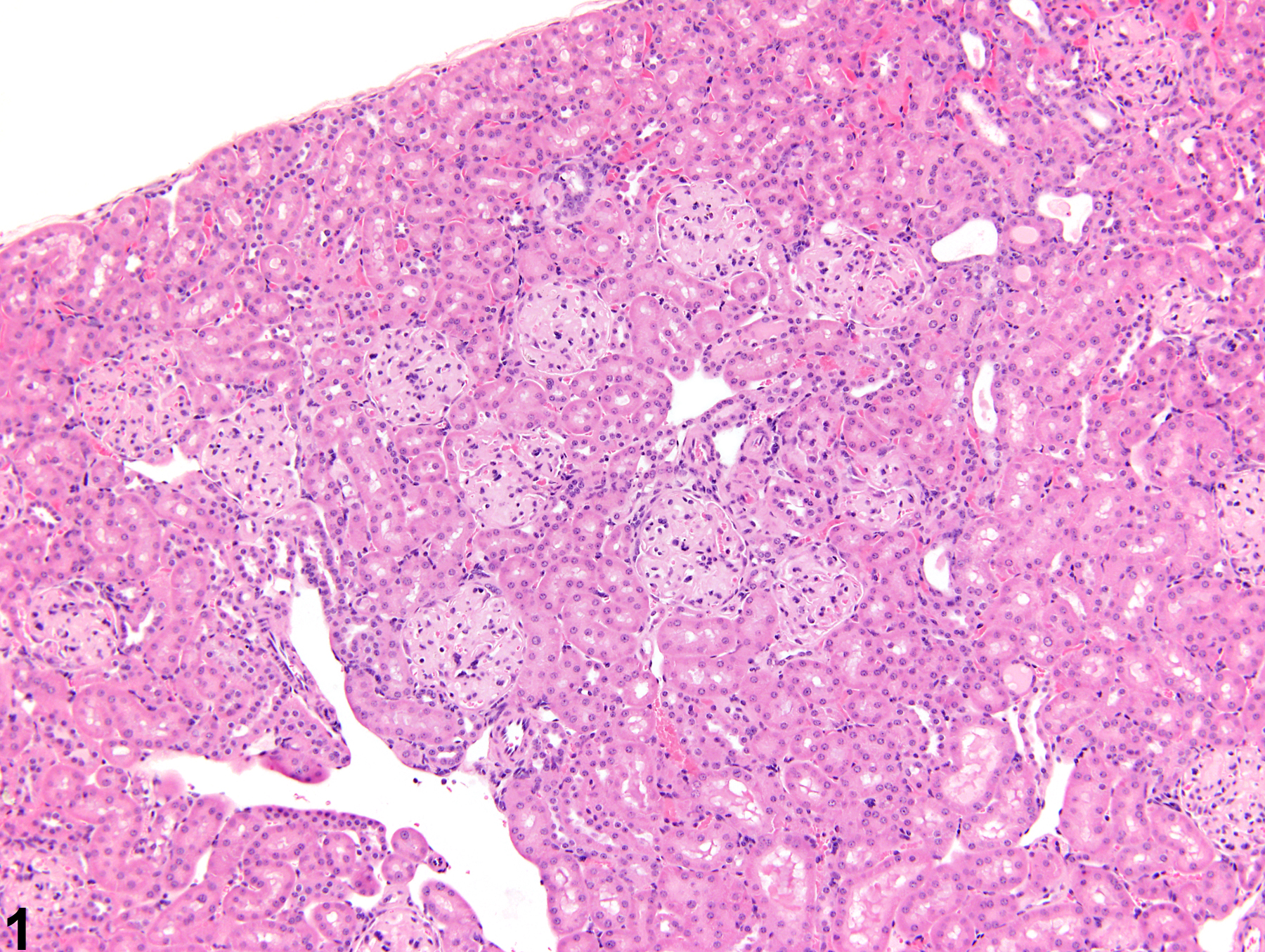
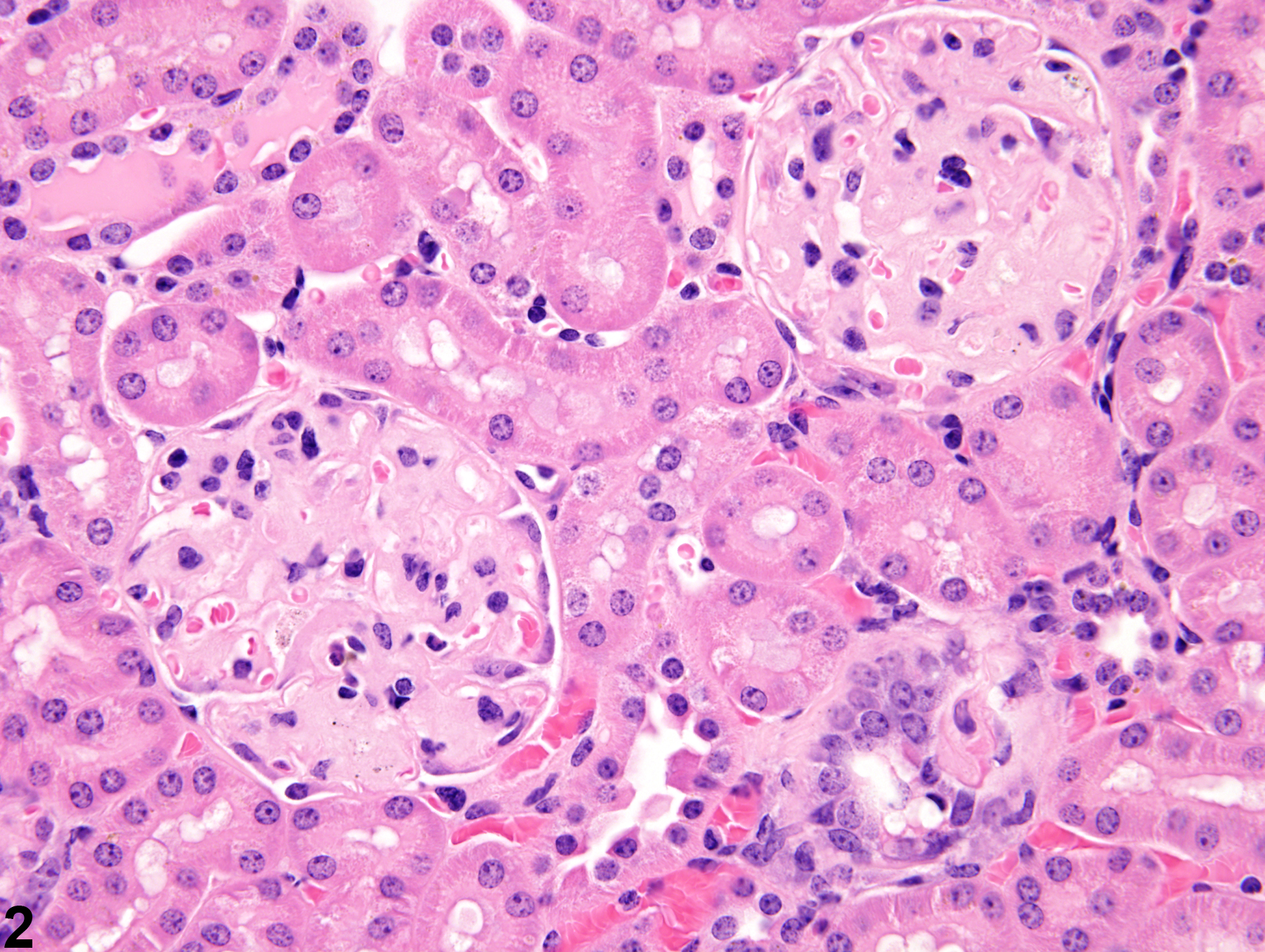
Kidney, Glomerulus - Amyloid in a female B6C3F1 mouse from a chronic study. Glomeruli contain a pale, amorphous, eosinophilic material identified as amyloid.
Figure 1 of 6
Image of amyloid in the kidney from a female B6C3F1 mouse in a chronic study
Kidney, Glomerulus - Amyloid in a female B6C3F1 mouse from a chronic study. Increased amounts of pale-staining eosinophilic glomerular deposits of amyloid are present.
Figure 2 of 6
Image of amyloid in the kidney from a  B6C3F1 mouse in a chronic study
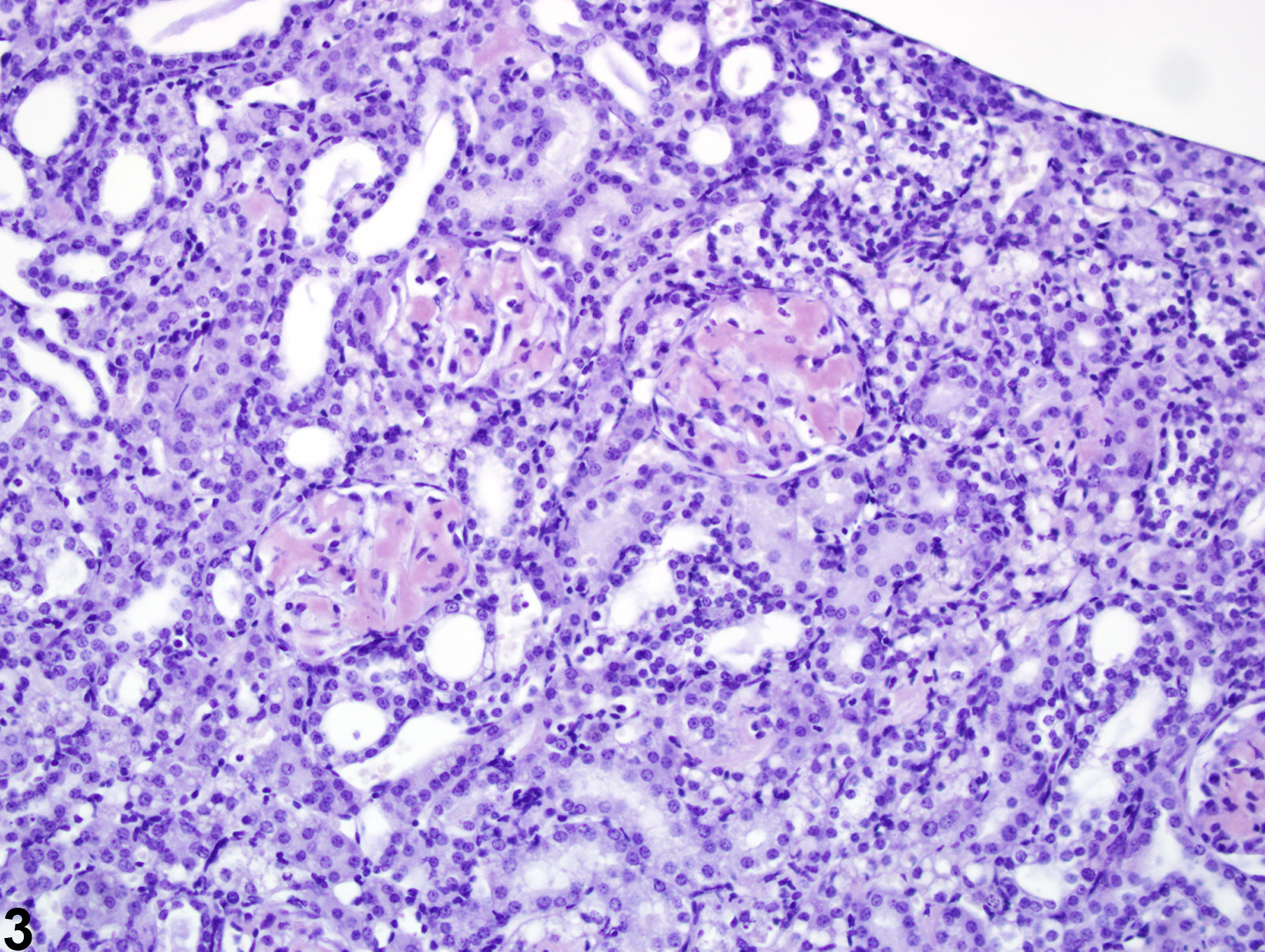
Kidney, Glomerulus - Amyloid in a B6C3F1 mouse from a chronic study. Positive Congo red staining of glomerular amyloid deposits are present in the glomeruli.
Figure 3 of 6
Image of amyloid in the kidney from a  B6C3F1 mouse in a chronic study
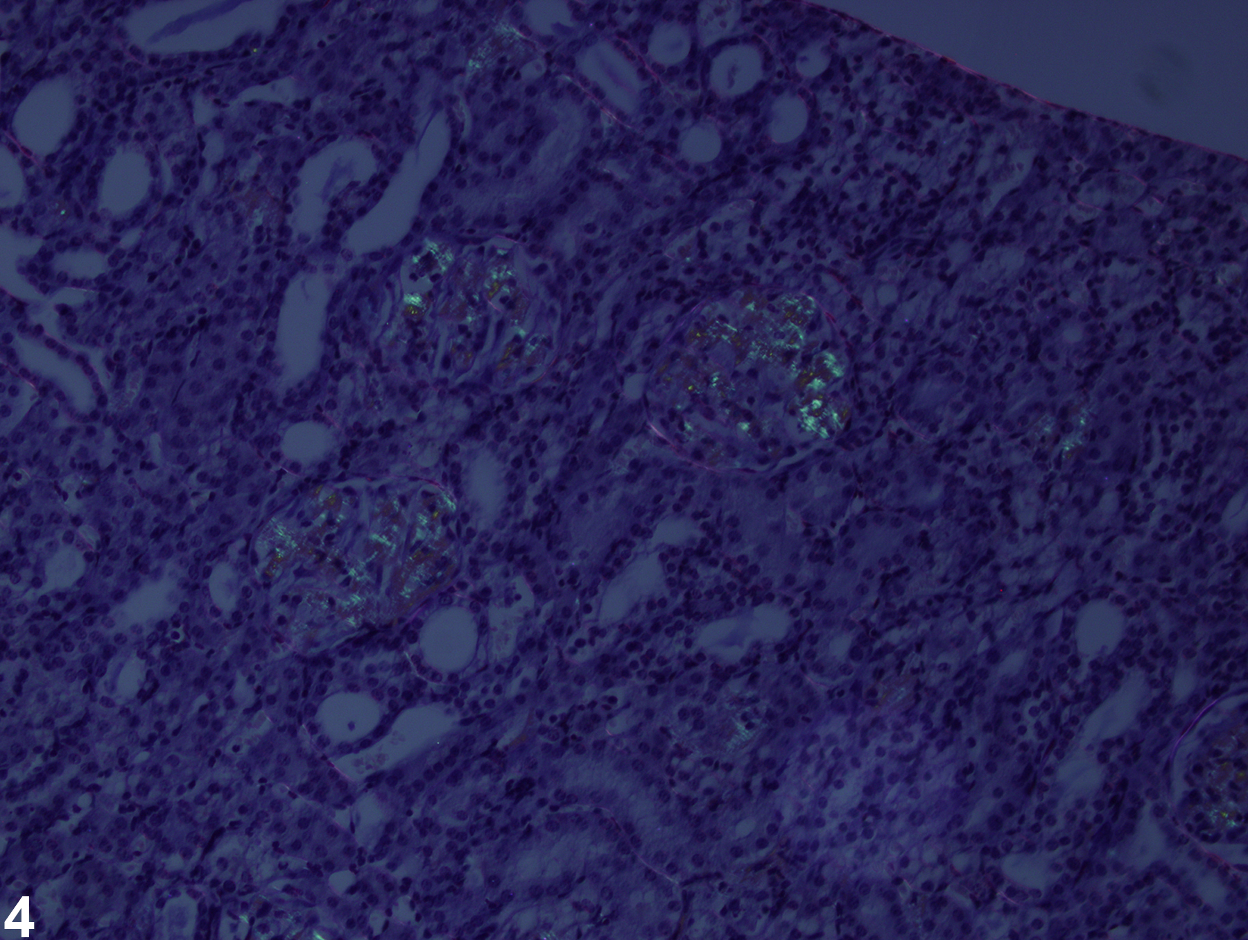
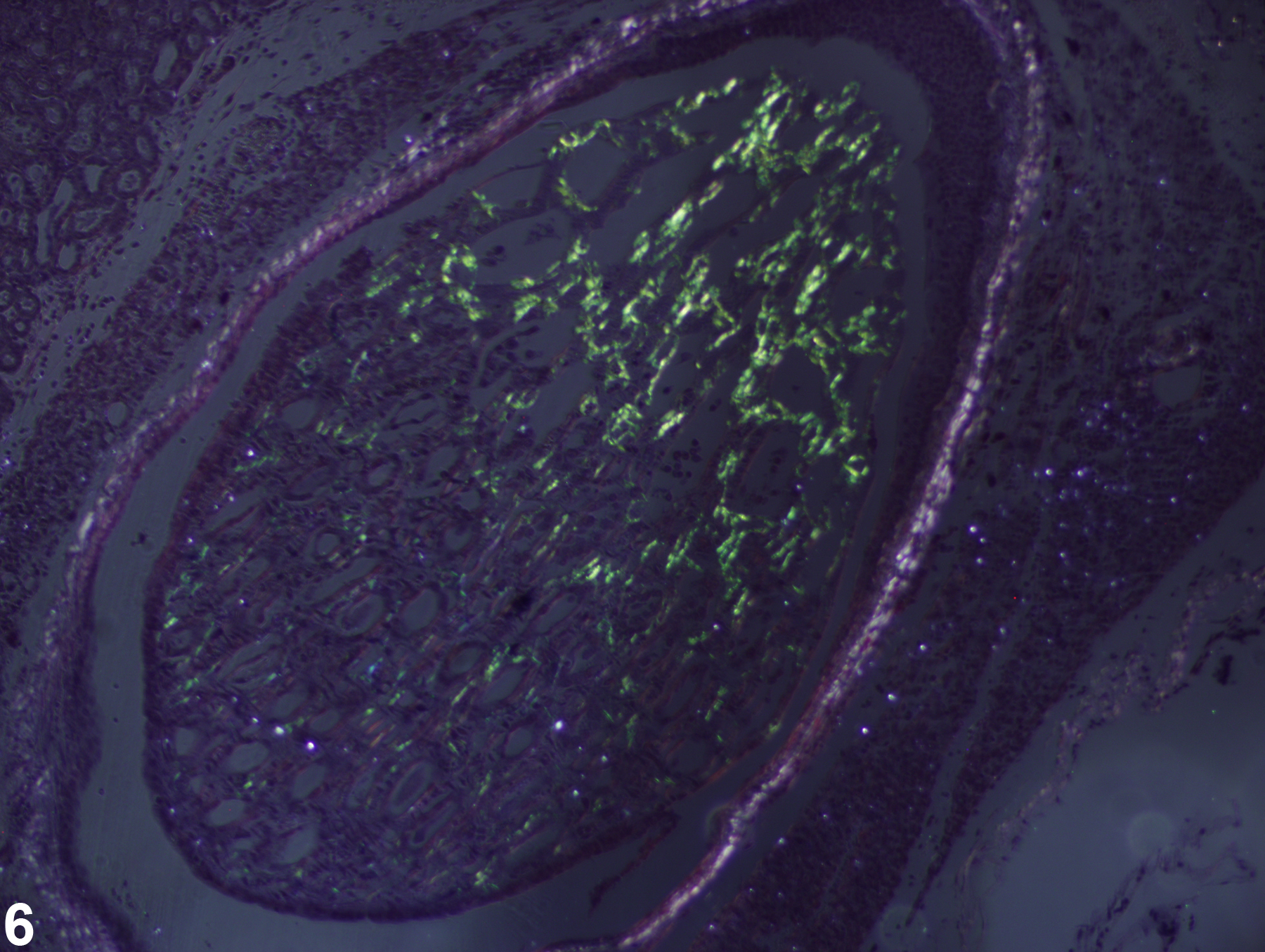
Kidney, Glomerulus - Amyloid in a B6C3F1 mouse from a chronic study (same mouse as in Figure 3). Congo red staining with polarization of amyloid deposits shows the characteristic apple-green birefringence.
Figure 4 of 6
Image of amyloid in the kidney from a  B6C3F1 mouse in a chronic study
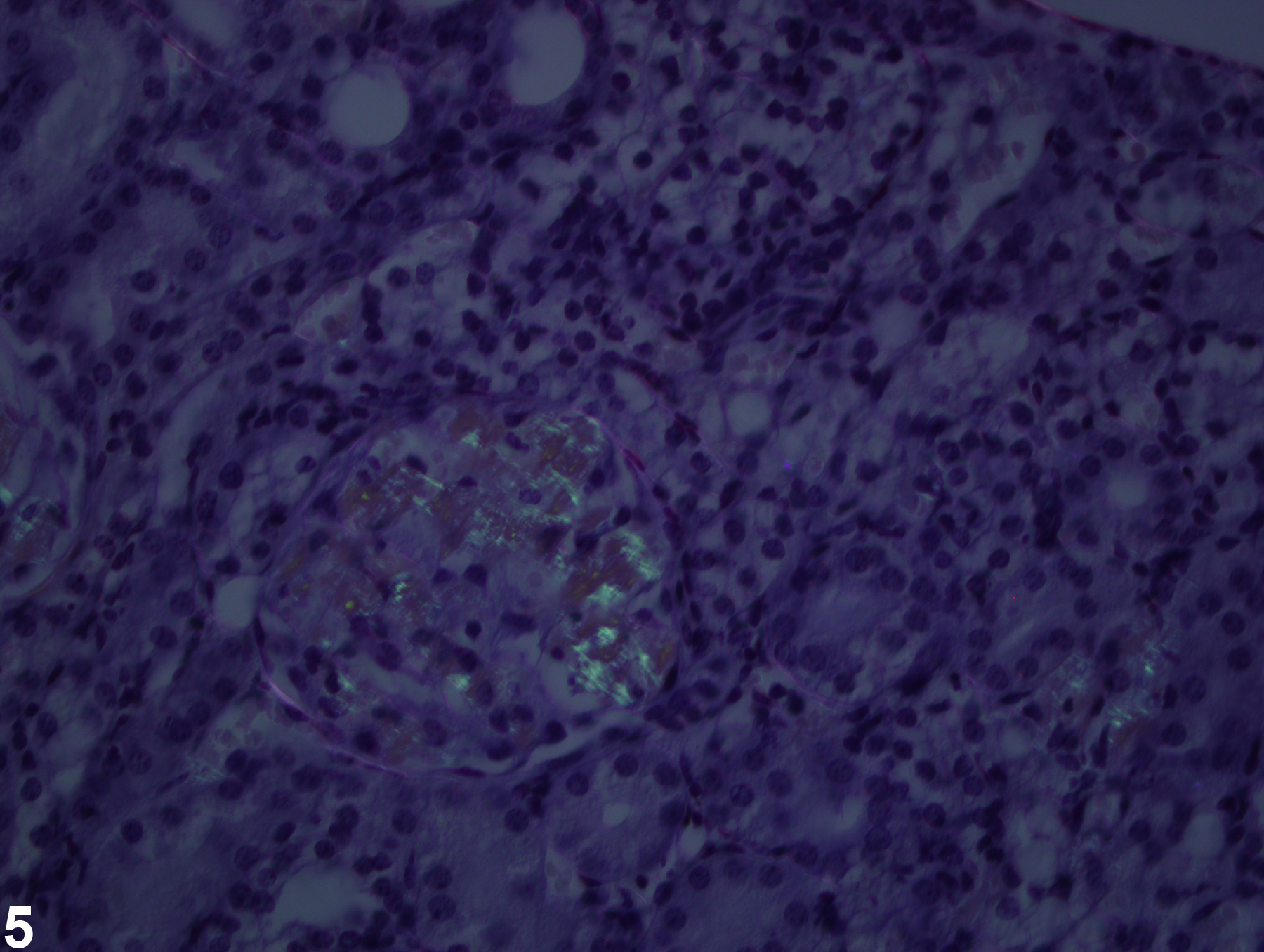
Kidney, Glomerulus - Amyloid in a B6C3F1 mouse from a chronic study (higher magnification of Figure 4). Congo red staining with polarization shows the characteristic apple-green birefringence of amyloid deposits.
Figure 5 of 6
Image of amyloid in the kidney from a  B6C3F1 mouse in a chronic study
Kidney, Papilla - Amyloid in a B6C3F1 mouse from a chronic study (same mouse as in Figure 3). Congo red staining with polarization of amyloid deposits in the renal papilla shows the characteristic apple-green birefringence.
Figure 6 of 6
next arrow

comment:

Amyloid deposition is a spontaneous and age-related disease that occasionally can be related to test item administration. It is commonly noted in some strains of mouse, particularly the CD-1 mouse, but is rarely reported or observed in the rat. Amyloid is most commonly noted as an extracellular deposition of pale, homogeneous, slightly eosinophilic material in renal glomeruli ( Figure 1image opens in a pop-up window , Figure 2image opens in a pop-up window , and Figure 3image opens in a pop-up window ). However, amyloid may also be identified in peritubular interstitial spaces. Special stains are often used to identify amyloid, including Congo red positivity with apple green birefringence when polarized ( Figure 4image opens in a pop-up window , Figure 5image opens in a pop-up window , and Figure 6image opens in a pop-up window ) and periodic acid-Schiff negativity. Amyloid deposits in mice seem to be faintly positive with Congo red compared with other species, where apple green birefringence is more pronounced. Amyloid must be distinguished from hyaline glomerulopathy, a morphologically similar disease of mice with a different pathogenesis and staining pattern (see Kidney - Hyaline Glomerulopathy). Amyloid may result in papillary necrosis. Concurrently, amyloid deposits may be present in other organs, such as the spleen, liver, and gastrointestinal tract.

recommendation:

Amyloid should be diagnosed and graded. The location of the amyloid deposits should be indicated in the diagnosis by using a site modifier. Any secondary lesions, such as necrosis or degeneration, should not be diagnosed separately unless warranted by severity. Secondary lesions should, however, be described in the pathology narrative.

references:

Frith CH, Chandra M. 1991. Incidence, distribution, and morphology of amyloidosis in Charles Rivers CD-1 mice. Toxicol Pathol 19:123-127.
Abstract: http://www.ncbi.nlm.nih.gov/pubmed/1771365

Higuchi K, Naiki H, Kitagawa K, Hosokawa M, Takeda T. 1991. Mouse senile amyloidosis. ASSAM amyloidosis in mice presents universally as a systemic age-associated amyloidosis. Virchows Arch B Cell Pathol Incl Mol Pathol 60:231-238.
Abstract: http://www.ncbi.nlm.nih.gov/pubmed/1681611

Wojcinski ZW, Albassam MA, Smith GS. 1992. Hyaline glomerulopathy in B6C3F1 mice. Toxicol Pathol 19:224-229.