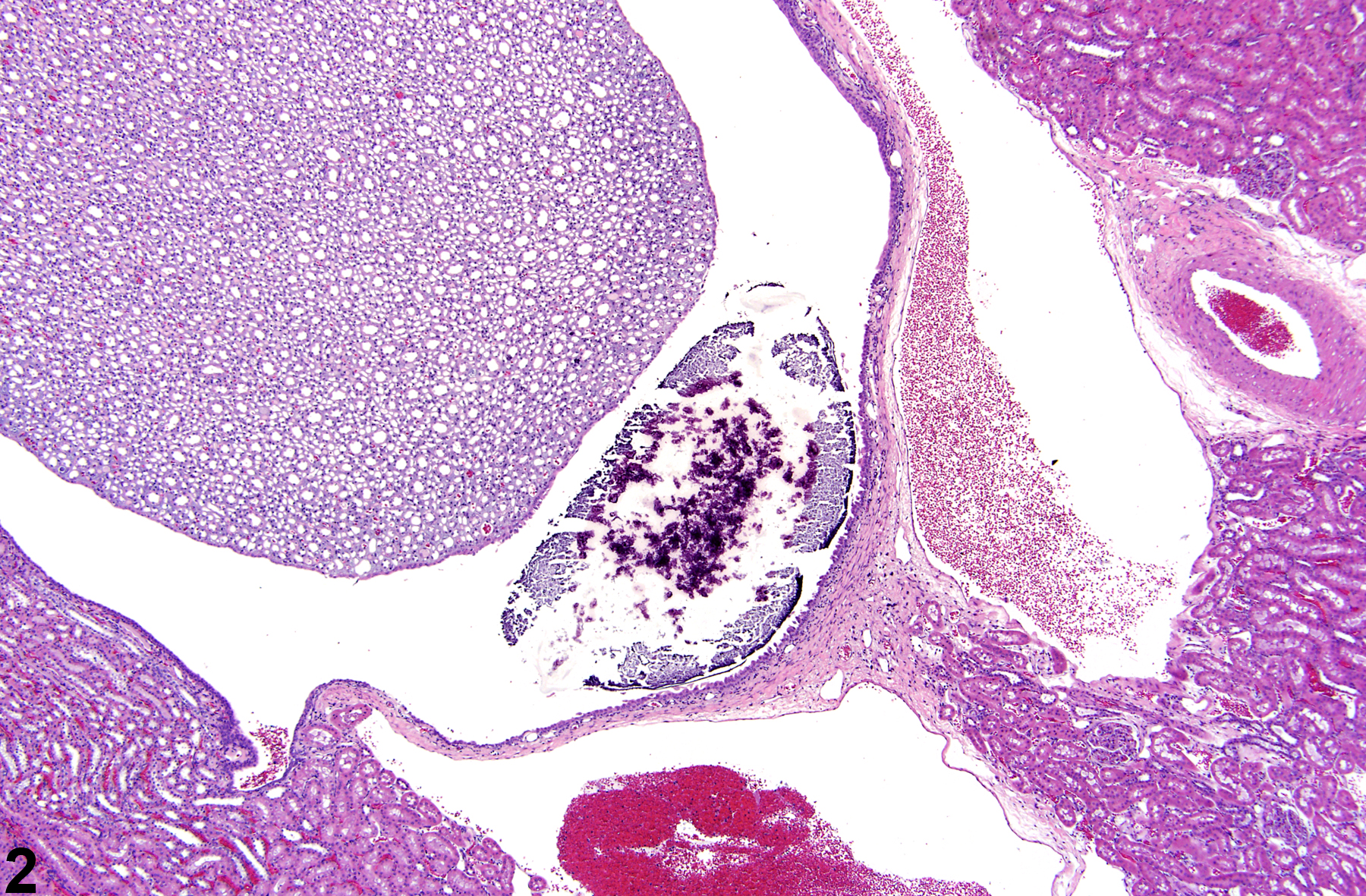
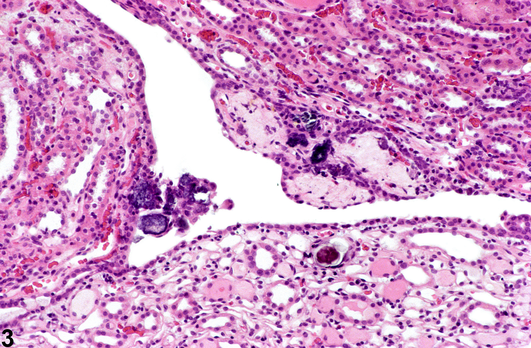
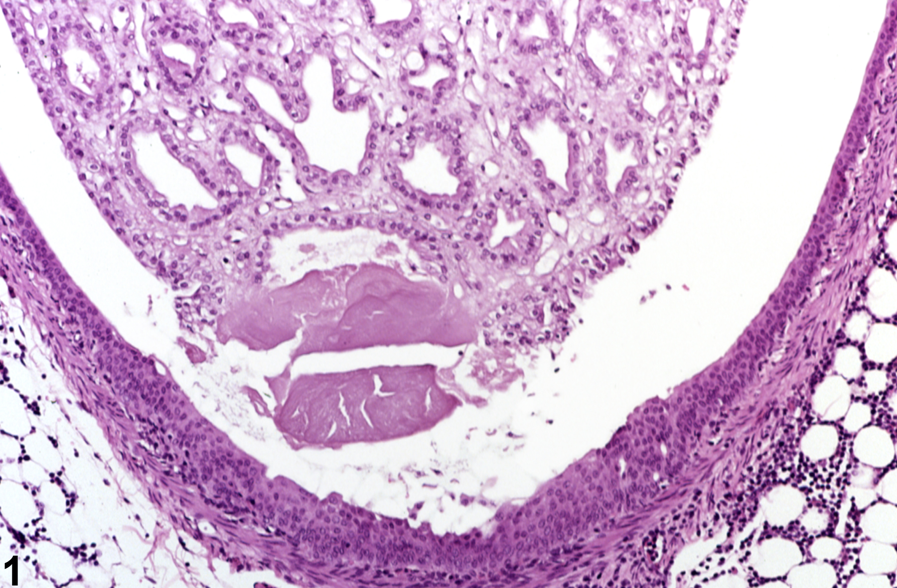
U.S. flag

An official website of the United States government

Dot gov

The .gov means it's official.
Federal government websites often end in .gov or .mil. Before sharing sensitive information, make sure you're on a federal government site.

Https

The site is secure.
The https:// ensures that you are connecting to the official website and that any information you provide is encrypted and transmitted securely.

Skip to Main Navigation
Skip to Page Content
Skip to Atlas Navigation

Kidney - Calculus

Image of calculus in the kidney from a female F344/N rat in a chronic study
Kidney - Calculus in a female F344/N rat from a chronic study. A small calculus is present near the tip of the renal papilla.
Figure 1 of 3
Image of calculus in the kidney from a male Harlan Sprague-Dawley rat in a lifetime study
Kidney - Calculus in a rat. A calculus is present within the renal pelvis.
Figure 2 of 3
Image of calculus in the kidney from a female F344/N rat in a chronic study
Kidney - Calculus in a male F344/N rat. Small calculi are present in the renal fornix.
Figure 3 of 3
next arrow

comment:

Spontaneous cases of calculi are sporadically seen in the renal pelvis of rodents. Calculi may also be associated with xenobiotic administration and may even contain the administered chemical or compound. Calculi tend to be basophilic concretions of variable size, which may result in inflammation, obstruction, or urothelial hyperplasia ( Figure 1image opens in a pop-up window and Figure 2image opens in a pop-up window ). Calculi can be differentiated from mineralization in that calculi are generally free within the urinary space whereas mineralization occurs in tissue. Small calculi are often observed in the renal fornices, where they often do not result in any secondary changes ( Figure 3image opens in a pop-up window ).

recommendation:

Calculi should be diagnosed and given a severity grade. The pathologist shoulduse his or her judgment in deciding if small calculi in the renal pelvis or pelvic fornices without other secondary changes are prominent enough to warrant a diagnosis, or if changes secondary to the calculi, such as inflammation or epithelial hyperplasia, are prominent enough to warrant a separate diagnosis.

references:

Montgomery CA, Seely JC. 1990. Kidney. In: Pathology of the Fischer Rat: Reference and Atlas (Boorman GA, Eustis SL, Elwell MR, Montgomery CA, MacKenzie WF, eds). Academic Press, San Diego, 127-153.
Abstract: http://www.ncbi.nlm.nih.gov/nlmcatalog/9002563

Seely JC. 1999. Kidney. In: Pathology of the Mouse: Reference and Atlas (Maronpot RR, Boorman GA, Gaul BW, eds). Cache River Press, Vienna, IL, 207-234.
Abstract: http://www.cacheriverpress.com/books/pathmouse.htm