

Urinary System
Kidney - Crystals
Narrative
Frazier KS, Seely JC, Hard GC, Betton G, Burnett R, Nakatsuji S, Nishikawa A, Durchfeld-Meyer B, Bube A. 2012. Proliferative and non-proliferative lesions in the rat and mouse urinary system. Toxicol Pathol 40:14S-86S.
Abstract: http://www.ncbi.nlm.nih.gov/pubmed/22637735Hagiwara A, Asakawa E, Kurata Y, Sano M, Hirose M, Ito N. 1992. Dose-dependent renal tubular toxicity of harman and norharman in male F344 rats. Toxicol Pathol 20:197-204.
Abstract: http://www.ncbi.nlm.nih.gov/pubmed/1475580Yarlagadda SG, Perazella MA. 2008. Drug-induced crystal nephropathy: An update. Expert Opin Drug Safety 7:147-158.
Abstract: http://www.ncbi.nlm.nih.gov/pubmed/18324877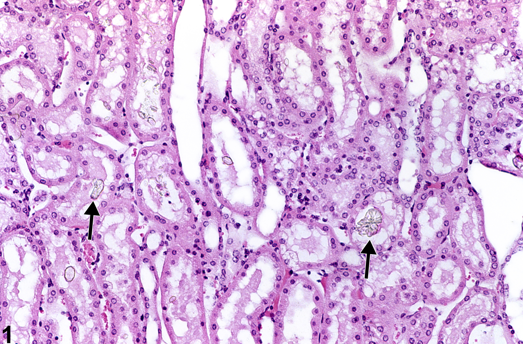
Kidney, Renal tubule - Crystals in a male B6C3F1 mouse from a chronic study. There are crystals (arrows) within tubule lumens.
All Images

Kidney, Renal tubule - Crystals in a male B6C3F1 mouse from a chronic study. There are crystals (arrows) within tubule lumens.

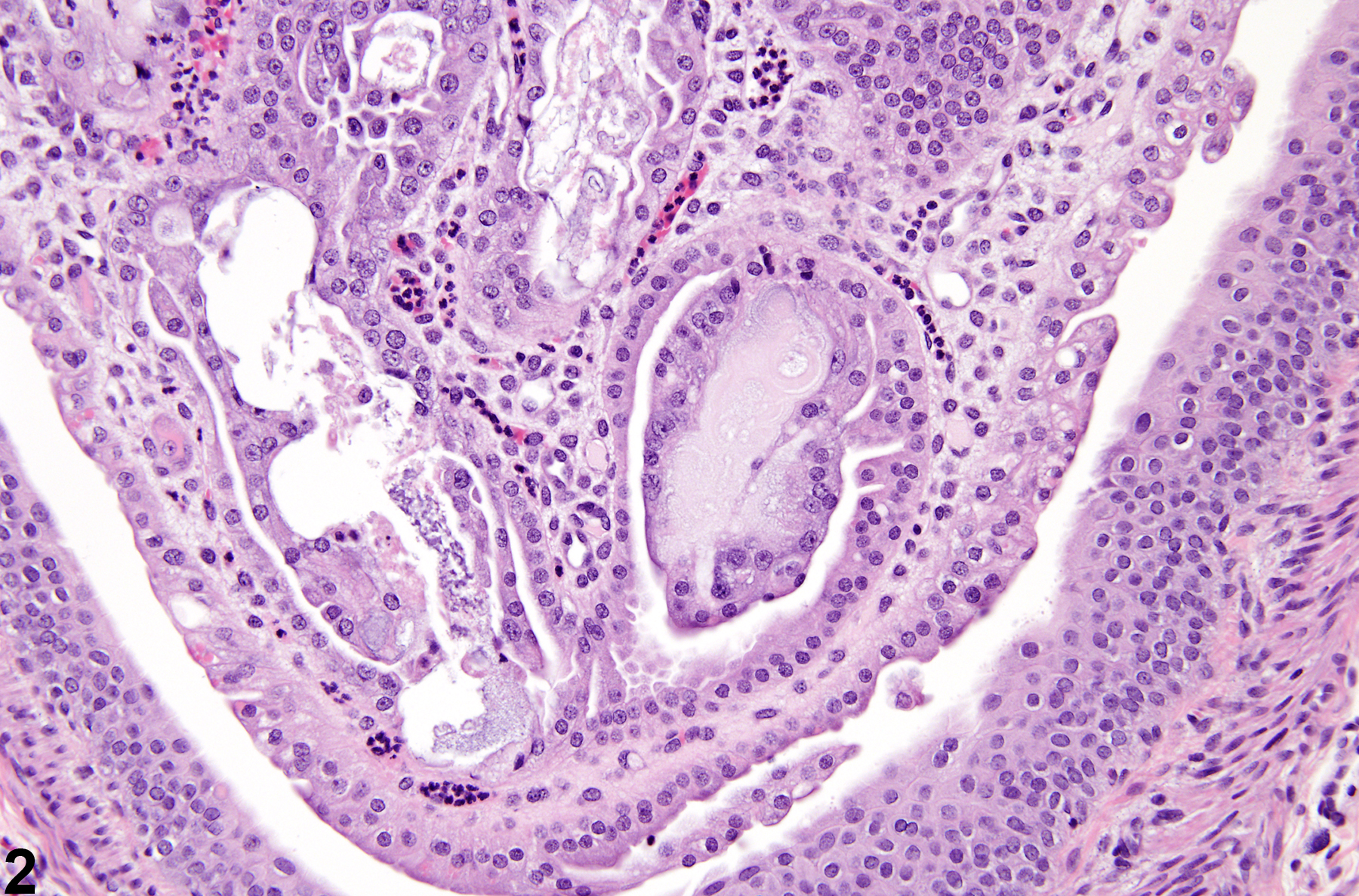
Kidney, Renal tubule - Crystals in a male rat from an acute study. Crystal deposition with secondary inflammation is present in the renal papilla.

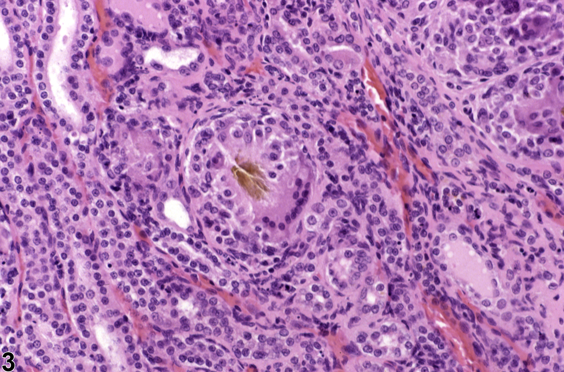
Kidney, Renal tubule - Crystals in a female F344/N rat from a chronic study. A pigmented crystal is surrounded by multinucleated giant cells.