U.S. flag

An official website of the United States government

Dot gov

The .gov means it's official.
Federal government websites often end in .gov or .mil. Before sharing sensitive information, make sure you're on a federal government site.

Https

The site is secure.
The https:// ensures that you are connecting to the official website and that any information you provide is encrypted and transmitted securely.

Skip to Main Navigation
Skip to Page Content
Skip to Atlas Navigation

Kidney - Cyst

Image of cyst in the kidney from a male Tg.Ac (FVB/N) hemizygous mouse in a subchronic study
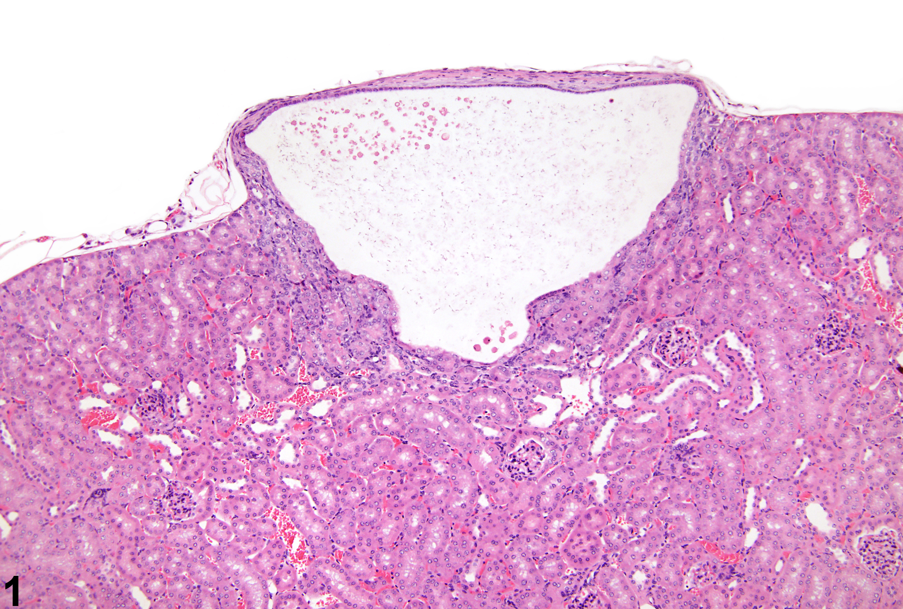
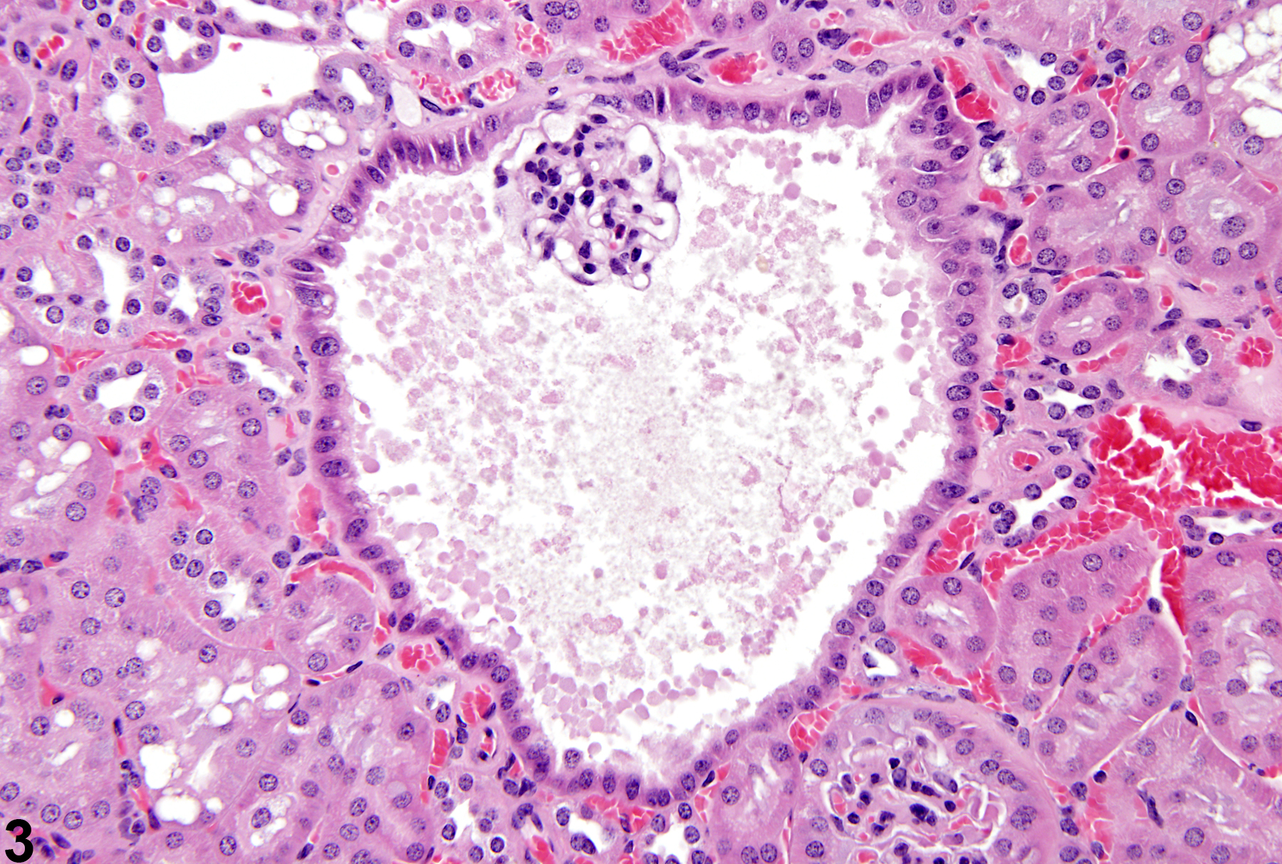
Kidney, Renal tubule - Cyst in a male Tg.Ac (FVB/N) hemizygous mouse from a subchronic study. A spontaneous cortical cyst lined by flattened cells is present.
Figure 1 of 5
Image of cyst in the kidney from a male Tg.Ac (FVB/N) hemizygous mouse in a subchronic study
Kidney, Renal tubule - Cyst in a male Tg.Ac (FVB/N) hemizygous mouse from a subchronic study. A small tubule cyst is lined by cuboidal cells.
Figure 2 of 5
Image of cyst in the kidney from a male B6C3F1 mouse in a chronic study
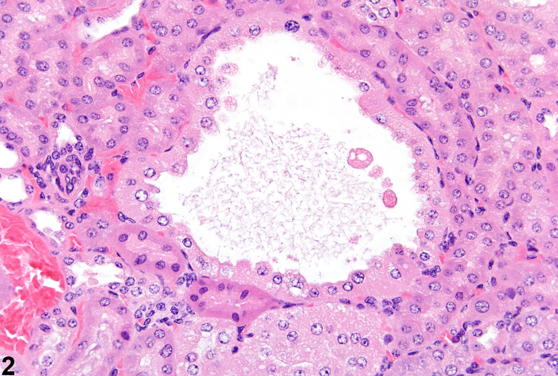
Kidney, Glomerulus - Cyst in a male B6C3F1 mouse from a chronic study. A glomerular cyst is present, with the glomerular tuft visible.
Figure 3 of 5
Image of cyst in the kidney from a  F344/N rat
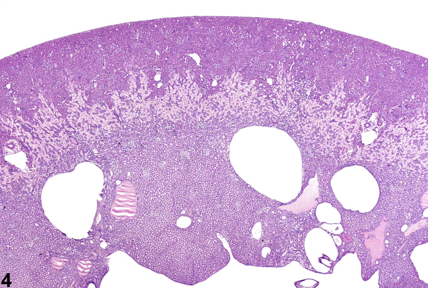
Kidney, Renal tubule - Cyst in an F344/N rat. These medullary cysts are the result of a toxic effect on the kidney.
Figure 4 of 5
Image of cyst in the kidney from a rat
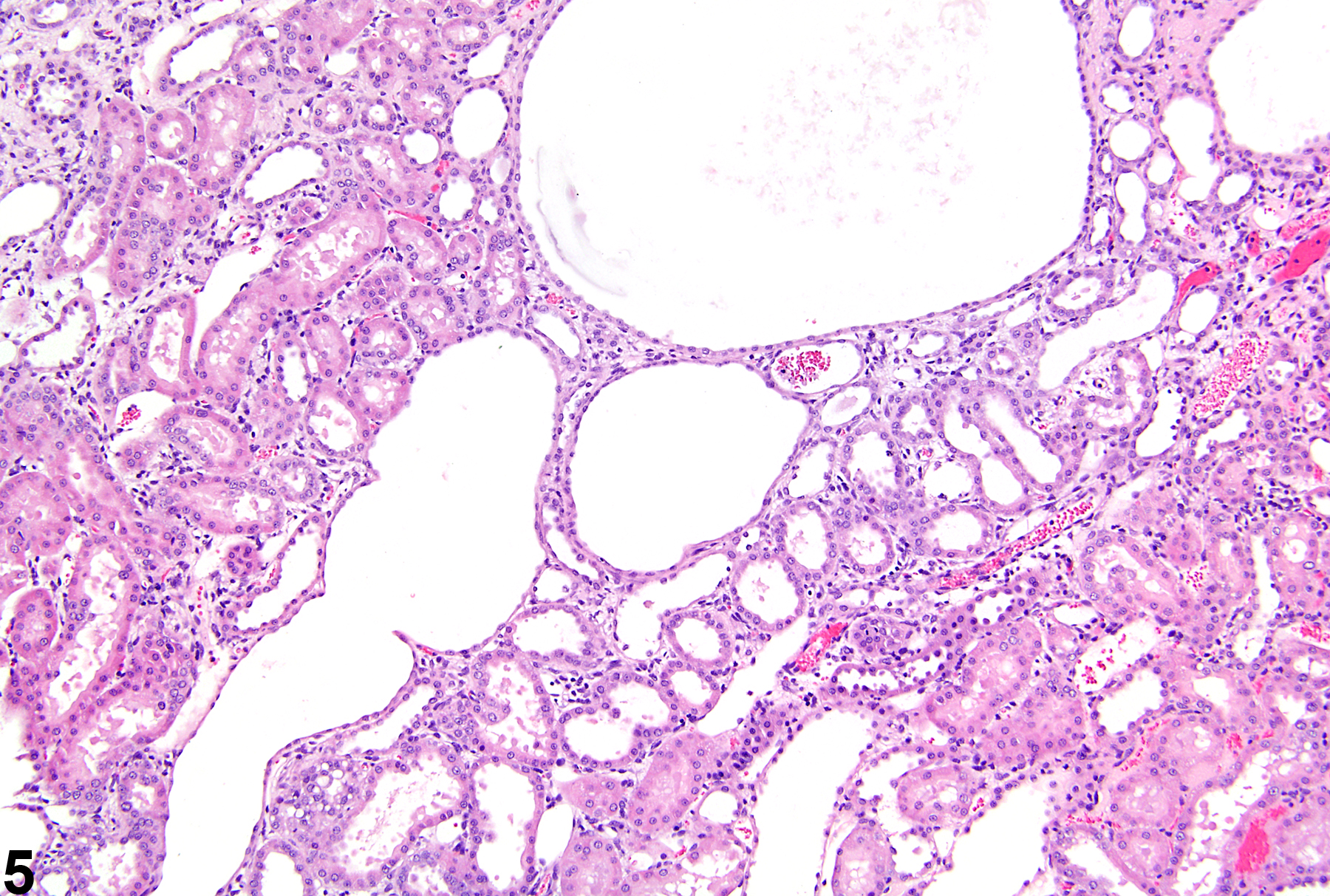
Kidney, Renal tubule - Cyst in a rat. Multiple cysts are present in this polycystic kidney.
Figure 5 of 5
next arrow

comment:

A variety of renal cysts can be seen in rodent kidneys. Cysts may be congenital, acquired, or associated with chronic progressive nephropathy or chemical administration and may be noted as isolated findings in the absence of other renal pathology ( Figure 1image opens in a pop-up window and Figure 2image opens in a pop-up window ). They can be solitary or multiple and can occur anywhere in the renal parenchyma, cortex, medulla, or papilla. Cysts can arise from tubules or glomeruli ( Figure 3image opens in a pop-up window ). Some cysts may arise from progressive dilation, whereas others have been postulated to arise from tubules undergoing epithelial cell proliferation. Most cysts are lined by flattened to cuboidal epithelium and appear empty or may contain eosinophilic or amphophilic homogeneous fluid. Larger cysts may be associated with pericystic and/or tubulointerstitial inflammation and fibrosis. Cysts may arise from chemical administration ( Figure 4image opens in a pop-up window ). Hereditary conditions manifesting as polycystic kidneys have been reported in rats and mice ( Figure 5image opens in a pop-up window ).

recommendation:

Primary renal cysts should be diagnosed. Their location should be indicated in the diagnosis with a site modifier (e.g., renal tubule, glomerulus). Cysts need not be given a severity grade unless grading would highlight a toxic effect. Cysts arising as secondary changes associated with a primary lesion such as with increasing severities of chronic progressive nephropathy, interstitial fibrosis, and/or retrograde nephropathy need not be recorded separately. However, the presence of cysts arising as a secondary mechanism needs to be carefully interpreted and diagnosed when appropriate with respect to chemical administration. Inflammation associated with renal cysts should be recorded if warranted by severity or if the pathologist deems the inflammatory reaction important to the overall understanding of the cystic change.

references:

Cowley BD, Gudapaty S, Kraybill AL, Barish BD, Harding MA, Calvet JP, Gattone VH. 1993. Autosomal-dominant polycystic kidney disease in the rat. Kidney Int 43:522-534.
Abstract: http://www.ncbi.nlm.nih.gov/pubmed/8455352

Flaherty L, Bryda EC, Collins D, Rudofsky U, Montgomery JC. 1995. New mouse model for polycystic kidney disease with both recessive and dominant gene effects. Kidney Int 47:552-558.
Abstract: http://www.ncbi.nlm.nih.gov/pubmed/7723240

Kai K, Sato N, Watanabe A, Shiraiwa K, Ogawa S, Kobayashi Y. 2001. Polycystic disease of the kidney and liver in Crj:CD (SD) rats. J Toxicol Pathol 14:51-55.
Full Text: https://www.jstage.jst.go.jp/article/tox/14/1/14_1_51/_pdf

Nakazawa T, Kasahara K, Ikezaki S, Yamaguchi Y, Edamoto H, Nishimura N, Yahata M, Tamura K, Kamata E, Ema M, Hasegawa R. 2009. Renal tubular cyst formation in newborn rats treated with p- cumylphenol. J Toxicol Pathol 22:125-131.
Abstract: http://www.ncbi.nlm.nih.gov/pubmed/22271985

Ricker JL, Gattone VH, Calvet JP, Rankin CA. 2000. Development of autosomal recessive polycystic kidney disease in BALB/c-cpk/cpk mice. J Am Soc Nephrol 11:1837-1847.
Abstract: http://www.ncbi.nlm.nih.gov/pubmed/11004214