U.S. flag

An official website of the United States government

Dot gov

The .gov means it's official.
Federal government websites often end in .gov or .mil. Before sharing sensitive information, make sure you're on a federal government site.

Https

The site is secure.
The https:// ensures that you are connecting to the official website and that any information you provide is encrypted and transmitted securely.

Skip to Main Navigation
Skip to Page Content
Skip to Atlas Navigation

Kidney - Fibrosis

Image of fibrosis in the kidney from a female F344/N rat in a chronic study
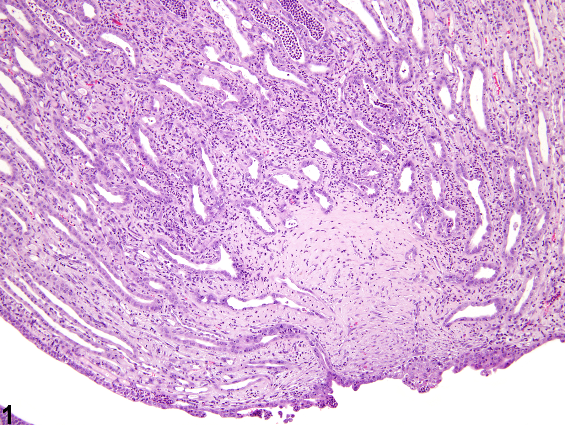
Kidney - Fibrosis in a female F344/N rat from a chronic study. A focal area of fibrosis is present in the distal renal papilla.
Figure 1 of 2
Image of fibrosis in the kidney from a male F344/N rat in a chronic study
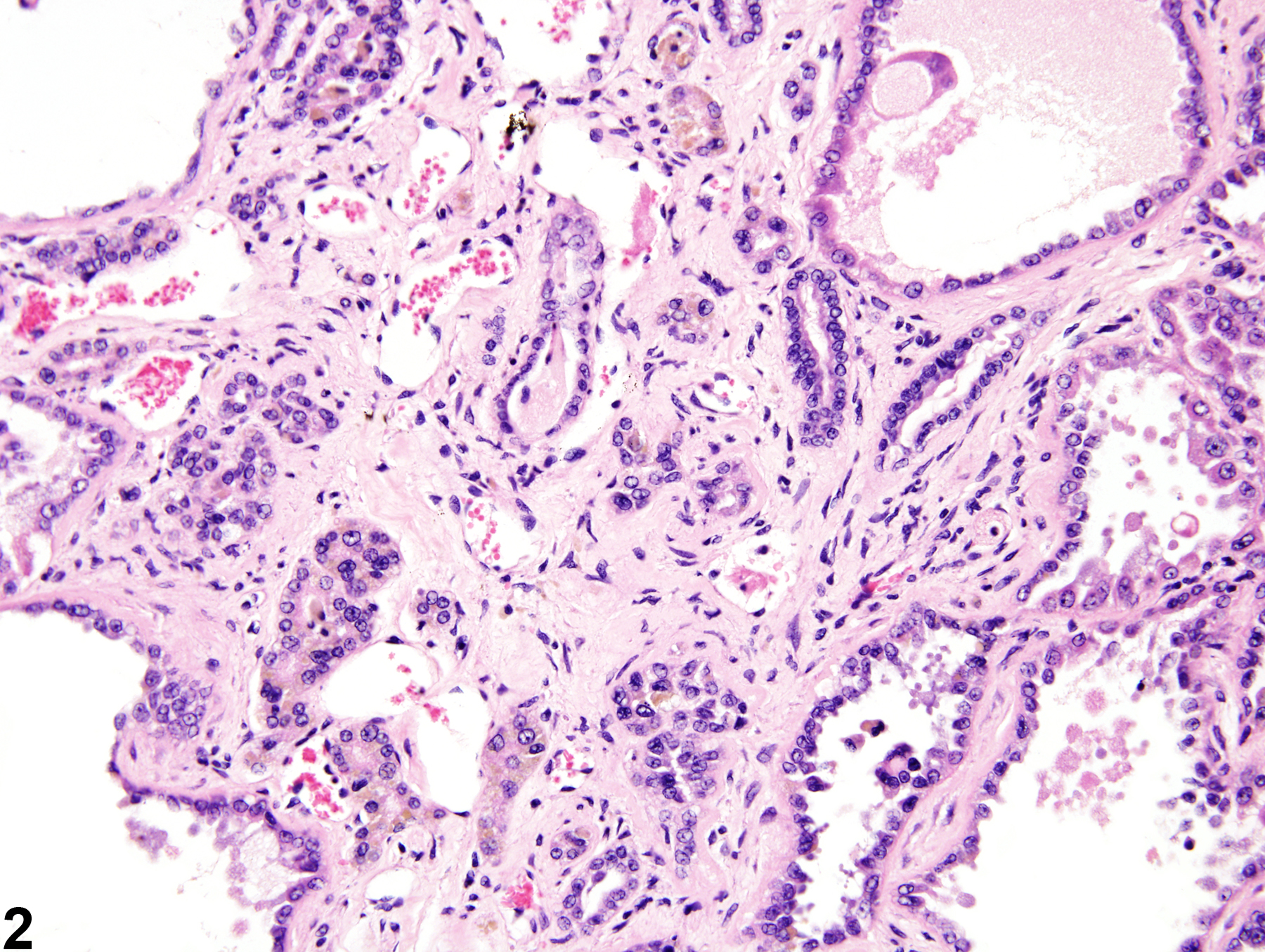
Kidney - Fibrosis in a male F344/N rat from a chronic study. Interstitial fibrosis separates renal tubules.
Figure 2 of 2
next arrow

comment:

Renal (interstitial) fibrosis is associated with previous or ongoing injury to the renal parenchyma,such as that associated with chronic interstitial inflammation. The amount of fibrosis dependson the inciting condition, the amount of damage to the renal parenchyma, and the chronicity of the lesion. Fibrosis results from a complex interaction involving cytokines released by inflammatory cells, thedebatable influence of tubule epithelial-mesenchyme transition, and decreased removal of collagen due to inhibition of collagenolytic enzymes. Fibrosis appears as a slightly fibrillar, eosinophilic material in the interstitium ( Figure 1image opens in a pop-up window and Figure 2image opens in a pop-up window ). Fibrosis may result in progressive atrophy of the nephron in affected areas and/or dilation of renal tubules and Bowman’s space.

recommendation:

If fibrosis is a primary change or is present with little or no inflammation, then it should be diagnosed and given a severity grade. When fibrosis is secondary to another process, such as inflammation, and both are present concurrently, the pathologist should use his or her judgment in deciding whether or not the fibrosis is prominent enough to warrant a separate diagnosis.

references:

Cavallo T. 1998. Tubulointerstitial nephritis. In: Heptinstall’s Pathology of the Kidney, 5th ed (Jennette JC, Olson JL, Schwartz MM, Silva FG, eds). Lippincott-Raven, Philadelphia, 667-723.

Eddy AA. 1996. Molecular insights into renal interstitial fibrosis. J Am Soc Nephrol 7:2495-2508.
Abstract: http://www.ncbi.nlm.nih.gov/pubmed/8989727

Frazier KS, Dube P, Paredes A, Styer E. 2000. Connective tissue growth factor expression in the rat remnant kidney model and association with tubular epithelial cells undergoing transdifferentiation. Vet Pathol 37:328-335.
Full Text: http://vet.sagepub.com/content/37/4/328.full.pdf

Yamate J, Sato K, Machida Y, Ide M, Sato S, Nakatsuji S, Kuwamura M, Kotani T, Sakuma S. 2000. Cisplatin-induced rat renal interstitial fibrosis: A possible pathogenesis based on the data. J Toxicol Pathol 13:237-247.
Full Text: http://tpx.sagepub.com/content/33/2/207.long

Yang J, Liu Y. 2001. Dissection of key events in tubular epithelial to myofibroblast transition and its implications in renal interstitial fibrosis. Am J Pathol 159:1465-1475.
Full Text: http://www.ncbi.nlm.nih.gov/pmc/articles/PMC1850509/