U.S. flag

An official website of the United States government

Dot gov

The .gov means it's official.
Federal government websites often end in .gov or .mil. Before sharing sensitive information, make sure you're on a federal government site.

Https

The site is secure.
The https:// ensures that you are connecting to the official website and that any information you provide is encrypted and transmitted securely.

Skip to Main Navigation
Skip to Page Content
Skip to Atlas Navigation

Kidney - Infarct

Image of infarct in the kidney from a female F344/N rat in a chronic study
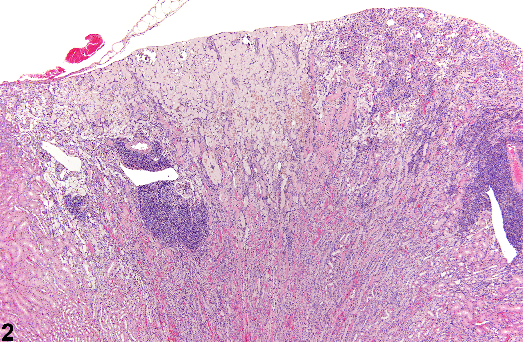
Kidney - Infarct in a female F344/N rat from a chronic study. A wedged-shaped cortical area of acute coagulative necrosis is present.
Figure 1 of 3
Image of infarct in the kidney from a female B6C3F1 mouse in a chronic study
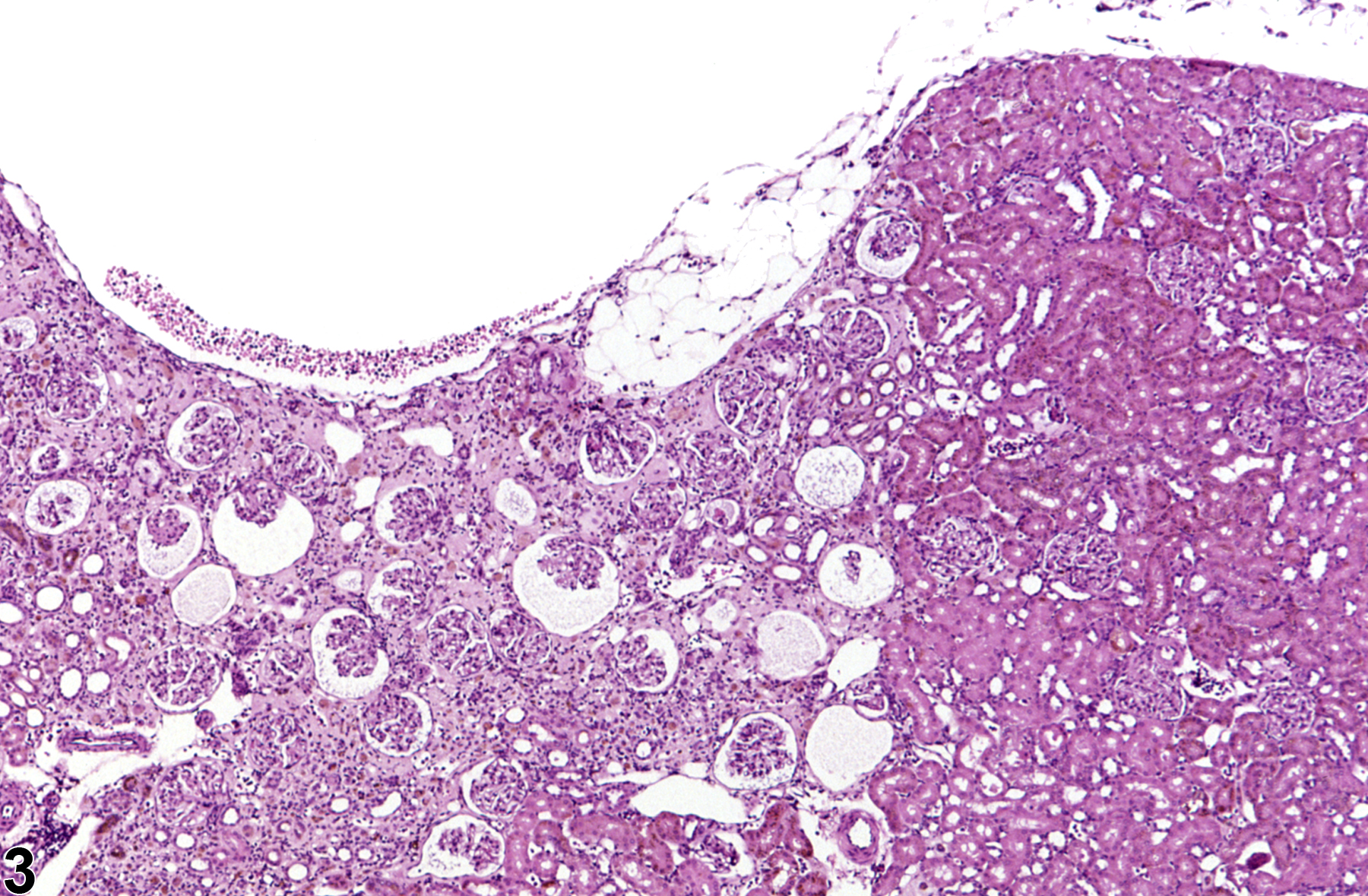
Kidney - Infarct in a female B6C3F1 mouse from a chronic study. This infarct has a prominent area of basophilic inflammatory cellularity associated with an infarcted area.
Figure 2 of 3
Image of infarct in the kidney from a female B6C3F1 mouse in a chronic study
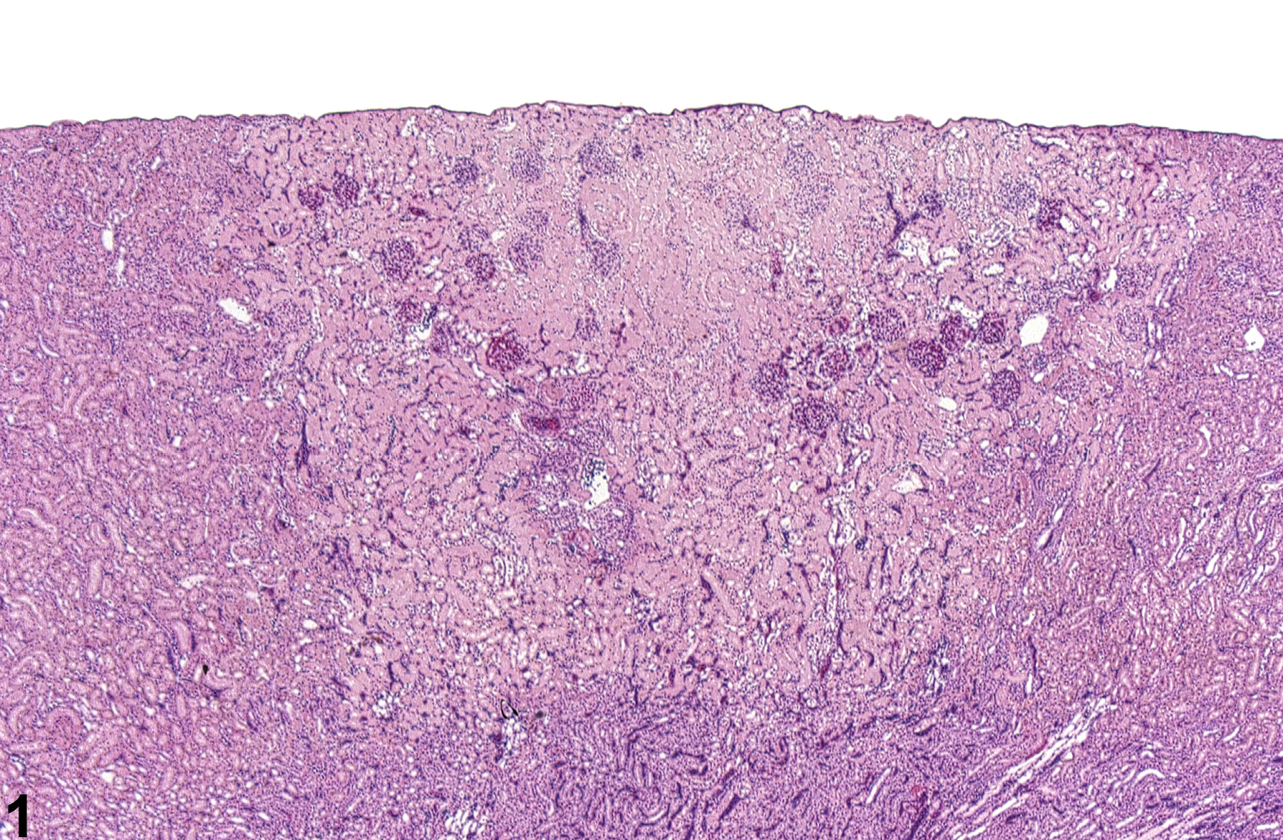
Kidney - Infarct in a female B6C3F1 mouse from a chronic study. This chronic or healed infarct is characterized by cortical depression and interstitial fibrosis.
Figure 3 of 3
next arrow

comment:

Renal infarcts usually appear as well-demarcated, wedge-shaped or triangular areas of coagulative necrosis that extend from the capsular surface into the medulla. The characteristic shape results from the kidney’s unique vascular supply. Infarcts can arise spontaneously from a number of causes that compromise the vascular supply, such as neoplastic infiltrates or nephrotoxicants. Acute infarcts have a central area of necrosis with little peripheral inflammation ( Figure 1image opens in a pop-up window ). As the lesion progresses, neutrophilic to mononuclear infiltrates become evident near the area of necrosis ( Figure 2image opens in a pop-up window ). Chronic infarcts are characterized by renal parenchymal interstitial fibrosis and capsule surface depression ( Figure 3image opens in a pop-up window ). However, ascribing an infarct to all areas with cortical depression and fibrosis may not be warranted since some might represent areas of previous interstitial inflammation that are resolving.

recommendation:

Infarcts should be diagnosed as infarcts rather than diagnosing the individual components (e.g., necrosis, inflammation, fibrosis). They should be graded according to the extent of renal involvement. In the pathology narrative, infarcts should be designated as either acute or chronic, and if possible, the cause of the infarct should be identified.

references:

Hard GC, Alden CL, Bruner RH, Frith CH, Lewis RM, Owen RA, Krieg K, Durchfeld-Meyer B. 1999. Non-proliferative lesions of the kidney and lower urinary tract in rats. In: Guides for Toxicologic Pathology. STP/ARP/AFIP, Washington, DC, 1-32.

Montgomery CA, Seely JC. 1990. Kidney. In: Pathology of the Fischer Rat: Reference and Atlas (Boorman GA, Eustis SL, Elwell MR, Montgomery CA, MacKenzie WF, eds). Academic Press, San Diego, 127-153.
Abstract: http://www.ncbi.nlm.nih.gov/nlmcatalog/9002563

Seely JC. 1999. Kidney. In: Pathology of the Mouse: Reference and Atlas (Maronpot RR, Boorman GA, Gaul BW, eds). Cache River Press, Vienna, IL, 207-234.
Abstract: http://www.cacheriverpress.com/books/pathmouse.htm